Infundibular Widening of Angiographically Invisible Duplicate Anterior Choroidal Artery Mimicking Typical Anterior Choroidal Artery Aneurysm
- Affiliations
-
- 1Department of Neurosurgery, Kyungpook National University, Daegu, Korea
- 2Department of Neurosurgery, Samsung Medical Center, Sungkyunkwan University, Seoul, Korea
- KMID: 2537854
- DOI: http://doi.org/10.3340/jkns.2022.0046
Abstract
- A diagnosis of an intracranial aneurysm depends on the angiographic configuration and should be cautiously differentiated from aneurysm mimics. In cases of duplicate anterior choroidal arteries (AChAs), infundibular widening of the distal minor AChA can be an aneurysm mimic. If the minor AChA with a smaller diameter is obscured angiographically due to poor contrast filling, an associated infundibular widening beside the proximal large AChA can misinterpreted as a typical AChA aneurysm in angiograms. The authors report on two such cases of duplicate AChAs with infundibular widening presenting like a typical AChA aneurysm in angiograms. Surgical exploration revealed a perforating artery emitting from the dome of the saccular lesion, confirming infundibular widening of a duplicate AChA. No reparative procedure was applied to the infundibular widening in a 48-year-old man, while two vascular outpouchings from the infundibular widening were clipped preserving the duplicate AChA in a 55-year-old woman.
Keyword
Figure
-
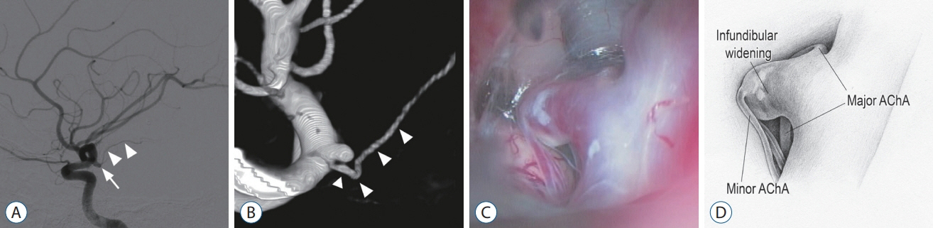
Fig. 1. case 1. A : Two-dimensional digital subtraction angiography (2d-dSA) image suggesting a saccular aneurysm (arrow) arising from the internal carotid artery (IcA) at the origin of the anterior choroidal artery (AchA) (arrowheads). b : Volume-rendered three-dimensional (3d) rotational angiography image showing the origin of the AchA (arrowheads) at the base of the saccular lesion. c : Intraoperative photograph revealing a minor AchA emitting from infundibular widening with the major AchA at its base. d : Illustration corresponding to panel (c).
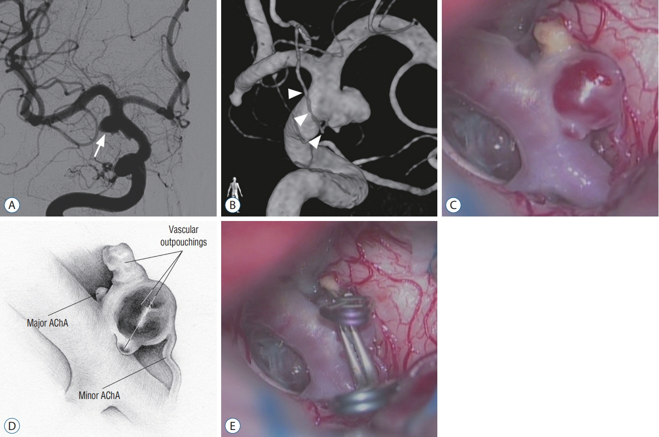
Fig. 2. case 2. A : Two-dimensional digital subtraction angiography image showing an anterior choroidal artery (AchA) aneurysm (arrow). b : Volumerendered 3-dimensional rotational angiography image showing the origin of the AchA (arrowheads) at the proximal base of the saccular lesion. c : Intraoperative photograph revealing a minor AchA emitting from the dome of the saccular lesion with the major AchA at its base. Vascular outpouchings are seen in two areas. d : Illustration corresponding to panel (c). e : Intraoperative photograph showing the aneurysm clips repairing the aneurysmal changes.
Reference
-
References
1. Chen CJ, Moosa S, Ding D, Raper DM, Burke RM, Lee CC, et al. Infundibular dilations of the posterior communicating arteries: pathogenesis, anatomical variants, aneurysm formation, and subarachnoid hemorrhage. J Neurointerv Surg. 8:791–795. 2016.2. Cho MS, Kim MS, Chang CH, Kim SW, Kim SH, Choi BY. Analysis of clipinduced ischemic complication of anterior choroidal artery aneurysms. J Korean Neurosurg Soc. 43:131–134. 2008.3. Chung J, Park W, Hong SH, Park JC, Ahn JS, Kwun BD, et al. Intraoperative use of transcranial motor/sensory evoked potential monitoring in the clipping of intracranial aneurysms: evaluation of false-positive and false-negative cases. J Neurosurg. 130:936–948. 2018.4. Endo S, Furuichi S, Takaba M, Hirashima Y, Nishijima M, Takaku A. Clinical study of enlarged infundibular dilation of the origin of the posterior communicating artery. J Neurosurg. 83:421–425. 1995.5. Epstein F, Ransohoff J, Budzilovich GN. The clinical significance of junctional dilatation of the posterior communicating artery. J Neurosurg. 33:529–531. 1970.6. Flamm ES. Other aneurysms of the internal carotid artery in Wilkins RH, Rengachary SS (eds). Neurosurgery. New York: McGraw-Hill;1996. p. 2301.7. Friedman JA, Pichelmann MA, Piepgras DG, Atkinson JL, Maher CO, Meyer FB, et al. Ischemic complications of surgery for anterior choroidal artery aneurysms. J Neurosurg. 94:565–572. 2001.8. Furtado SV, Venkatesh PK, Hegde AS. Neurological complications and surgical outcome in patients with anterior choroidal segment aneurysms. Int J Neurosci. 120:291–297. 2010.9. Irie T, Yoshitani K, Ohnishi Y, Shinzawa M, Miura N, Kusaka Y, et al. The efficacy of motor-evoked potentials on cerebral aneurysm surgery and new-onset postoperative motor deficits. J Neurosurg Anesthesiol. 22:247–251. 2010.10. Ito A, Sato K, Niizuma K, Endo H, Matsumoto Y, Tominaga T. Intraoperative motor-evoked potential monitoring during coil embolization for anterior choroidal artery aneurysms. Neuroradiology. 64:1221–1229. 2022.11. Kang HS, Kwon BJ, Kwon OK, Jung C, Kim JE, Oh CW, et al. Endovascular coil embolization of anterior choroidal artery aneurysms. Clinical article. J Neurosurg. 111:963–969. 2009.12. Kim BM, Kim DI, Chung EC, Kim SY, Shin YS, Park SI, et al. Endovascular coil embolization for anterior choroidal artery aneurysms. Neuroradiology. 50:251–257. 2008.13. Kim BM, Kim DI, Shin YS, Chung EC, Kim DJ, Suh SH, et al. Clinical outcome and ischemic complication after treatment of anterior choroidal artery aneurysm: comparison between surgical clipping and endovascular coiling. AJNR Am J Neuroradiol. 29:286–290. 2008.14. Koike G, Seguchi K, Kyoshima K, Kobayashi S. Subarachnoid hemorrhage due to rupture of infundibular dilation of a circumflex branch of the posterior cerebral artery: case report. Neurosurgery. 34:1075–1077. 1994.15. Kucukay F, Okten RS, Tekiner A, Dagli M, Gocek C, Bayar MA, et al. Three-dimensional volume rendering digital subtraction angiography in comparison with two-dimensional digital subtraction angiography and rotational angiography for detecting aneurysms and their morphological properties in patients with subarachnoid hemorrhage. Eur J Radiol. 81:2794–2800. 2012.16. Lee YS, Park J. Anterior choroidal artery aneurysm surgery: ischemic complications and clinical outcomes revisited. J Korean Neurosurg Soc. 54:86–92. 2013.17. Li J, Mukherjee R, Lan Z, Liu Y, He M. Microneurosurgical management of anterior choroidal artery aneurysms: a 16-year institutional experience of 102 patients. Neurol Res. 34:272–280. 2012.18. Morris P. Practical Neuroangiography. ed 2. Philadelphia: Lippincott Williams & Wilkins;2007. p. p338–339.19. Nakagawa I, Park H, Kotsugi M, Motoyama Y, Myochin K, Takeshima Y, et al. Diagnostic impact of monitoring transcranial motor-evoked potentials to prevent ischemic complications during endovascular treatment for intracranial aneurysms. Neurosurg Rev. 44:1493–1501. 2021.20. Osborn AG. Diagnostic Cerebral Angiography. ed 2. Philadelphia: Lippincott Williams & Wilkins;1999. p. 272–273.21. Papke K, Kuhl CK, Fruth M, Haupt C, Schlunz-Hendann M, Sauner D, et al. Intracranial aneurysms: role of multidetector CT angiography in diagnosis and endovascular therapy planning. Radiology. 244:532–540. 2007.22. Park J, Kang DH. Infundibular widening mimicking anterior communicating artery aneurysm: report of 2 cases. J Neurosurg. 119:243–246. 2013.23. Piotin M, Mounayer C, Spelle L, Williams MT, Moret J. Endovascular treatment of anterior choroidal artery aneurysms. AJNR Am J Neuroradiol. 25:314–318. 2004.24. Saltzman GF. Infundibular widening of the posterior communicating artery studied by carotid angiography. Acta Radiol. 51:415–421. 1959.25. Senturk C, Bandeira A, Bruneau M, Dewindt A, Balériaux D, De Witte O, et al. Endovascular treatment of anterior choroidal artery aneurysms. J Neuroradiol. 36:228–232. 2009.26. Shi WY, Li YD, Li MH, Gu BX, Gu JP. Differential diagnosis of infundibular dilation versus a small aneurysm of the internal carotid artery: assessment by three-dimensional rotational angiography with volume rendering. Neurol Sci. 34:1065–1070. 2013.27. Song J, Lang L, Zhu W, Gu Y, Xu B, Cai J, et al. Application of intraoperative motor evoked potential monitoring during giant internal carotid artery aneurysm surgery using prolonged temporary occlusion. Acta Neurochir (Wien). 157:1833–1840. 2015.28. Suzuki K, Kodama N, Sasaki T, Matsumoto M, Konno Y, Sakuma J, et al. Intraoperative monitoring of blood flow insufficiency in the anterior choroidal artery during aneurysm surgery. J Neurosurg. 98:507–514. 2003.29. Trasi S, Vincent LM, Zingesser LH. Development of aneurysm from infundibulum of posterior communicating artery with documentation of prior hemorrhage. AJNR Am J Neuroradiol. 2:368–370. 1981.30. Wollschlaeger G, Wollschlaegerr PB. The circle of Willis in Newton TH, Potts DG (eds). Radiology of the Skull and Brain. St. Louis: CV Mosby;1974. Vol 2. p. 1171–1201.31. Yasargil MG. Clinical considerations, surgery of intracranial aneurysms and results. New York: Thieme;1984. p. 99–108.32. Yasargil MG, Yonas H, Gasser JC. Anterior choroidal artery aneurysms: their anatomy and surgical significance. Surg Neurol. 9:129–138. 1978.33. Yeon JY, Seo DW, Hong SC, Kim JS. Transcranial motor evoked potential monitoring during the surgical clipping of unruptured intracranial aneurysms. J Neurol Sci. 293:29–34. 2010.
- Full Text Links
-
- Actions
-
Cited
- CITED
-
- Close
- Share
-
- Similar articles
-
- Transposition of Anterior Choroidal Artery and Posterior Communicating Artery Origin
- Clinical Experiences of Anterior Choroidal Artery Aneurysm
- Oculomotor Nerve Palsy due to Ruptured Multiple Anterior Choroidal Artery Aneurysms
- Blood Blister-Like Aneurysm with Rupture Point Close to Origin of Anterior Choroidal Artery
- Flow recovery after posterior clinoidectomy for surgical clipping of anterior choroidal aneurysm
