Anat Cell Biol.
2022 Dec;55(4):441-451. 10.5115/acb.22.061.
The impact of age on the morphology of the 12th thoracic vertebral endplates
- Affiliations
-
- 1Department of Animal and Human Physiology, Faculty of Biology, School of Sciences, National and Kapodistrian University of Athens, Athens, Greece
- 2Section of Forensic Pathology, Department of Forensic Medicine, University of Copenhagen, Copenhagen, Denmark
- KMID: 2537463
- DOI: http://doi.org/10.5115/acb.22.061
Abstract
- The current article explores the aging effects on the overall morphology of the endplates of the 12th thoracic vertebra (T12), while screening for sex differences. It further evaluates the suitability of T12 for estimating age-at-death in bioarcheaological contexts. We captured the morphology of the vertebral endplates, including the formation of osteophytes, in a novel continuous quantitative manner using digital photography. 168 Greek adults from the Athens Collection were used for modeling the aging effects and another 107 individuals from two Danish archaeological assemblages for evaluation. Regression analysis is based on generalized additive models for correlating age-at-death and morphological variation. Our proposed measurement method is highly reliable (R>0.98) and the main differences observed between sexes are size related. Aging has considerable effect on the endplate morphology of the T12 with the total area of the endplate, the area of the epiphyseal rim, and the shape irregularities of the endplate’s external boundary being mostly affected. Multivariate regression shows that aging effects account up to 46% of the observed variation, although with differential expression between sexes. Correct age prediction on archaeological remains reached 33% with a prominent tendency for overestimation. The morphology of the T12 endplates is influenced by age and it can provide some insight with respect to the age-at-death of unidentified individuals, especially when other skeletal age markers are unavailable. Our proposed method provides an ageestimation framework for bioarchaeological settings, especially for estimating broader age ranges, such as discriminating between young and old adults.
Keyword
Figure
-
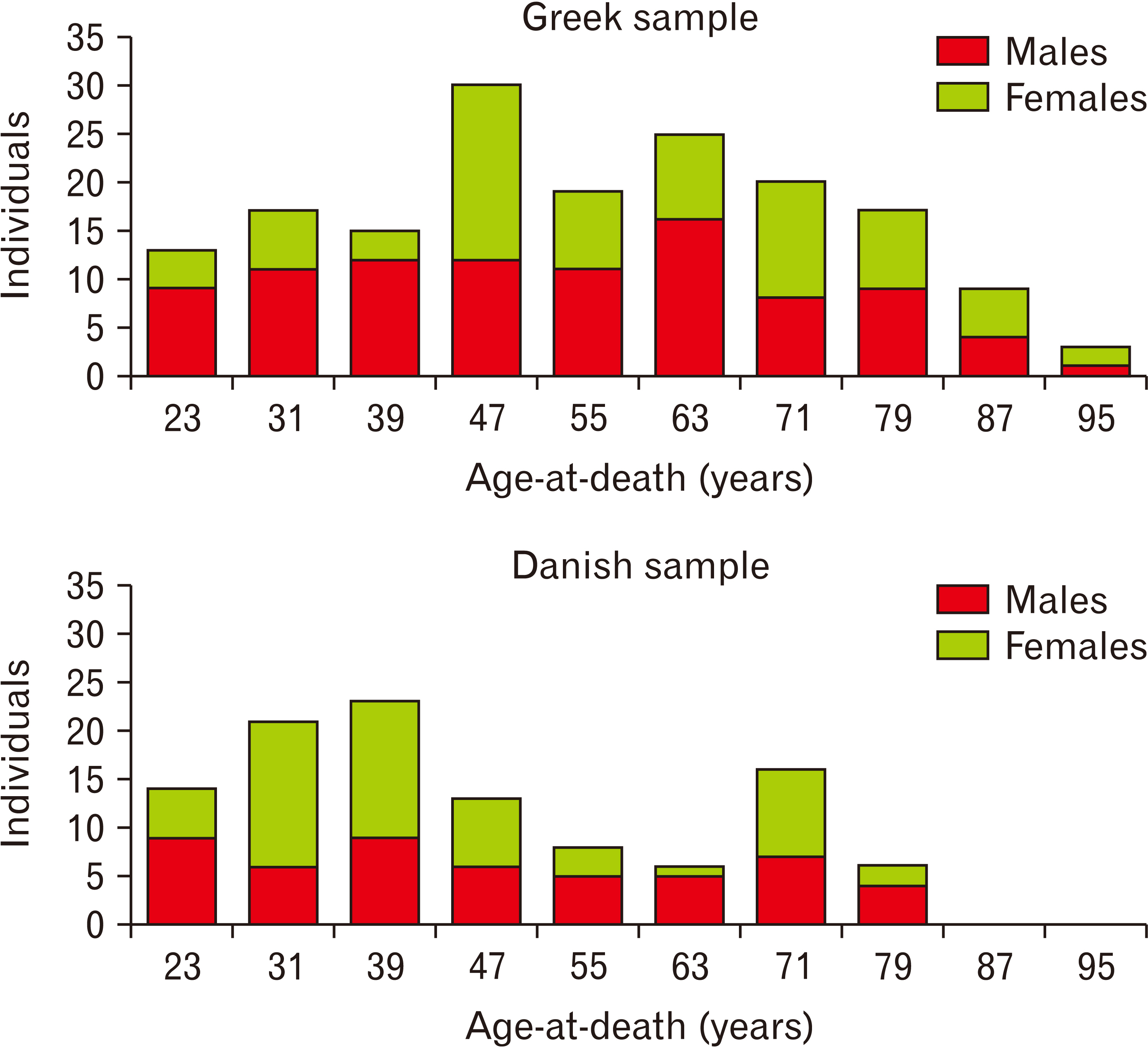
Fig. 1 Age and sex distributions of Greek and Danish samples.
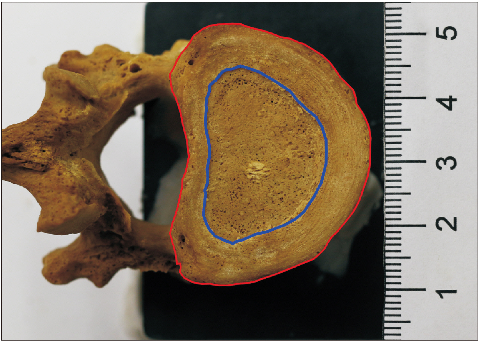
Fig. 2 Polyline tracing of external (red) and internal (blue) endplate boundaries on the endplate’s surface.

Fig. 3 Regression of age against selected morphometric variables with statistically significant models.

Fig. 4 Evaluation of multivariate models on age prediction of the archaeological sample.
Reference
-
References
1. Cunha E, Baccino E, Martrille L, Ramsthaler F, Prieto J, Schuliar Y, Lynnerup N, Cattaneo C. 2009; The problem of aging human remains and living individuals: a review. Forensic Sci Int. 193:1–13. DOI: 10.1016/j.forsciint.2009.09.008. PMID: 19879075.2. İşcan MY, Steyn M. 2013. The human skeleton in forensic medicine. 3rd ed. Charles C Thomas Publisher;Springfield: p. 493. DOI: 10.1016/j.forsciint.2009.09.008.3. Meindl RS, Lovejoy CO. 1985; Ectocranial suture closure: a revised method for the determination of skeletal age at death based on the lateral-anterior sutures. Am J Phys Anthropol. 68:57–66. DOI: 10.1002/ajpa.1330680106. PMID: 4061602.4. Smith SL, Buschang PH. 2004; Variation in longitudinal diaphyseal long bone growth in children three to ten years of age. Am J Hum Biol. 16:648–57. DOI: 10.1002/ajhb.20077. PMID: 15495231.5. Kagerer P, Grupe G. 2001; Age-at-death diagnosis and determination of life-history parameters by incremental lines in human dental cementum as an identification aid. Forensic Sci Int. 118:75–82. DOI: 10.1016/S0379-0738(00)00382-0. PMID: 11343858.6. Lovejoy CO. 1985; Dental wear in the Libben population: its functional pattern and role in the determination of adult skeletal age at death. Am J Phys Anthropol. 68:47–56. DOI: 10.1002/ajpa.1330680105. PMID: 4061601.7. Parra RC, Suárez-Ponce DG, Escalante-Flórez KJ, Condori LA, Calcina-Mendoza O, Peralta-Cerro LM, Rosas-Moyano GA. 2021; Age-at-death estimation in adults and verification of a forensic international methodology using single-rooted teeth: an approach for a Peruvian context. Forensic Sci Int Rep. 3:100176. DOI: 10.1016/j.fsir.2021.100176.8. Buckberry JL, Chamberlain AT. 2002; Age estimation from the auricular surface of the ilium: a revised method. Am J Phys Anthropol. 119:231–9. DOI: 10.1002/ajpa.10130. PMID: 12365035.9. Lovejoy CO, Meindl RS, Pryzbeck TR, Mensforth RP. 1985; Chronological metamorphosis of the auricular surface of the ilium: a new method for the determination of adult skeletal age at death. Am J Phys Anthropol. 68:15–28. DOI: 10.1002/ajpa.1330680103. PMID: 4061599.10. Meindl RS, Lovejoy CO, Mensforth RP, Walker RA. 1985; A revised method of age determination using the os pubis, with a review and tests of accuracy of other current methods of pubic symphyseal aging. Am J Phys Anthropol. 68:29–45. DOI: 10.1002/ajpa.1330680104. PMID: 4061600.11. DiGangi EA, Bethard JD, Kimmerle EH, Konigsberg LW. 2009; A new method for estimating age-at-death from the first rib. Am J Phys Anthropol. 138:164–76. DOI: 10.1002/ajpa.20916. PMID: 18711740.12. İşcan MY, Loth SR, Wright RK. 1984; Metamorphosis at the sternal rib end: a new method to estimate age at death in white males. Am J Phys Anthropol. 65:147–56. DOI: 10.1002/ajpa.1330650206. PMID: 6507605.13. Nikita E. 2013; Quantitative assessment of the sternal rib end morphology and implications for its application in aging human remains. J Forensic Sci. 58:324–9. DOI: 10.1111/1556-4029.12047. PMID: 23278810.14. Milner GR, Boldsen JL, Ousley SD, Getz SM, Weise S, Tarp P. 2022. Transition Analysis 3, version 0.8.4. [Software]. Available from: https://statsmachine.net/software/TA3/. accessed 2022 Mar 15.15. Milner GR, Boldsen JL. 2012; Transition analysis: a validation study with known-age modern American skeletons. Am J Phys Anthropol. 148:98–110. DOI: 10.1002/ajpa.22047. PMID: 22419394.16. Snodgrass JJ. 2004; Sex differences and aging of the vertebral column. J Forensic Sci. 49:458–63. DOI: 10.1520/JFS2003198. PMID: 15171159.17. Stewart TD. 1958. Rate of development of vertebral osteoarthritis in American whites and its significance in skeletal age identification. Leech;Tallahassee: DOI: 10.1520/jfs2003198.18. Watanabe S, Terazawa K. 2006; Age estimation from the degree of osteophyte formation of vertebral columns in Japanese. Leg Med (Tokyo). 8:156–60. DOI: 10.1016/j.legalmed.2006.01.001. PMID: 16621650.19. Van der Merwe AE, Işcan MY, L'Abbè EN. 2006; The pattern of vertebral osteophyte development in a South African population. Int J Osteoarchaeol. 16:459–64. DOI: 10.1002/oa.841.20. Kim DK, Kim MJ, Kim YS, Oh CS, Shin DH. 2012; Vertebral osteophyte of pre-modern Korean skeletons from Joseon tombs. Anat Cell Biol. 45:274–81. DOI: 10.5115/acb.2012.45.4.274. PMID: 23301195. PMCID: PMC3531591.21. Praneatpolgrang S, Prasitwattanaseree S, Mahakkanukrauh P. 2019; Age estimation equations using vertebral osteophyte formation in a Thai population: comparison and modified osteophyte scoring method. Anat Cell Biol. 52:149–160. DOI: 10.5115/acb.2019.52.2.149. PMID: 31338232. PMCID: PMC6624338.22. Prescher A. 1998; Anatomy and pathology of the aging spine. Eur J Radiol. 27:181–95. DOI: 10.1016/S0720-048X(97)00165-4. PMID: 9717634.23. Goh S, Price RI, Song S, Davis S, Singer KP. 2000; Magnetic resonance-based vertebral morphometry of the thoracic spine: age, gender and level-specific influences. Clin Biomech (Bristol, Avon). 15:417–25. DOI: 10.1016/S0268-0033(99)00100-X. PMID: 10771120.24. Rühli FJ, Müntener M, Henneberg M. 2005; Age-dependent changes of the normal human spine during adulthood. Am J Hum Biol. 17:460–9. DOI: 10.1002/ajhb.20403. PMID: 15981187.25. Saadat Mostafavi SR, Memarian A, Motamedi O, Mohamadi nejad khanamani M, Khaleghi M, Habibi S. 2021; Fourth lumbar vertebral parameters in predicting the gender, height and age in Iranian population. Forensic Sci Int Rep. 3:100175. DOI: 10.1016/j.fsir.2021.100175.26. Eliopoulos C, Lagia A, Manolis S. 2007; A modern, documented human skeletal collection from Greece. Homo. 58:221–8. DOI: 10.1016/j.jchb.2006.10.003. PMID: 17574249.27. Buikstra JE, Ubelaker DH. 1994. Standards for data collection from human skeletal remains: proceedings of a Seminar at The Field Museum of Natural History. Arkansas Archeological Survey;Fayetteville: DOI: 10.1016/j.jchb.2006.10.003.28. Perini TA, de Oliveira GL, dos Santos Ornellas J, de Oliveira FP. 2005; Technical error of measurement in anthropometry. Rev Bras Med Esporte. 11:86–90. DOI: 10.1590/S1517-86922005000100009.29. Garoufi N, Bertsatos A, Chovalopoulou ME, Villa C. 2020; Forensic sex estimation using the vertebrae: an evaluation on two European populations. Int J Legal Med. 134:2307–18. DOI: 10.1007/s00414-020-02430-w. PMID: 32940842.30. Prentice A, Schoenmakers I, Laskey MA, de Bono S, Ginty F, Goldberg GR. 2006; Nutrition and bone growth and development. Proc Nutr Soc. 65:348–60. DOI: 10.1079/PNS2006519. PMID: 17181901. PMCID: PMC2039894.31. Ruff C. 2018. Skeletal variation and adaptation in Europeans: upper Paleolithic to the Twentieth Century. John Wiley & Sons;Hoboken: DOI: 10.1002/9781118628430.32. Wood SN. 2020; Inference and computation with generalized additive models and their extensions. TEST. 29:307–39. DOI: 10.1007/s11749-020-00711-5.33. İşcan MY, Kennedy KAR. 1989. Reconstruction of life from the skeleton. Liss;New York: p. 315. DOI: 10.1007/s11749-020-00711-5.34. Junghanns H, Schmorl G. 1971. The human spine in health and disease. 2nd ed. Grune & Stratton;New York: p. 504. DOI: 10.1007/s11749-020-00711-5.
- Full Text Links
-
- Actions
-
Cited
- CITED
-
- Close
- Share
-
- Similar articles
-
- A Study on the Vertebral Body and Intervertebral Disk Indices of the Normal Korean People (A Preliminary Report)
- Analysis of the Cement Distribution Pattern and Other Risk Factors that Affect the Incidence of Recompression Fractures of Vertebral Bodies after Vertebroplasty or Kyphoplasty
- The Factors between the Progression of the Compression Rate and Magnetic Resonance Imaging Findings in Osteoporotic Vertebral Fracture Patients Treated with Teriparatide
- Effect of Bone Mineral Density and Endplate Thickness on the Compressive Strength in the Cervical Spine
- Variation of canine vertebral bone architecture in computed tomography
