Peroxisomal Fitness: A Potential Protective Mechanism of Fenofibrate against High Fat Diet-Induced Non-Alcoholic Fatty Liver Disease in Mice
- Affiliations
-
- 1Graduate School of Pharmaceutical Sciences, Ewha Womans University, College of Pharmacy, Seoul, Korea
- 2Department of Life Sciences, Ewha Womans University, Seoul, Korea
- KMID: 2536142
- DOI: http://doi.org/10.4093/dmj.2021.0274
Abstract
- Background
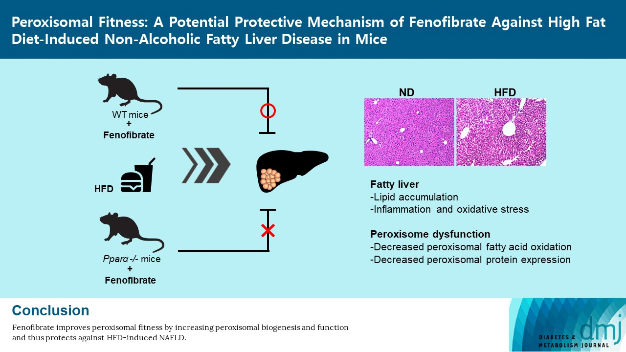
Non-alcoholic fatty liver disease (NAFLD) has been increasing in association with the epidemic of obesity and diabetes. Peroxisomes are single membrane-enclosed organelles that play a role in the metabolism of lipid and reactive oxygen species. The present study examined the role of peroxisomes in high-fat diet (HFD)-induced NAFLD using fenofibrate, a peroxisome proliferator-activated receptor α (PPARα) agonist.
Methods
Eight-week-old male C57BL/6J mice were fed either a normal diet or HFD for 12 weeks, and fenofibrate (50 mg/kg/day) was orally administered along with the initiation of HFD.
Results
HFD-induced liver injury as measured by increased alanine aminotransferase, inflammation, oxidative stress, and lipid accumulation was effectively prevented by fenofibrate. Fenofibrate significantly increased the expression of peroxisomal genes and proteins involved in peroxisomal biogenesis and function. HFD-induced attenuation of peroxisomal fatty acid oxidation was also significantly restored by fenofibrate, demonstrating the functional significance of peroxisomal fatty acid oxidation. In Ppara deficient mice, fenofibrate failed to maintain peroxisomal biogenesis and function in HFD-induced liver injury.
Conclusion
The present data highlight the importance of PPARα-mediated peroxisomal fitness in the protective effect of fenofibrate against NAFLD.
Figure
-

Fig. 1. Fenofibrate (FF) ameliorates high-fat diet (HFD)-induced liver dysfunction in wild-type (WT) mice. (A) Liver sections were immunofluorescence (IF) stained for adipose differentiation-related protein (ADFP; red) and 4´,6-diamidino-2-phenylindole dihydrochloride (DAPI) nuclear counterstaining (blue). Original magnification, 200×; scale bar, 100 μm. (A, B, C, D, E) Liver sections were also stained with anti-F4/80, 8-hydroxyguanine (8-oxo-dG), nitrotyrosine (NT), and 4-hydroxynonena (4-HNE) antibodies and were quantified. Original magnification, 100×; scale bar, 200 μm, n=4. (F) Interleukin 1β (Il1b), Il6, F4/80, and monocyte chemoattractant protein 1 (Mcp1) were measured by real-time polymerase chain reaction, and the results were normalized to the 18S rRNA levels. (G, H) The protein levels of phospho-nuclear factor kappa B (p-NF-κB) and total-NF-κB (t-NF-κB) were measured by Western blotting. (I) Plasma alanine aminotransferase (ALT) levels were measured using an enzyme-linked immunosorbent assay (ELISA) kit. Data are expressed as the mean±standard error of 6 mice/group. ND, normal diet. aP<0.05 vs. ND mice, bP<0.05 vs. HFD mice.
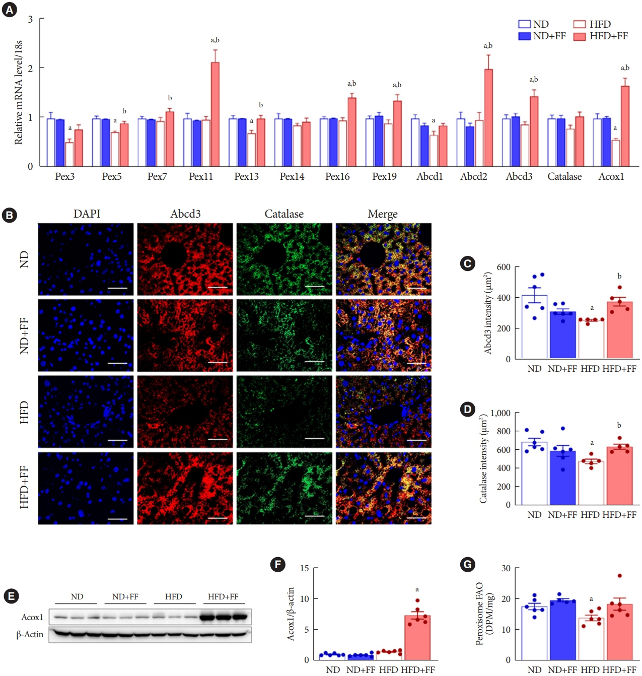
Fig. 2. Fenofibrate (FF) improves liver peroxisomal function in high-fat diet (HFD)-fed wild-type (WT) mice. (A) Peroxisome-related genes were analyzed by real-time polymerase chain reaction, and the results were normalized to 18S rRNA levels. (B, C, D) Liver sections were used for immunofluorescence (IF) staining of ATP binding cassette subfamily D member 3 (ABCD3; red), catalase (green), and 4´,6-diamidino-2-phenylindole dihydrochloride (DAPI) counterstaining (blue) and were quantified. Original magnification, 200×; scale bar, 200 μm. (E, F) Protein levels of acyl-CoA oxidase 1 (ACOX1) were measured by Western blotting, and the results were normalized to β-actin levels. (G) Peroxisomal fatty acid oxidation (FAO) was measured in liver tissue. Data are expressed as the mean±standard error of 6 mice/group. ND, normal diet; Pex, peroxisomal biogenesis factor; DPM, disintegration per minute. aP<0.05 vs. ND mice, bP<0.05 vs. HFD mice.
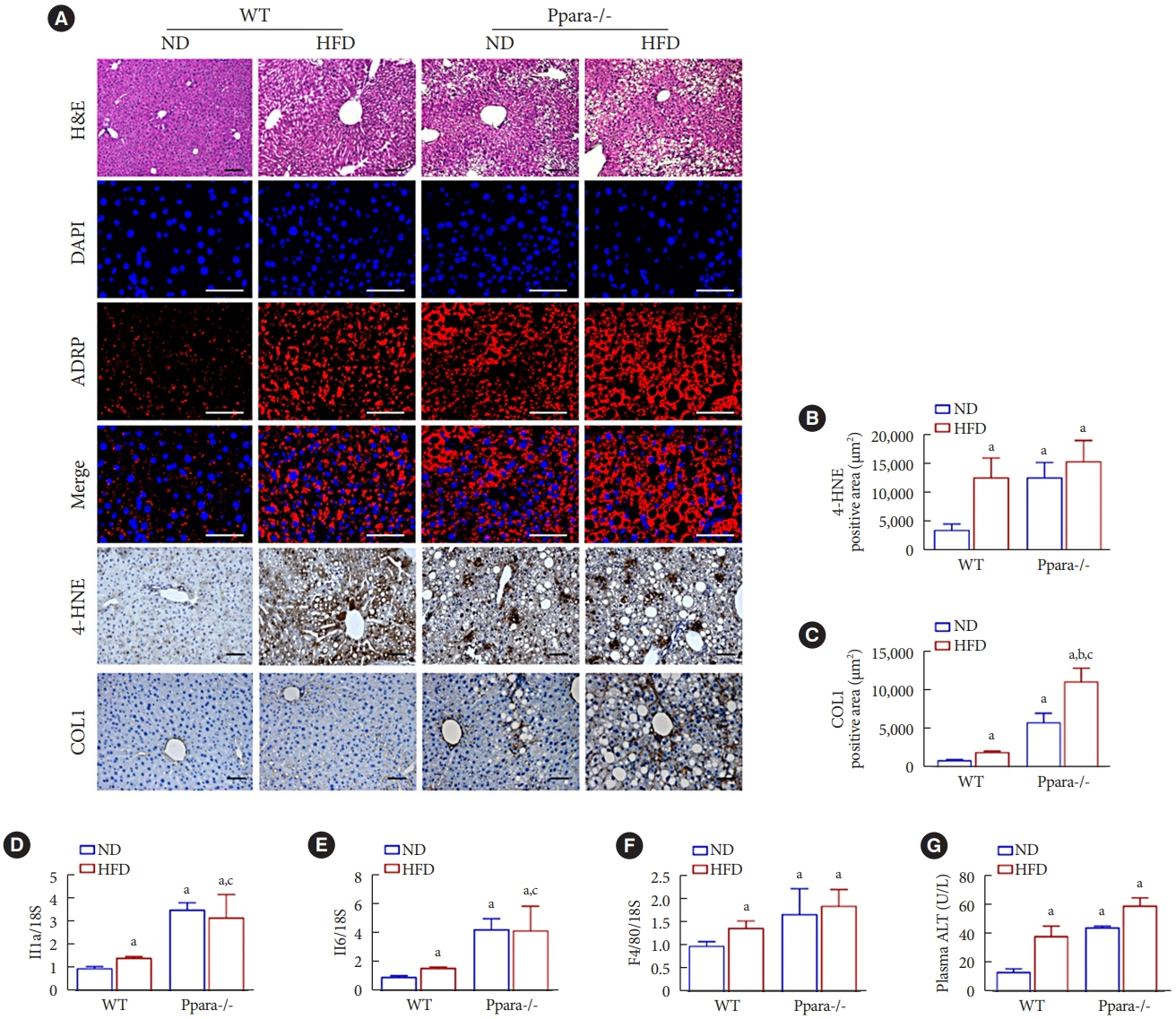
Fig. 3. Role of peroxisome proliferator-activated receptor α (PPARα) in maintaining liver homeostasis in Ppara-/- mice. (A, B, C) Liver morphology was detected by H&E staining. Original magnification, 100×; scale bar, 200 μm. Liver sections were immunofluorescence (IF) stained for adipose differentiation-related protein (ADFP; red) and 4´,6-diamidino-2-phenylindole dihydrochloride (DAPI) nuclear counterstaining (blue). Original magnification, 200×; scale bar, 200 μm. Liver sections were also stained with anti-4-hydroxynonena (4-HNE) and collagen 1 (COL1) antibody and the positive area were quantified. Original magnification, 100×; scale bar, 200 μm. (D, E, F) Interleukin 1β (Il1b), Il6, and F4/80 were measured by real-time polymerase chain reaction, and the results were normalized to the 18S rRNA levels. (G) Plasma alanine aminotransferase (ALT) levels were measured using an enzyme-linked immunosorbent assay (ELISA) kit. Data are expressed as the mean±standard error of 6 mice/group. ND, normal diet; WT, wild-type; HFD, high-fat diet; ADRP, adipose differentiation-related protein. aP<0.05 vs. WT mice with ND, bP<0.05 vs. Ppara-/- mice with ND, cP<0.05 vs. WT mice with HFD.
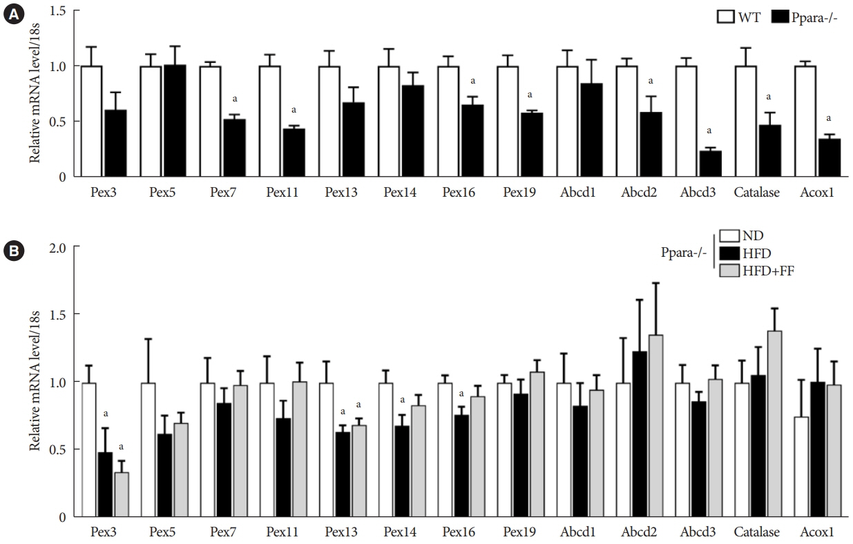
Fig. 4. Fenofibrate fails to maintain peroxisomal biogenesis in Ppara-/- mice. (A) The expression of peroxisome-related genes was decreased in Ppara-/- mice compared to wild-type (WT) mice. (B) Peroxisome-related genes in Ppara-/- mice were analyzed by real-time polymerase chain reaction, and the results were normalized to 18S rRNA levels. Data are expressed as the mean±standard error of 6 mice/group. PPAR, peroxisome proliferator-activated receptor; Pex, peroxisomal biogenesis factor; Abcd, ATP binding cassette subfamily D member; Acox1, acyl-CoA oxidase 1; ND, normal diet; HFD, high-fat diet; FF, fenofibrate. aP<0.05 vs. WT mice or Ppara-/- mice with ND.
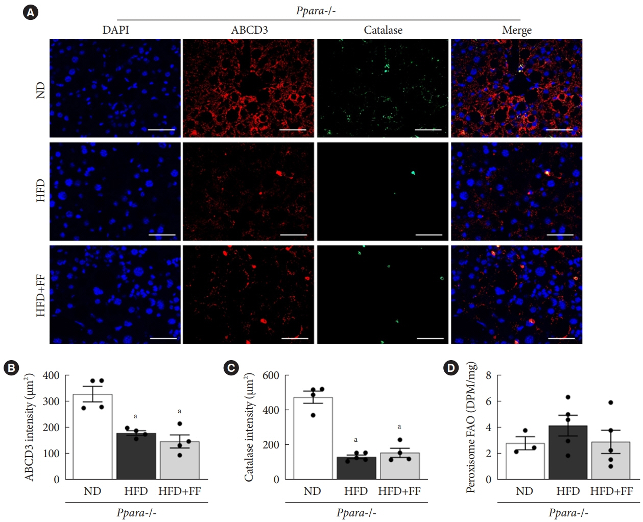
Fig. 5. Fenofibrate fails to improve peroxisomal function in Ppara-/- mice. (A, B, C) Liver sections were immunofluorescence stained for ATP binding cassette subfamily D member 3 (ABCD3; red), catalase (green), and 4´,6-diamidino-2-phenylindole dihydrochloride (DAPI) nuclear counterstaining (blue) and intensities were quantified. Original magnification, 200×; scale bar, 200 μm. (D) Peroxisomal fatty acid oxidation (FAO) was measured in liver tissue. Data are expressed as the mean±standard error of 6 mice/group. PPAR, peroxisome proliferator-activated receptor; ND, normal diet; HFD, high-fat diet; FF, fenofibrate; DPM, disintegration per minute. aP<0.05 vs. Ppara-/- mice with ND.
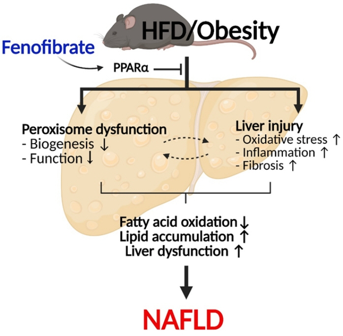
Fig. 6. Suggested model of fenofibrate-mediated peroxisomal fitness against high-fat diet (HFD)-induced non-alcoholic fatty liver disease (NAFLD). HFD or obesity decreases peroxisomal biogenesis and function and increases liver injury, including oxidative stress, inflammation, and fibrosis, due to inhibition of the peroxisome proliferator-activated receptor α (PPARα) pathway. Subsequently, it results in decreased hepatic fatty acid oxidation, increased lipid accumulation, and the induction of liver dysfunction, which leads to the development of NAFLD. Fenofibrate maintains peroxisomal biogenesis and function through activation of the PPARα pathway. It also attenuates liver injury and increases hepatic fatty acid oxidation. Thus, fenofibrate may mediate protective effects against NAFLD by maintaining peroxisomal biogenesis and function.
Cited by 1 articles
-
Role of Fenofibrate Use in Dyslipidemia and Related Comorbidities in the Asian Population: A Narrative Review
Chaicharn Deerochanawong, Sin Gon Kim, Yu-Cheng Chang
Diabetes Metab J. 2024;48(2):184-195. doi: 10.4093/dmj.2023.0168.
Reference
-
1. Younossi ZM, Koenig AB, Abdelatif D, Fazel Y, Henry L, Wymer M. Global epidemiology of nonalcoholic fatty liver disease-Meta-analytic assessment of prevalence, incidence, and outcomes. Hepatology. 2016; 64:73–84.
Article2. Ng M, Fleming T, Robinson M, Thomson B, Graetz N, Margono C, et al. Global, regional, and national prevalence of overweight and obesity in children and adults during 1980-2013: a systematic analysis for the Global Burden of Disease Study 2013. Lancet. 2014; 384:766–81.3. Tiniakos DG, Vos MB, Brunt EM. Nonalcoholic fatty liver disease: pathology and pathogenesis. Annu Rev Pathol. 2010; 5:145–71.
Article4. Bril F, Barb D, Portillo-Sanchez P, Biernacki D, Lomonaco R, Suman A, et al. Metabolic and histological implications of intrahepatic triglyceride content in nonalcoholic fatty liver disease. Hepatology. 2017; 65:1132–44.
Article5. Chen Z, Tian R, She Z, Cai J, Li H. Role of oxidative stress in the pathogenesis of nonalcoholic fatty liver disease. Free Radic Biol Med. 2020; 152:116–41.
Article6. Zelber-Sagi S, Ivancovsky-Wajcman D, Fliss-Isakov N, Hahn M, Webb M, Shibolet O, et al. Serum malondialdehyde is associated with non-alcoholic fatty liver and related liver damage differentially in men and women. Antioxidants (Basel). 2020; 9:578.
Article7. Sekiya M, Hiraishi A, Touyama M, Sakamoto K. Oxidative stress induced lipid accumulation via SREBP1c activation in HepG2 cells. Biochem Biophys Res Commun. 2008; 375:602–7.
Article8. Ferramosca A, Di Giacomo M, Zara V. Antioxidant dietary approach in treatment of fatty liver: new insights and updates. World J Gastroenterol. 2017; 23:4146–57.
Article9. Germain K, Kim PK. Pexophagy: a model for selective autophagy. Int J Mol Sci. 2020; 21:578.
Article10. Sugiura A, Mattie S, Prudent J, McBride HM. Newly born peroxisomes are a hybrid of mitochondrial and ER-derived preperoxisomes. Nature. 2017; 542:251–4.
Article11. Smith JJ, Aitchison JD. Peroxisomes take shape. Nat Rev Mol Cell Biol. 2013; 14:803–17.12. Lodhi IJ, Semenkovich CF. Peroxisomes: a nexus for lipid metabolism and cellular signaling. Cell Metab. 2014; 19:380–92.
Article13. Kovacs WJ, Charles KN, Walter KM, Shackelford JE, Wikander TM, Richards MJ, et al. Peroxisome deficiency-induced ER stress and SREBP-2 pathway activation in the liver of newborn Pex2 knock-out mice. Biochim Biophys Acta. 2012; 1821:895–907.
Article14. Li X, Baumgart E, Morrell JC, Jimenez-Sanchez G, Valle D, Gould SJ. PEX11 beta deficiency is lethal and impairs neuronal migration but does not abrogate peroxisome function. Mol Cell Biol. 2002; 22:4358–65.
Article15. Hwang I, Lee J, Huh JY, Park J, Lee HB, Ho YS, et al. Catalase deficiency accelerates diabetic renal injury through peroxisomal dysfunction. Diabetes. 2012; 61:728–38.
Article16. Hwang I, Uddin MJ, Pak ES, Kang H, Jin EJ, Jo S, et al. The impaired redox balance in peroxisomes of catalase knockout mice accelerates nonalcoholic fatty liver disease through endoplasmic reticulum stress. Free Radic Biol Med. 2020; 148:22–32.
Article17. Piao L, Dorotea D, Jiang S, Koh EH, Oh GT, Ha H. Impaired peroxisomal fitness in obese mice, a vicious cycle exacerbating adipocyte dysfunction via oxidative stress. Antioxid Redox Signal. 2019; 31:1339–51.18. Islam S, Won J, Khan M, Chavin KD, Singh I. Peroxisomal footprint in the pathogenesis of nonalcoholic steatohepatitis. Ann Hepatol. 2020; 19:466–71.
Article19. Farnier M. Update on the clinical utility of fenofibrate in mixed dyslipidemias: mechanisms of action and rational prescribing. Vasc Health Risk Manag. 2008; 4:991–1000.
Article20. Montagner A, Polizzi A, Fouche E, Ducheix S, Lippi Y, Lasserre F, et al. Liver PPARα is crucial for whole-body fatty acid homeostasis and is protective against NAFLD. Gut. 2016; 65:1202–14.
Article21. Jo SH, Nam H, Lee J, Park S, Lee J, Kyoung DS. Fenofibrate use is associated with lower mortality and fewer cardiovascular events in patients with diabetes: results of 10,114 patients from the Korean National Health Insurance Service Cohort. Diabetes Care. 2021; 44:1868–76.
Article22. Elam MB, Ginsberg HN, Lovato LC, Corson M, Largay J, Leiter LA, et al. Association of fenofibrate therapy with long-term cardiovascular risk in statin-treated patients with type 2 diabetes. JAMA Cardiol. 2017; 2:370–80.
Article23. Sohn M, Kim K, Uddin MJ, Lee G, Hwang I, Kang H, et al. Delayed treatment with fenofibrate protects against high-fat dietinduced kidney injury in mice: the possible role of AMPK autophagy. Am J Physiol Renal Physiol. 2017; 312:F323–34.
Article24. Weng H, Ji X, Endo K, Iwai N. Pex11a deficiency is associated with a reduced abundance of functional peroxisomes and aggravated renal interstitial lesions. Hypertension. 2014; 64:1054–60.
Article25. Lee JN, Dutta RK, Kim SG, Lim JY, Kim SJ, Choe SK, et al. Fenofibrate, a peroxisome proliferator-activated receptor α ligand, prevents abnormal liver function induced by a fastingrefeeding process. Biochem Biophys Res Commun. 2013; 442:22–7.
Article26. Araki H, Tamada Y, Imoto S, Dunmore B, Sanders D, Humphrey S, et al. Analysis of PPARalpha-dependent and PPARalpha-independent transcript regulation following fenofibrate treatment of human endothelial cells. Angiogenesis. 2009; 12:221–9.
Article27. Hua H, Yang J, Lin H, Xi Y, Dai M, Xu G, et al. PPARα-independent action against metabolic syndrome development by fibrates is mediated by inhibition of STAT3 signalling. J Pharm Pharmacol. 2018; 70:1630–42.
Article28. Akiyama TE, Nicol CJ, Fievet C, Staels B, Ward JM, Auwerx J, et al. Peroxisome proliferator-activated receptor-alpha regulates lipid homeostasis, but is not associated with obesity: studies with congenic mouse lines. J Biol Chem. 2001; 276:39088–93.29. Kondo K, Sugioka T, Tsukada K, Aizawa M, Takizawa M, Shimizu K, et al. Fenofibrate, a peroxisome proliferator-activated receptor alpha agonist, improves hepatic microcirculatory patency and oxygen availability in a high-fat-diet-induced fatty liver in mice. Adv Exp Med Biol. 2010; 662:77–82.30. Kostapanos MS, Kei A, Elisaf MS. Current role of fenofibrate in the prevention and management of non-alcoholic fatty liver disease. World J Hepatol. 2013; 5:470–8.
Article31. Walton PA, Brees C, Lismont C, Apanasets O, Fransen M. The peroxisomal import receptor PEX5 functions as a stress sensor, retaining catalase in the cytosol in times of oxidative stress. Biochim Biophys Acta Mol Cell Res. 2017; 1864:1833–43.
Article32. Imanaka T, Aihara K, Suzuki Y, Yokota S, Osumi T. The 70- kDa peroxisomal membrane protein (PMP70), an ATP-binding cassette transporter. Cell Biochem Biophys. 2000; 32:131–8.33. Pawlak M, Lefebvre P, Staels B. Molecular mechanism of PPARα action and its impact on lipid metabolism, inflammation and fibrosis in non-alcoholic fatty liver disease. J Hepatol. 2015; 62:720–33.
Article34. Regnier M, Polizzi A, Smati S, Lukowicz C, Fougerat A, Lippi Y, et al. Hepatocyte-specific deletion of Pparα promotes NAFLD in the context of obesity. Sci Rep. 2020; 10:6489.
Article35. Jain MR, Giri SR, Bhoi B, Trivedi C, Rath A, Rathod R, et al. Dual PPARα/γ agonist saroglitazar improves liver histopathology and biochemistry in experimental NASH models. Liver Int. 2018; 38:1084–94.
Article36. Rakhshandehroo M, Knoch B, Muller M, Kersten S. Peroxisome proliferator-activated receptor alpha target genes. PPAR Res. 2010; 2010:612089.
Article37. Cheng S, Liang S, Liu Q, Deng Z, Zhang Y, Du J, et al. Diosgenin prevents high-fat diet-induced rat non-alcoholic fatty liver disease through the AMPK and LXR signaling pathways. Int J Mol Med. 2018; 41:1089–95.
Article38. Chen WL, Chen YL, Chiang YM, Wang SG, Lee HM. Fenofibrate lowers lipid accumulation in myotubes by modulating the PPARα/AMPK/FoxO1/ATGL pathway. Biochem Pharmacol. 2012; 84:522–31.
Article39. Yan F, Wang Q, Xu C, Cao M, Zhou X, Wang T, et al. Peroxisome proliferator-activated receptor α activation induces hepatic steatosis, suggesting an adverse effect. PLoS One. 2014; 9:e99245.
Article40. Oosterveer MH, Grefhorst A, van Dijk TH, Havinga R, Staels B, Kuipers F, et al. Fenofibrate simultaneously induces hepatic fatty acid oxidation, synthesis, and elongation in mice. J Biol Chem. 2009; 284:34036–44.
Article41. Cortes-Lopez F, Sanchez-Mendoza A, Centurion D, CervantesPerez LG, Castrejon-Tellez V, Del Valle-Mondragon L, et al. Fenofibrate protects cardiomyocytes from hypoxia/reperfusion- and high glucose-induced detrimental effects. PPAR Res. 2021; 2021:8895376.
Article42. Ning LJ, He AY, Lu DL, Li JM, Qiao F, Li DL, et al. Nutritional background changes the hypolipidemic effects of fenofibrate in Nile tilapia (Oreochromis niloticus). Sci Rep. 2017; 7:41706.
Article43. Honsho M, Tamura S, Shimozawa N, Suzuki Y, Kondo N, Fujiki Y. Mutation in PEX16 is causal in the peroxisome-deficient Zellweger syndrome of complementation group D. Am J Hum Genet. 1998; 63:1622–30.44. Sacksteder KA, Jones JM, South ST, Li X, Liu Y, Gould SJ. PEX19 binds multiple peroxisomal membrane proteins, is predominantly cytoplasmic, and is required for peroxisome membrane synthesis. J Cell Biol. 2000; 148:931–44.
Article45. Ghaedi K, Tamura S, Okumoto K, Matsuzono Y, Fujiki Y. The peroxin pex3p initiates membrane assembly in peroxisome biogenesis. Mol Biol Cell. 2000; 11:2085–102.
Article46. Delille HK, Dodt G, Schrader M. Pex11pβ-mediated maturation of peroxisomes. Commun Integr Biol. 2011; 4:51–4.
Article47. Weng H, Ji X, Naito Y, Endo K, Ma X, Takahashi R, et al. Pex11α deficiency impairs peroxisome elongation and division and contributes to nonalcoholic fatty liver in mice. Am J Physiol Endocrinol Metab. 2013; 304:E187–96.
Article48. Walker CL, Pomatto L, Tripathi DN, Davies K. Redox regulation of homeostasis and proteostasis in peroxisomes. Physiol Rev. 2018; 98:89–115.
Article49. Piao L, Choi J, Kwon G, Ha H. Endogenous catalase delays high-fat diet-induced liver injury in mice. Korean J Physiol Pharmacol. 2017; 21:317–25.
Article50. Shin SK, Cho HW, Song SE, Bae JH, Im SS, Hwang I, et al. Ablation of catalase promotes non-alcoholic fatty liver via oxidative stress and mitochondrial dysfunction in diet-induced obese mice. Pflugers Arch. 2019; 471:829–43.
Article51. Koepke JI, Wood CS, Terlecky LJ, Walton PA, Terlecky SR. Progeric effects of catalase inactivation in human cells. Toxicol Appl Pharmacol. 2008; 232:99–108.
Article52. Hwang I, Uddin MJ, Lee G, Jiang S, Pak ES, Ha H. Peroxiredoxin 3 deficiency accelerates chronic kidney injury in mice through interactions between macrophages and tubular epithelial cells. Free Radic Biol Med. 2019; 131:162–72.
Article53. Wang YX. PPARs: diverse regulators in energy metabolism and metabolic diseases. Cell Res. 2010; 20:124–37.
Article54. Schrader M, Grille S, Fahimi HD, Islinger M. Peroxisome interactions and cross-talk with other subcellular compartments in animal cells. Subcell Biochem. 2013; 69:1–22.
Article55. Li X, Gould SJ. PEX11 promotes peroxisome division independently of peroxisome metabolism. J Cell Biol. 2002; 156:643–51.
Article56. Chen Z, Yu R, Xiong Y, Du F, Zhu S. A vicious circle between insulin resistance and inflammation in nonalcoholic fatty liver disease. Lipids Health Dis. 2017; 16:203.
Article
- Full Text Links
-
- Actions
-
Cited
- CITED
-
- Close
- Share
-
- Similar articles
-
- An Activation of Peroxisome Proliferator-Activated Receptor delta Attenuate Alcoholic Liver Disease and Nonalcoholic Fatty Liver Disease in Rats
- PIK3R3 regulates PPARα expression to stimulate fatty acid β-oxidation and decrease hepatosteatosis
- Association between a High-fat Low-carbohydrate Diet and Non-alcoholic Fatty Liver Disease: Truth or Myth?
- Statins Increase Mitochondrial and Peroxisomal Fatty Acid Oxidation in the Liver and Prevent Non-Alcoholic Steatohepatitis in Mice
- Effects of an aqueous extract of purple sweet potato on nonalcoholic fatty liver in high fat/cholesterol-fed mice
