J Stroke.
2022 Sep;24(3):433-435. 10.5853/jos.2022.02054.
Automated Composition Analysis of Thrombus from Endovascular Treatment in Acute Ischemic Stroke Using Computer Vision
- Heo J1
- Seog Y1
- Lee H1
- Lee IH1
- Kim S1
- Baek JH2
- Park H3
- Seo KD4
- Kim GS4
- Cho HJ5
- Baik M6
- Yoo J6
- Kim J6
- Lee J7
- Chang YK8
- Song TJ9
- Seo JH10
- Ahn SH11
- Lee HW1
- Kwon I1,12
- Park E12
- Kim YD1,12
- Nam HS
 1,12
1,12
- Affiliations
-
- 1Department of Neurology, Yonsei University College of Medicine, Seoul, Korea
- 2Department of Neurology, Kangbuk Samsung Hospital, Sungkyunkwan University School of Medicine, Seoul, Korea
- 3Department of Neurology, Brain Research Institute, Keimyung University School of Medicine, Daegu, Korea
- 4Department of Neurology, National Health Insurance Service Ilsan Hospital, Goyang, Korea
- 5Department of Neurology, Pusan National University School of Medicine, Busan, Korea
- 6Department of Neurology, Yongin Severance Hospital, Yonsei University College of Medicine, Yongin, Korea
- 7Department of Neurology, Yeungnam University College of Medicine, Daegu, Korea
- 8Department of Neurology, Ewha Womans University Mokdong Hospital, Ewha Womans University School of Medicine, Seoul, Korea
- 9Department of Neurology, Ewha Womans University Seoul Hospital, Ewha Womans University School of Medicine, Seoul, Korea
- 10Department of Neurology, Inje University Busan Paik Hospital, College of Medicine, Inje University, Busan, Korea
- 11Department of Neurology, Chosun University Hospital, Chosun University College of Medicine, Gwangju, Korea
- 12Integrative Research Center for Cerebrovascular and Cardiovascular Diseases, Seoul, Korea
- KMID: 2534275
- DOI: http://doi.org/10.5853/jos.2022.02054
Figure
-
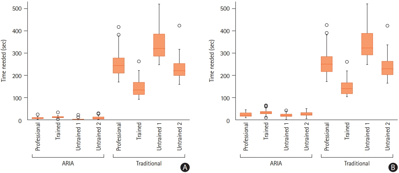
Figure 1. Comparison of (A) focused time and (B) total time needed for analysis using Automated Region-of-interest based Image Analysis (ARIA) and traditional methodology
Reference
-
References
1. Heo JH, Nam HS, Kim YD, Choi JK, Kim BM, Kim DJ, et al. Pathophysiologic and therapeutic perspectives based on thrombus histology in stroke. J Stroke. 2020; 22:64–75.2. Schuhmann MK, Gunreben I, Kleinschnitz C, Kraft P. Immunohistochemical analysis of cerebral thrombi retrieved by mechanical thrombectomy from patients with acute ischemic stroke. Int J Mol Sci. 2016; 17:298.3. Simons N, Mitchell P, Dowling R, Gonzales M, Yan B. Thrombus composition in acute ischemic stroke: a histopathological study of thrombus extracted by endovascular retrieval. J Neuroradiol. 2015; 42:86–92.4. Schneider CA, Rasband WS, Eliceiri KW. NIH Image to ImageJ: 25 years of image analysis. Nat Methods. 2012; 9:671–675.5. Office Adobe Photoshop. Photo and design software. Adobe;https://www.adobe.com/products/photoshop.html. Accessed September 7, 2022.6. Ahn SH, Hong R, Choo IS, Heo JH, Nam HS, Kang HG, et al. Histologic features of acute thrombi retrieved from stroke patients during mechanical reperfusion therapy. Int J Stroke. 2016; 11:1036–1044.7. Borovec J, Kybic J, Arganda-Carreras I, Sorokin DV, Bueno G, Khvostikov AV, et al. ANHIR: automatic non-rigid histological image registration challenge. IEEE Trans Med Imaging. 2020; 39:3042–3052.8. Canny J. A computational approach to edge detection. IEEE Trans Pattern Anal Mach Intell. 1986; 8:679–698.9. Otsu N. A threshold selection method from gray-level histograms. IEEE Trans Syst Man Cybern. 1979; 9:62–66.10. Goode A, Gilbert B, Harkes J, Jukic D, Satyanarayanan M. OpenSlide: a vendor-neutral software foundation for digital pathology. J Pathol Inform. 2013; 4:27.
- Full Text Links
-
- Actions
-
Cited
- CITED
-
- Close
- Share
-
- Similar articles
-
- Traditional Thrombus Composition and Related Endovascular Outcomes: Catching up with the Recent Evidence
- Pathophysiologic and Therapeutic Perspectives Based on Thrombus Histology in Stroke
- Endovascular Treatment of Acute Ischemic Stroke
- Intravenous Thrombolysis and Endovascular Thrombectomy in Acute Ischemic Stroke with Minor Symptom
- Imaging in Acute Anterior Circulation Ischemic Stroke: Current and Future
