Hepatocellular carcinoma diagnosed in a patient who had Fontan operation 30 years ago: a case report
- Affiliations
-
- 1Department of Internal Medicine and Liver Research Institute, Seoul National University College of Medicine, Seoul, Korea
- 2Department of Pathology, Seoul National University College of Medicine, Seoul, Korea
- KMID: 2534246
- DOI: http://doi.org/10.17998/jlc.2022.08.17
Abstract
- The Fontan operation is performed in patients with a single ventricle. As the systemic venous return is directly connected to the pulmonary circulation during this procedure, chronic hepatic congestion is induced, leading to Fontan-associated liver disease (FALD) including liver cirrhosis and hepatocellular carcinoma (HCC). In this report, we present a case of HCC diagnosed in a patient who underwent the Fontan operation 30 years ago. The patient underwent regular surveillance for FALD, which revealed a 4 cm-sized hepatic mass with elevated serum alpha-fetoprotein. After surgical treatment, there was no evidence of HCC recurrence during 3 years of follow-up. As the risk of HCC and Fontan-associated liver cirrhosis increases with the duration elapsed since the operation, regular surveillance should be emphasized. Serial follow-up of serum alpha-fetoprotein levels and abdominal imaging are necessary to achieve early and accurate diagnosis of HCC in post-Fontan patients.
Figure
-
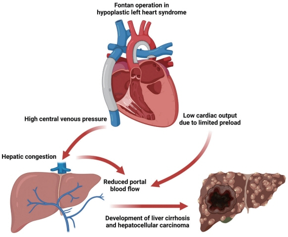
Figure 1. Effect of Fontan operation on the development of Fontan-associated liver disease. In Fontan circulation, the systemic veins and pulmonary arteries are directly connected. Elevated central venous pressure induced by passive filling of the pulmonary circulation without the support of the right ventricle results in chronic hepatic congestion. In addition, low cardiac output due to limited preload reduces portal blood flow, which contributes to ischemic insults. Consequently, accumulated liver damage can lead to liver cirrhosis and even hepatocellular carcinoma.
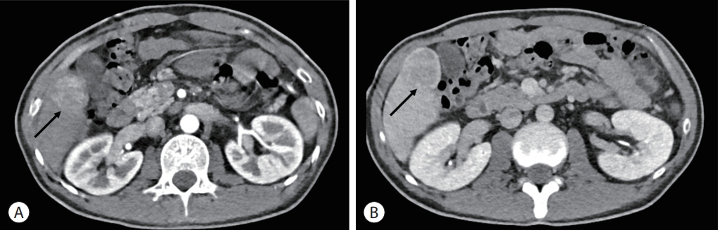
Figure 2. Computed tomography image showing a 4 cm-sized hepatic mass in segment V (arrows). The mass is seen as hyperenhancement in the arterial phase (A) and washout in the portal venous phase (B).

Figure 3. Contrast-enhanced liver magnetic resonance imaging showing a well-circumscribed mass (arrow) with arterial phase hyperenhancement (A) and hepatobiliary phase defect (arrow) (B). In a T2-weighted image, it shows a high signal intensity (arrow) (C). The hepatic mass exhibited significant hypermetabolism in the positron emission tomography-computed tomography image (arrow) (D).
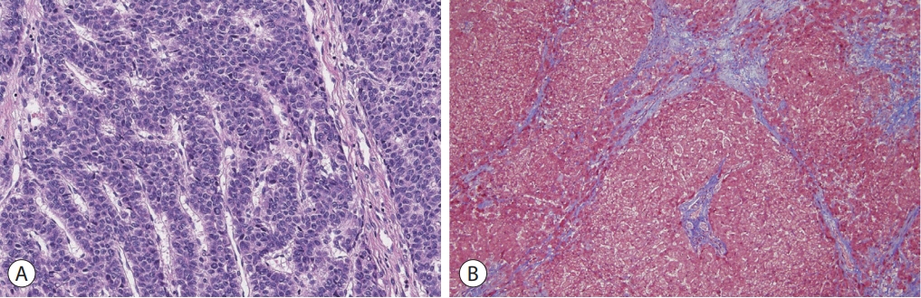
Figure 4. On histological examination, a moderately differentiated hepatocellular carcinoma with a trabecular pattern was observed (A, hematoxylin and eosin stain, ×200). The background liver shows micronodular venocentric cirrhosis (B, Masson’s trichrome stain, ×100).
Reference
-
1. Fontan F, Baudet E. Surgical repair of tricuspid atresia. Thorax. 1971; 26:240–248.2. Gewillig M. The Fontan circulation. Heart. 2005; 91:839–846.3. Gordon-Walker TT, Bove K, Veldtman G. Fontan-associated liver disease: a review. J Cardiol. 2019; 74:223–232.4. Munsterman ID, Duijnhouwer AL, Kendall TJ, Bronkhorst CM, Ronot M, van Wettere M, et al. The clinical spectrum of Fontanassociated liver disease: results from a prospective multimodality screening cohort. Eur Heart J. 2019; 40:1057–1068.5. Yoon JS, Lee DH, Cho EJ, Song MK, Choi YH, Kim GB, et al. Risk of liver cirrhosis and hepatocellular carcinoma after Fontan operation: a need for surveillance. Cancers (Basel). 2020; 12:1805.6. Gersony WM. Fontan operation after 3 decades: what we have learned. Circulation. 2008; 117:13–15.7. Khairy P, Fernandes SM, Mayer JE Jr, Triedman JK, Walsh EP, Lock JE, et al. Long-term survival, modes of death, and predictors of mortality in patients with Fontan surgery. Circulation. 2008; 117:85–92.8. Emamaullee J, Zaidi AN, Schiano T, Kahn J, Valentino PL, Hofer RE, et al. Fontan-associated liver disease: screening, management, and transplant considerations. Circulation. 2020; 142:591–604.9. Olsen AL, Bloomer SA, Chan EP, Gaça MD, Georges PC, Sackey B, et al. Hepatic stellate cells require a stiff environment for myofibroblastic differentiation. Am J Physiol Gastrointest Liver Physiol. 2011; 301:G110–G118.10. Iwakiri Y, Shah V, Rockey DC. Vascular pathobiology in chronic liver disease and cirrhosis - current status and future directions. J Hepatol. 2014; 61:912–924.11. Baek JS, Bae EJ, Ko JS, Kim GB, Kwon BS, Lee SY, et al. Late hepatic complications after Fontan operation; non-invasive markers of hepatic fibrosis and risk factors. Heart. 2010; 96:1750–1755.12. Shimizu M, Miyamoto K, Nishihara Y, Izumi G, Sakai S, Inai K, et al. Risk factors and serological markers of liver cirrhosis after Fontan procedure. Heart Vessels. 2016; 31:1514–1521.13. Asrani SK, Warnes CA, Kamath PS. Hepatocellular carcinoma after the Fontan procedure. N Engl J Med. 2013; 368:1756–1757.14. Yamada K, Shinmoto H, Kawamura Y, Wakamatsu H, Kawauchi T, Soga S, et al. Transarterial embolization for pediatric hepatocellular carcinoma with cardiac cirrhosis. Pediatr Int. 2015; 57:766–770.15. Kogiso T, Tokushige K. Fontan-associated liver disease and hepatocellular carcinoma in adults. Sci Rep. 2020; 10:21742.16. Possner M, Gordon-Walker T, Egbe AC, Poterucha JT, Warnes CA, Connolly HM, et al. Hepatocellular carcinoma and the Fontan circulation: clinical presentation and outcomes. Int J Cardiol. 2021; 322:142–148.17. Rodriguez De Santiago E, Téllez L, Guerrero A, Albillos A. Hepatocellular carcinoma after Fontan surgery: a systematic review. Hepatol Res. 2021; 51:116–134.18. Egbe AC, Poterucha JT, Warnes CA, Connolly HM, Baskar S, Ginde S, et al. Hepatocellular carcinoma after Fontan operation: multicenter case series. Circulation. 2018; 138:746–748.19. Asrani SK, Asrani NS, Freese DK, Phillips SD, Warnes CA, Heimbach J, et al. Congenital heart disease and the liver. Hepatology. 2012; 56:1160–1169.20. Wells ML, Hough DM, Fidler JL, Kamath PS, Poterucha JT, Venkatesh SK. Benign nodules in post-Fontan livers can show imaging features considered diagnostic for hepatocellular carcinoma. Abdom Radiol (NY). 2017; 42:2623–2631.21. Wells ML, Fenstad ER, Poterucha JT, Hough DM, Young PM, Araoz PA, et al. Imaging findings of congestive hepatopathy. Radiographics. 2016; 36:1024–1037.22. Nandwana SB, Olaiya B, Cox K, Sahu A, Mittal P. abdominal imaging surveillance in adult patients after fontan procedure: risk of chronic liver disease and hepatocellular carcinoma. Curr Probl Diagn Radiol. 2018; 47:19–22.23. Téllez L, Rodríguez de Santiago E, Minguez B, Payance A, Clemente A, Baiges A, et al. Prevalence, features and predictive factors of liver nodules in Fontan surgery patients: The VALDIG Fonliver prospective cohort. J Hepatol. 2020; 72:702–710.
- Full Text Links
-
- Actions
-
Cited
- CITED
-
- Close
- Share
-
- Similar articles
-
- Liver transplantation in an adult patient with hepatocellular carcinoma following liver cirrhosis as a complication of the Fontan procedure -A case report-
- The Extracardiac Fontan Operation in Adult: A case report
- Unusual Semimembranosus Muscle Metastasis from Hepatocellular Carcinoma
- clinical Evaluation for the Progrosis after the Fontan Operation
- Treatment of Protein-losing Enteropathy After Fontan Procedure by Conversion to the Total Cavopulmonary Connection with Fenestration
