Sclerosing polycystic adenosis arising in the parotid gland with trismus: a case report and literature review
- Affiliations
-
- 1Department of Oral and Maxillofacial surgery, Oral Science Research Center, Yonsei University College of Dentistry, Seoul, Korea
- 2Department of Oral Pathology, Oral Cancer Research Institute, Yonsei University College of Dentistry, Seoul, Korea
- 3Department of Oral and Maxillofacial Radiology, Yonsei University College of Dentistry, Seoul, Korea
- KMID: 2532830
- DOI: http://doi.org/10.5125/jkaoms.2022.48.4.237
Abstract
- Sclerosing polycystic adenosis (SPA) is a rare, asymptomatic disease that occurs mainly in the salivary glands. We report the case of a 51-year-old man who presented with trismus and pain upon mouth opening. Magnetic resonance imaging revealed a 2-cm mass located in the anterior portion of the left parotid gland. SPA was diagnosed based on histopathological examination of the surgical specimen. In pathologic findings, there was a well-circumscribed multicystic nodule in the parenchyma. Dense fibrosis and chronic non-specific inflammatory cells were observed in the stroma. In 13 previous reports on SPA, the most preferred treatment was superficial or total parotidectomy. This report suggests that simple excision of SPA preserves facial nerve function and facial volume.
Keyword
Figure
-

Fig. 1 Magnetic resonance imaging revealed a 2-cm-sized mass in the anterior portion of the left parotid gland, compressing the masseter muscle anteriorly (A). The lesion is mainly T2-hyperintense; it is a rim-enhancing lesion with a small localized enhancing area (A, C). The lesion in T1-hypointense (B, D). (Asterisks: cystic area, Hollow arrow: masseter muscle, Arrowhead: solid area of the tumor)
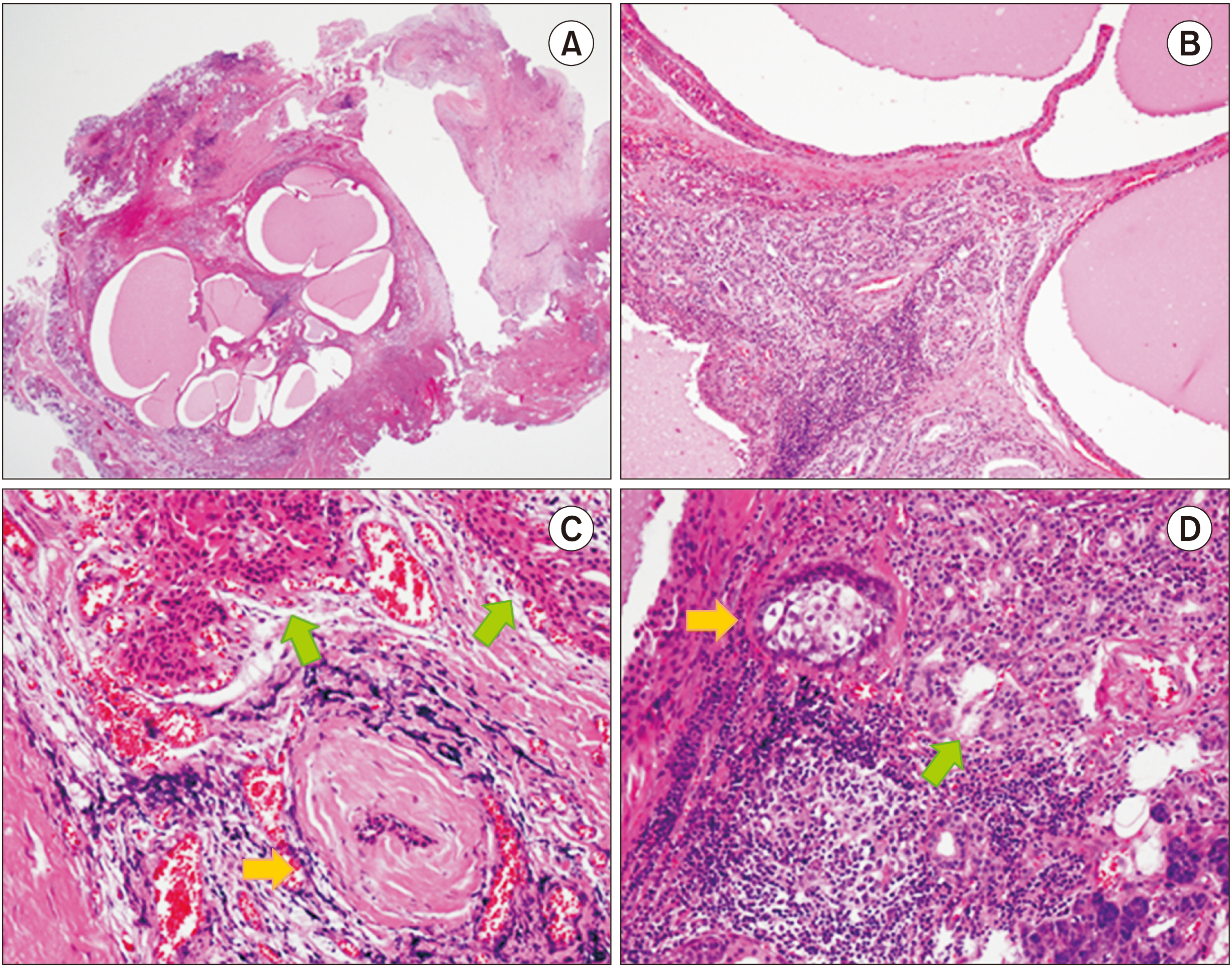
Fig. 2 Histopathological examination of the surgical specimen through H&E staining. (A). A multicystic nodular mass within the parotid parenchyma was observed under low magnification. Original magnification, ×12.5. (B). The cysts lining the epithelium show simple or stratified cuboidal cells with oncocytic cytoplasm. Ductal hyperplasia, chronic non-specific inflammation, and dense fibrosis are present. Original magnification, ×100. (C). A characteristic hyalinized nodule surrounding the degenerative duct is observed (yellow arrow). Some acini and ducts show eosinophilic cytoplasmic changes (green arrows). Original magnification, ×200. (D). Some glandular ducts show sebaceous metaplasia (yellow arrow). Prominent lymphocytic infiltration and ductal hyperplasia are also observed (green arrow). Original magnification, ×200.
Reference
-
References
1. Petersson F. 2013; Sclerosing polycystic adenosis of salivary glands: a review with some emphasis on intraductal epithelial proliferations. Head Neck Pathol. 7(Suppl 1):S97–106. https://doi.org/10.1007/s12105-013-0465-9. DOI: 10.1007/s12105-013-0465-9. PMID: 23821217. PMCID: PMC3712090.
Article2. Kennedy RA. 2018; WHO is in and WHO is out of the mouth, salivary glands, and jaws sections of the 4th edition of the WHO classification of head and neck tumours. Br J Oral Maxillofac Surg. 56:90–5. https://doi.org/10.1016/j.bjoms.2017.12.009. DOI: 10.1016/j.bjoms.2017.12.009. PMID: 29352614.
Article3. Espinosa CA, Rua L, Torres HE, Fernández Del Valle Á, Fernandes RP, Devicente JC. 2017; Sclerosing polycystic adenosis of the parotid gland: a systematic review and report of 2 new cases. J Oral Maxillofac Surg. 75:984–93. https://doi.org/10.1016/j.joms.2016.10.031. DOI: 10.1016/j.joms.2016.10.031. PMID: 27888671.
Article4. Bharadwaj G, Nawroz I, O'Regan B. 2007; Sclerosing polycystic adenosis of the parotid gland. Br J Oral Maxillofac Surg. 45:74–6. https://doi.org/10.1016/j.bjoms.2005.05.018. DOI: 10.1016/j.bjoms.2005.05.018. PMID: 16054736.
Article5. Eliot CA, Smith AB, Foss RD. 2012; Sclerosing polycystic adenosis. Head Neck Pathol. 6:247–9. https://doi.org/10.1007/s12105-011-0317-4. DOI: 10.1007/s12105-011-0317-4. PMID: 22183766. PMCID: PMC3370019.
Article6. Tang CG, Fong JB, Axelsson KL, Gurushanthaiah D. 2016; Sclerosing polycystic adenosis: a rare tumor of the salivary glands. Perm J. 20:e113–4. https://doi.org/10.7812/TPP/15-147. DOI: 10.7812/TPP/15-147. PMID: 27043832. PMCID: PMC4867840.
Article7. Kim BC, Yang DH, Kim J, Samayoa SR, Na HY, Choi EJ, et al. 2012; Sclerosing polycystic adenosis of the parotid gland. J Craniofac Surg. 23:e451–2. https://doi.org/10.1097/SCS.0b013e318262d2a5. DOI: 10.1097/SCS.0b013e318262d2a5. PMID: 22976703.
Article8. Perottino F, Barnoud R, Ambrun A, Poupart M, Pignat JC, Merrot O. 2010; Sclerosing polycystic adenosis of the parotid gland: diagnosis and management. Eur Ann Otorhinolaryngol Head Neck Dis. 127:20–2. https://doi.org/10.1016/j.anorl.2010.02.005. DOI: 10.1016/j.anorl.2010.02.005. PMID: 20822751.
Article9. Kawai M, Inoue T, Yonaga T, Mochizuki K, Nakazawa T, Masuyama K, et al. 2019; Juvenile sclerosing polycystic adenosis cytologically mimicking Warthin tumor. Diagn Cytopathol. 47:1208–12. https://doi.org/10.1002/dc.24283. DOI: 10.1002/dc.24283. PMID: 31329351.
Article10. Petersson F, Tan PH, Hwang JS. 2011; Sclerosing polycystic adenosis of the parotid gland: report of a bifocal, paucicystic variant with ductal carcinoma in situ and pronounced stromal distortion mimicking invasive carcinoma. Head Neck Pathol. 5:188–92. https://doi.org/10.1007/s12105-011-0242-6. DOI: 10.1007/s12105-011-0242-6. PMID: 21286874. PMCID: PMC3098337.
Article11. Matsumoto NM, Umezawa H, Ohashi R, Peng WX, Naito Z, Ogawa R. 2016; Surgical treatment of rare sclerosing polycystic adenosis of the deep parotid gland. Plast Reconstr Surg Glob Open. 4:e645. https://doi.org/10.1097/GOX.0000000000000614. DOI: 10.1097/GOX.0000000000000614. PMID: 27257575. PMCID: PMC4874289.
Article12. Kahraman D, Yalavac P, Akar E, Özen Ö, Günhan Ö. 2020; Coexistence of sclerosing polycystic adenosis and dysgenetic polycystic disease of parotid, report of a case. Indian J Pathol Microbiol. 63:109–11. https://doi.org/10.4103/IJPM.IJPM_502_18. DOI: 10.4103/IJPM.IJPM_502_18. PMID: 32031136.
Article13. Tokyol C, Aktepe F, Hastürk GS, Yıldız H, Miman MC. 2012; Sclerosing polycystic adenosis of the parotid gland presenting with a Warthin tumor. Kulak Burun Bogaz Ihtis Derg. 22:288–92. https://doi.org/10.5606/kbbihtisas.2012.055. DOI: 10.5606/kbbihtisas.2012.055. PMID: 22991990.
Article14. Fulciniti F, Losito NS, Ionna F, Longo F, Aversa C, Botti G, et al. 2010; Sclerosing polycystic adenosis of the parotid gland: report of one case diagnosed by fine-needle cytology with in situ malignant transformation. Diagn Cytopathol. 38:368–73. https://doi.org/10.1002/dc.21228. DOI: 10.1002/dc.21228. PMID: 19937766.
Article15. Gnepp DR, Wang LJ, Brandwein-Gensler M, Slootweg P, Gill M, Hille J. 2006; Sclerosing polycystic adenosis of the salivary gland: a report of 16 cases. Am J Surg Pathol. 30:154–64. https://doi.org/10.1097/01.pas.0000186394.64840.1d. DOI: 10.1097/01.pas.0000186394.64840.1d. PMID: 16434888.
Article16. Skálová A, Michal M, Simpson RH, Stárek I, Prádná J, Pfaltz M. 2002; Sclerosing polycystic adenosis of parotid gland with dysplasia and ductal carcinoma in situ. Report of three cases with immunohistochemical and ultrastructural examination. Virchows Arch. 440:29–35. https://doi.org/10.1007/s004280100481. DOI: 10.1007/s004280100481. PMID: 11942573.
Article17. Smith BC, Ellis GL, Slater LJ, Foss RD. 1996; Sclerosing polycystic adenosis of major salivary glands. A clinicopathologic analysis of nine cases. Am J Surg Pathol. 20:161–70. https://doi.org/10.1097/00000478-199602000-00004. DOI: 10.1097/00000478-199602000-00004. PMID: 8554105.
Article18. Bishop JA, Gagan J, Baumhoer D, McLean-Holden AL, Oliai BR, Couce M, et al. 2020; Sclerosing polycystic "adenosis" of salivary glands: a neoplasm characterized by PI3K pathway alterations more correctly named sclerosing polycystic adenoma. Head Neck Pathol. 14:630–6. https://doi.org/10.1007/s12105-019-01088-0. DOI: 10.1007/s12105-019-01088-0. PMID: 31605313. PMCID: PMC7413933.
Article19. Skálová A, Gnepp DR, Simpson RH, Lewis JE, Janssen D, Sima R, et al. 2006; Clonal nature of sclerosing polycystic adenosis of salivary glands demonstrated by using the polymorphism of the human androgen receptor (HUMARA) locus as a marker. Am J Surg Pathol. 30:939–44. https://doi.org/10.1097/00000478-200608000-00002. DOI: 10.1097/00000478-200608000-00002. PMID: 16861963.
Article
- Full Text Links
-
- Actions
-
Cited
- CITED
-
- Close
- Share
-
- Similar articles
-
- A Case of Sclerosing Polycystic Adenosis of Parotid Gland
- Sclerosing Polycystic Adenosis of the Parotid Gland: A Case Report
- Sclerosing Polycystic Adenosis of the Nasal Septum: The Risk of Misdiagnosis
- Sclerosing Mucoepidermoid Carcinoma in the Parotid Gland: Literature Review
- A Case of Intraductal Papilloma arising in the Parotid Gland
