Rare variant of type V choledochal cyst masquerading as a biliary cystadenoma
- Affiliations
-
- 1Department of Surgical Gastroenterology and MIS, Sahasra Hospitals, Jayanagar, Bangalore, India
- KMID: 2532648
- DOI: http://doi.org/10.14701/ahbps.21-167
Abstract
- Cystic lesions of the liver are commonly encountered in routine clinical practice with a reported prevalence of 15%–18%. They may range from a benign simple developmental cyst to a malignancy. Therefore, an accurate diagnosis is essential for adequate management. Cystic tumors of the liver are classified based on the content (mucin containing or not), presence of ovarian stroma, and biliary communication. Biliary cystadenoma are a group of hepatobiliary neoplasia which by definition must be multilocular, lined by a columnar epithelium, and have a densely cellular ovarian stroma. We report a case of a cystic lesion in the hilar region of the liver, which had features of biliary cystadenoma on the preoperative imaging. However, on exploration was found to be a diverticular variant of type V choledochal cyst arising from both hepatic ducts. We have discussed the preoperative imaging features, intraoperative cholangiogram, and the management of this cystic lesion.
Keyword
Figure
-
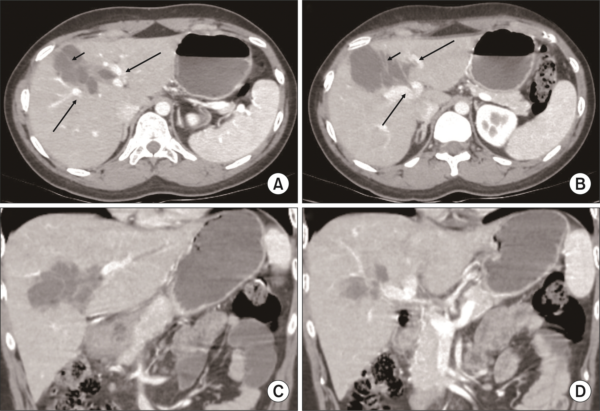
Fig. 1 Contrast enhanced computed tomography images. (A) Multiloculated cyst involving segment IVa and V of the liver with enhancing internal septa (short arrow), the cyst is in close proximity to the tributaries of the portal vein just above the confluence (long arrows). (B) At the level of the portal confluence (long arrows), cyst is seen extending into the segment IVb of the liver with multiple internal septations (short arrow). There are no other cystic lesions in the rest of the liver and other solid organs such as the kidney, pancreas and spleen. (C) Coronal image of the cyst anterior to the hilum in relation to the left portal pedicle. (D) Coronal image of the cyst near the hilum.
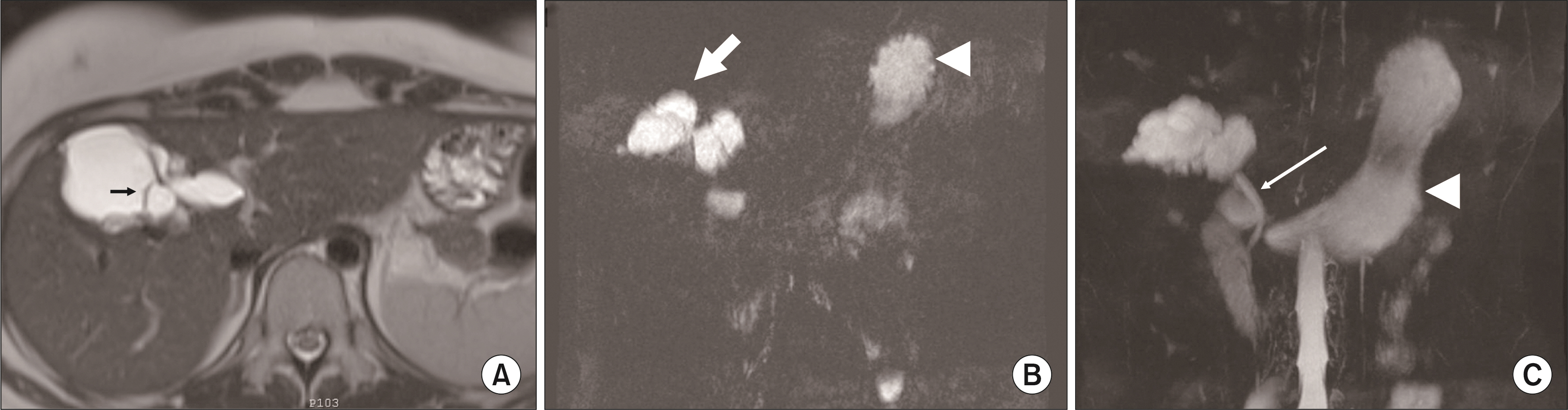
Fig. 2 Magnetic resonance images. (A) T2-weighted image showing a hyper-intense multilocular cyst with internal septa (short arrow). There is no biliary dilatation in the rest of the liver parenchyma. (B) Single-shot fast spin-echo sequence showing the bilobed butterfly like cystic structure (thick arrow), also seen is the fundic shadow (arrowhead). (C) Undilated distal common bile duct (long arrow), gastric shadow (arrowhead).
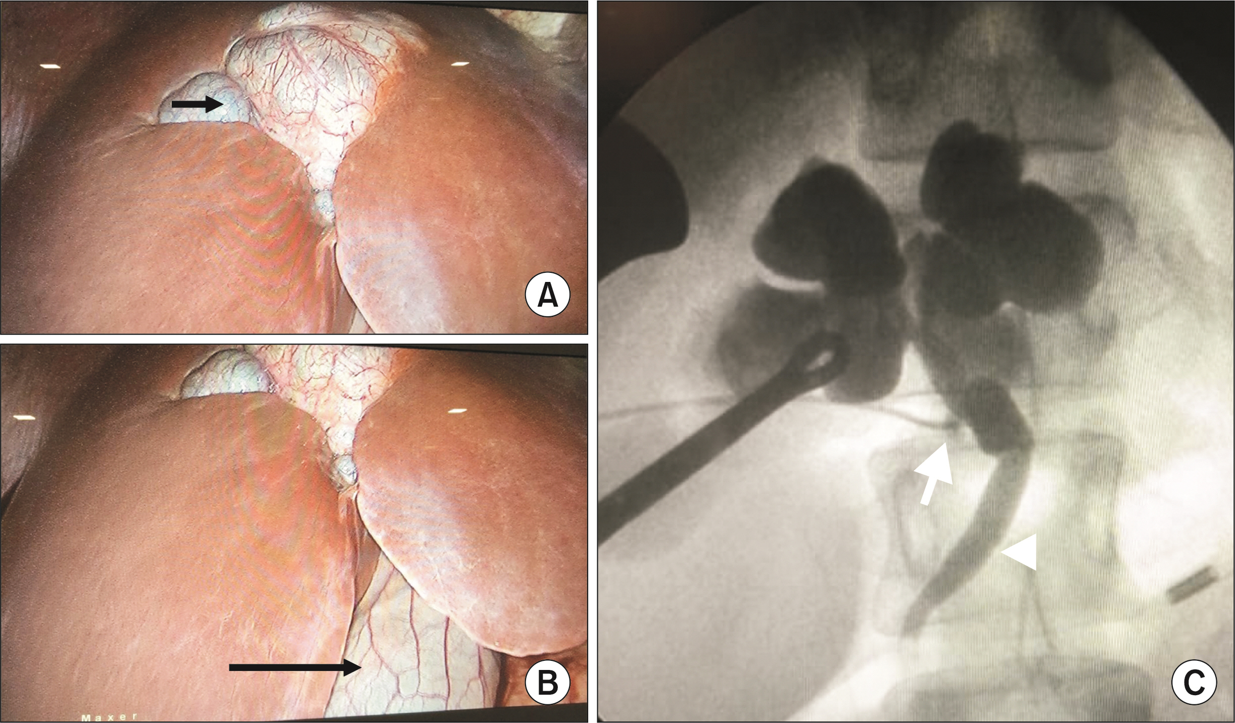
Fig. 3 (A) Laparoscopic image of the cyst (short arrow). (B) Gallbladder (long arrow) in relation to the cyst. Note a cleft in the liver at the region of the cyst. (C) Intraoperative cholangiogram done through the cystic duct (arrow) after cholecystectomy showing a bilobed structure in communication with the biliary system (arrowhead). The ducts distal to the cysts were not visualized.
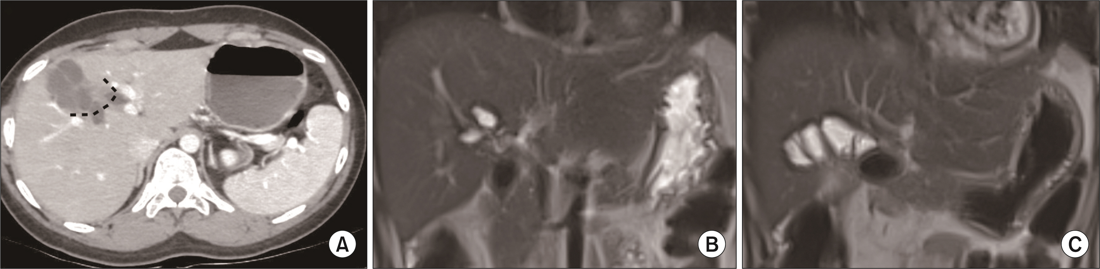
Fig. 4 (A) Representation of the extent of resection of the cyst on the computed tomography image (dashed lines). (B) Coronal image of the postoperative magnetic resonance imaging (MRI) shows the residual cystic structure near the hilum. (C) Coronal image of the postoperative MRI shows the cystojejunostomy.
Reference
-
1. Söreide K, Körner H, Havnen J, Söreide JA. 2004; Bile duct cysts in adults. Br J Surg. 91:1538–1548. DOI: 10.1002/bjs.4815. PMID: 15549778.2. Rawla P, Sunkara T, Muralidharan P, Raj JP. 2019; An updated review of cystic hepatic lesions. Clin Exp Hepatol. 5:22–29. DOI: 10.5114/ceh.2019.83153. PMID: 30915403. PMCID: PMC6431089.3. Wheeler DA, Edmondson HA. 1985; Cystadenoma with mesenchymal stroma (CMS) in the liver and bile ducts. A clinicopathologic study of 17 cases, 4 with malignant change. Cancer. 56:1434–1445. DOI: 10.1002/1097-0142(19850915)56:6<1434::AID-CNCR2820560635>3.0.CO;2-F. PMID: 4027877.4. Devaney K, Goodman ZD, Ishak KG. 1994; Hepatobiliary cystadenoma and cystadenocarcinoma. A light microscopic and immunohistochemical study of 70 patients. Am J Surg Pathol. 18:1078–1091. DOI: 10.1097/00000478-199411000-00002. PMID: 7943529.5. Gidi AD, González-Chávez MA, Villegas-Tovar E, Visag-Castillo V, Pantoja-Millan JP, Vélez-Pérez FM, et al. 2016; An unusual type of biliar cyst: a case report. Ann Hepatol. 15:788–794. DOI: 10.5604/16652681.1212617. PMID: 27493119.
- Full Text Links
-
- Actions
-
Cited
- CITED
-
- Close
- Share
-
- Similar articles
-
- A Case of a Choledochal Cyst with a Mucinous Cystadenoma of the Pancreas
- A case of type IVa choledochal cyst
- Type IV-A Choledochal Cyst with Intrahepatic Bile Duct Stricture
- A Case Report of an Unusual Type of Choledochal Cyst with Choledocholithiasis: Saccular Dilatation of the Confluent Portion of Both Intrahepatic Ducts
- Type IVB Choledochal Cyst : A case report
