A Case Report of an Unusual Type of Choledochal Cyst with Choledocholithiasis: Saccular Dilatation of the Confluent Portion of Both Intrahepatic Ducts
- Affiliations
-
- 1Department of Radiology, Eulji University Hospital, Daejeon, Korea. orionphil@hotmail.com
- KMID: 2068716
- DOI: http://doi.org/10.3348/jksr.2015.73.4.252
Abstract
- A choledochal cyst is a rare congenital anomaly of the biliary system manifested as the cystic dilatation of bile ducts, usually occurring in the common bile duct. Here, we describe an unusual type of choledochal cyst in a 45-year-old male that did not fit into the most widely accepted Todani classification of these cysts. The lesion mimicked duplication anomalies of the gallbladder and was finally diagnosed as a choledochal cyst involving the confluent portion of both intrahepatic ducts.
MeSH Terms
Figure
-
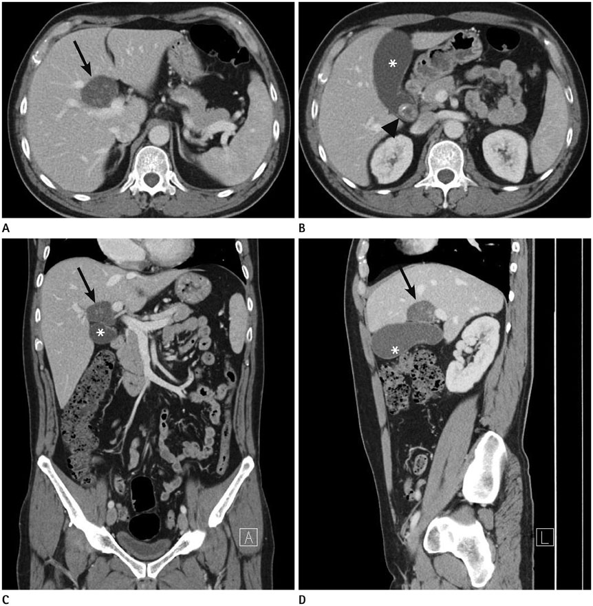
Fig. 1 Axial (A, B), coronal (C), and sagittal (D) views of a contrast-enhanced abdominal CT showing a round cystic lesion (arrow) with multiple internal calcified stones, at the porta hepatis (just superior to the gallbladder). Gallbladder (asterisk) showing a calcified stone (arrowhead) without mural thickening as an inflammation.
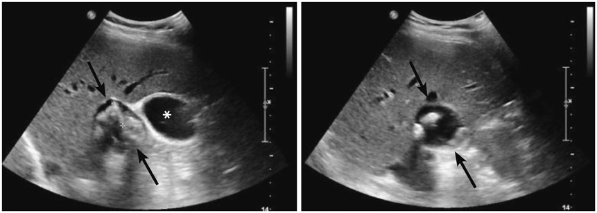
Fig. 2 Abdominal ultrasonography showing a thin-walled cystic lesion (arrows) abutting the gallbladder (asterisk) near the porta hepatis. It contains multiple echogenic foci with posterior shadowing, suggesting calcified stones. The lesion does not show the anatomical layering of its wall that is detected in the gallbladder.
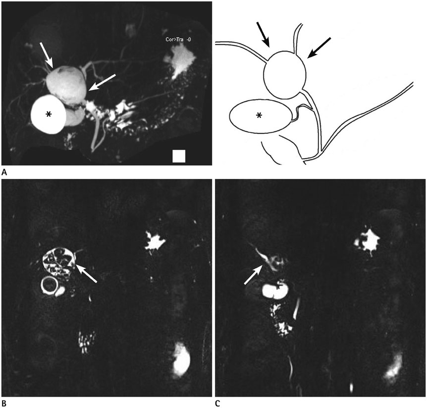
Fig. 3 Magnetic resonance cholangiopancreatography images. A. Maximal intensity projection image showing the gallbladder (asterisk) and a round cystic lesion (arrows) in the confluent portion of both intrahepatic ducts (IHDs). There is no anomalous pancreaticobiliary ductal union. A schematic representation is also shown (right). B, C. Left IHD (arrow in B) and right IHD (arrow in C) directly arose from the cyst, which is representative of the choledochal cyst. Multiple filling defects suggesting cholelithiasis and choledocholithiasis are shown in the gallbladder and choledochal cyst.

Fig. 4 Endoscopic retrograde cholangiopancreatography shows sequential contrast filling of the common bile duct, cystic lesion, and both intrahepatic ducts (IHDs) (arrows), which are consistent with a choledochal cyst occupying a confluent portion of both IHDs.
Reference
-
1. Michaelides M, Dimarelos V, Kostantinou D, Bintoudi A, Tzi-kos F, Kyriakou V, et al. A new variant of Todani type I choledochal cyst. Imaging evaluation. Hippokratia. 2011; 15:174–177.2. Babbitt DP, Starshak RJ, Clemett AR. Choledochal cyst: a concept of etiology. Am J Roentgenol Radium Ther Nucl Med. 1973; 119:57–62.3. Sadiq J, Nandi B, Lakhoo K. An unusual variant of choledochal cyst: a case report. J Med Case Rep. 2009; 3:54.4. Savader SJ, Benenati JF, Venbrux AC, Mitchell SE, Widlus DM, Cameron JL, et al. Choledochal cysts: classification and cholangiographic appearance. AJR Am J Roentgenol. 1991; 156:327–331.5. Yu J, Turner MA, Fulcher AS, Halvorsen RA. Congenital anomalies and normal variants of the pancreaticobiliary tract and the pancreas in adults: part 1, Biliary tract. AJR Am J Roentgenol. 2006; 187:1536–1543.6. Loke TK, Lam SH, Chan CS. Choledochal cyst: an unusual type of cystic dilatation of the cystic duct. AJR Am J Roentgenol. 1999; 173:619–620.7. Prekop I, Sevcik L, Jakubicka J, Bakos E, Bakos M, Durcansky D. Atypical cyst of the right ductus hepaticus. Bratisl Lek Listy. 2007; 108:474–476.8. Salles A, Kastenberg ZJ, Wall JK, Visser BC, Bruzoni M. Complete resection of a rare intrahepatic variant of a choledochal cyst. J Pediatr Surg. 2013; 48:652–654.9. Durgun AV, Gorgun E, Kapan M, Ozcelik MF, Eryilmaz R. Choledochal cysts in adults and the importance of differential diagnosis. J Hepatobiliary Pancreat Surg. 2002; 9:738–741.10. Sahoo MR, Kumar TA. Double gallbladder masquerading as a choledochal cyst: a case report. Int J Case Rep Image. 2013; 4:283–286.
- Full Text Links
-
- Actions
-
Cited
- CITED
-
- Close
- Share
-
- Similar articles
-
- Type IV-A Choledochal Cyst with Intrahepatic Bile Duct Stricture
- Postoperative Change of Intrahepatic Bile Duct Dilatation in Choledochal Cyst
- Choledochal Cysts in Children: Pre- and Postoperative Radiological Evaluation
- Choledochal Cyst Associated with Cystic Duct Dilatation: Report of Three Cases
- The diagnosis of choledochal cyst by ultrasound: report of two cases
