Anat Cell Biol.
2022 Jun;55(2):142-147. 10.5115/acb.22.037.
Three-dimensional linear and volumetric computed tomography analysis of the frontal sinus
- Affiliations
-
- 1Department of Anatomy, Research Institute of Medical Science, Konkuk University School of Medicine, Seoul, Korea
- KMID: 2531200
- DOI: http://doi.org/10.5115/acb.22.037
Abstract
- The frontal sinus is one of the four paranasal sinuses in humans, and knowledge of its anatomy is important when performing surgery involving the frontal bone or sinus. Although many studies have measured the frontal sinus using radiography and computed tomography (CT), few studies have evaluated by using three-dimensional (3D) analysis. The purpose of this study was to analyze the frontal sinus using 3D reconstruction analysis and determine the differences in linear and volumetric measurements between sexes, sides, and ages. The sample comprised 281 facial CT scans: 173 and 108 from males and females, respectively. The width, height, and length of each frontal sinus and total volume were all larger in males than in females. Almost all linear and volumetric measurements were larger in young adults than in older for both sexes, but not all of the differences were statistically significant. Linear and volumetric measurements were larger for males than females regardless of age group. There were no statistically significant differences between the right and left sides except the width in males. The size of the frontal sinus was strongly influenced by sex and age. The measurements reported here might be useful for improving surgical procedures involving the frontal sinus.
Keyword
Figure
-

Fig. 1 Computed tomography images and three-dimensionally reconstructed frontal sinuses. (A) Axial view of the frontal sinus, (B) sagittal view of the frontal sinus, (C) coronal view of the frontal sinus, (D) three-dimensional volume of the frontal sinus.
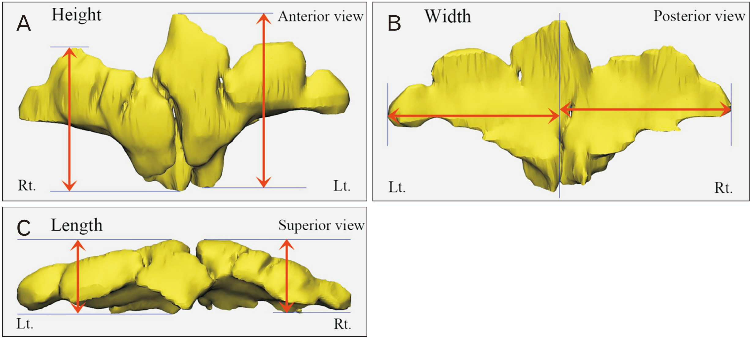
Fig. 2 Height, width, and length of the frontal sinus measured. (A) Left and right heights were respectively measured as the vertical distance between the lowest point and the highest point at the anterior view. (B) Left and right widths were respectively measured as the longest horizontal distance from the mid-sagittal plane. (C) Left and right lengths were respectively measured as the sagittal distance at the superior view.
Reference
-
References
1. Standring S. 2020. Gray's Anatomy: the anatomical basis of clinical practice. 42nd ed. Elsevier;London:2. Kountakis SE, Senior BA, Draf W. 2016. The frontal sinus. 2nd ed. Springer;Berlin: p. 21–31. DOI: 10.1007/978-3-662-48523-1.3. Yun IS, Kim YO, Lee SK, Rah DK. 2011; Three-dimensional computed tomographic analysis of frontal sinus in Asians. J Craniofac Surg. 22:462–7. DOI: 10.1097/SCS.0b013e3182074367. PMID: 21403576.
Article4. Zinreich SJ. 2006; Progress in sinonasal imaging. Ann Otol Rhinol Laryngol Suppl. 196:61–5. DOI: 10.1177/00034894061150S910. PMID: 17040020.
Article5. Sharma BN, Panta OB, Lohani B, Khanal U. 2015; Computed tomography in the evaluation of pathological lesions of paranasal sinuses. J Nepal Health Res Counc. 13:116–20. PMID: 26744195. PMID: 24af5dd0309d4f6e84eec9e64a736341.6. Choi IGG, Duailibi-Neto EF, Beaini TL, da Silva RLB, Chilvarquer I. 2018; The frontal sinus cavity exhibits sexual dimorphism in 3D cone-beam CT images and can be used for sex determination. J Forensic Sci. 63:692–8. DOI: 10.1111/1556-4029.13601. PMID: 28731499.
Article7. Sahlstrand-Johnson P, Jannert M, Strömbeck A, Abul-Kasim K. 2011; Computed tomography measurements of different dimensions of maxillary and frontal sinuses. BMC Med Imaging. 11:8. DOI: 10.1186/1471-2342-11-8. PMID: 21466703. PMCID: PMC3080316.
Article8. Hacl A, Costa ALF, Mayara Oliveira J, José Tucunduva M, Raul Girondi J, Nahás-Scocate ACR. 2017; Three-dimensional volumetric analysis of frontal sinus using medical software. J Forensic Radiol Imaging. 11:1–5. DOI: 10.1016/j.jofri.2017.08.004.
Article9. Shizuki K, Nameki H. 2014; Drainage pathways of the frontal sinus on oblique multi-planar reconstruction CT scans. Nihon Jibiinkoka Gakkai Kaiho. 117:1073–9. Japanese. DOI: 10.3950/jibiinkoka.117.1073. PMID: 25255645.10. Patel NS, Dearking AC, O'Brien EK, Pallanch JF. 2017; Virtual mapping of the frontal recess: guiding safe and efficient frontal sinus surgery. Otolaryngol Head Neck Surg. 156:946–51. DOI: 10.1177/0194599817699562. PMID: 28418817.
Article11. Kim DI, Lee UY, Park SO, Kwak DS, Han SH. 2013; Identification using frontal sinus by three-dimensional reconstruction from computed tomography. J Forensic Sci. 58:5–12. DOI: 10.1111/j.1556-4029.2012.02185.x. PMID: 22563883.
Article12. Gibelli D, Cellina M, Cappella A, Gibelli S, Panzeri MM, Oliva AG, Termine G, De Angelis D, Cattaneo C, Sforza C. 2019; An innovative 3D-3D superimposition for assessing anatomical uniqueness of frontal sinuses through segmentation on CT scans. Int J Legal Med. 133:1159–65. DOI: 10.1007/s00414-018-1895-4. PMID: 30039273.
Article13. Lee MK, Sakai O, Spiegel JH. 2010; CT measurement of the frontal sinus - gender differences and implications for frontal cranioplasty. J Craniomaxillofac Surg. 38:494–500. DOI: 10.1016/j.jcms.2010.02.001. PMID: 20335041.
Article14. Tatlisumak E, Ovali GY, Asirdizer M, Aslan A, Ozyurt B, Bayindir P, Tarhan S. 2008; CT study on morphometry of frontal sinus. Clin Anat. 21:287–93. DOI: 10.1002/ca.20617. PMID: 18428994.
Article15. Kirk NJ, Wood RE, Goldstein M. 2002; Skeletal identification using the frontal sinus region: a retrospective study of 39 cases. J Forensic Sci. 47:318–23. DOI: 10.1520/JFS15250J. PMID: 11908601.
Article16. Quatrehomme G, Fronty P, Sapanet M, Grévin G, Bailet P, Ollier A. 1996; Identification by frontal sinus pattern in forensic anthropology. Forensic Sci Int. 83:147–53. DOI: 10.1016/S0379-0738(96)02033-6. PMID: 9022276.
Article17. Pondé JM, Metzger P, Amaral G, Machado M, Prandini M. 2003; Anatomic variations of the frontal sinus. Minim Invasive Neurosurg. 46:29–32. DOI: 10.1055/s-2003-37956. PMID: 12640580.
Article18. Kanat A, Yazar U, Ozdemir B, Coskun ZO, Erdivanli O. 2015; Frontal sinus asymmetry: is it an effect of cranial asymmetry? X-ray analysis of 469 normal adult human frontal sinus. J Neurosci Rural Pract. 6:511–4. DOI: 10.4103/0976-3147.168436. PMID: 26752894. PMCID: PMC4692007.
Article19. Yüksel Aslier NG, Karabay N, Zeybek G, Keskinoğlu P, Kiray A, Sütay S, Ecevit MC. 2016; The classification of frontal sinus pneumatization patterns by CT-based volumetry. Surg Radiol Anat. 38:923–30. DOI: 10.1007/s00276-016-1644-7. PMID: 26884400.
Article20. Wanzeler AMV, Alves-Júnior SM, Ayres L, da Costa Prestes MC, Gomes JT, Tuji FM. 2019; Sex estimation using paranasal sinus discriminant analysis: a new approach via cone beam computerized tomography volume analysis. Int J Legal Med. 133:1977–84. DOI: 10.1007/s00414-019-02100-6. PMID: 31236677.
Article21. Kapakin S. 2016; The paranasal sinuses: three-dimensional reconstruction, photo-realistic imaging, and virtual endoscopy. Folia Morphol (Warsz). 75:326–33. DOI: 10.5603/FM.a2016.0006. PMID: 26916200.
Article22. Park IH, Song JS, Choi H, Kim TH, Hoon S, Lee SH, Lee HM. 2010; Volumetric study in the development of paranasal sinuses by CT imaging in Asian: a pilot study. Int J Pediatr Otorhinolaryngol. 74:1347–50. DOI: 10.1016/j.ijporl.2010.08.018. PMID: 20863577.
Article23. Michel J, Paganelli A, Varoquaux A, Piercecchi-Marti MD, Adalian P, Leonetti G, Dessi P. 2015; Determination of sex: interest of frontal sinus 3D reconstructions. J Forensic Sci. 60:269–73. DOI: 10.1111/1556-4029.12630. PMID: 25676659.
Article24. McLaughlin RB Jr, Rehl RM, Lanza DC. 2001; Clinically relevant frontal sinus anatomy and physiology. Otolaryngol Clin North Am. 34:1–22. DOI: 10.1016/S0030-6665(05)70291-7. PMID: 11344058.
Article
- Full Text Links
-
- Actions
-
Cited
- CITED
-
- Close
- Share
-
- Similar articles
-
- Post-Traumatic Pneumocele of the Frontal Sinus
- Endoscopic Frontal Sinus Surgery
- Pott's Puffy Tumor Arising from Frontal Sinusitis
- Volumetric growth analysis of maxillary sinus using computed tomography scan segmentation: a pilot study of Indonesian population
- Frontoethmoidal Cells on Computed Tomographic Analysis: The Prevalence and Relationship to Frontal Sinus/Recess Mucosal Thickening
