Endoscopic ultrasound-guided portal vein coiling: troubleshooting interventional endoscopic ultrasonography
- Affiliations
-
- 1Department of Gastroenterology, Aichi Cancer Center Hospital, Nagoya, Japan
- KMID: 2529970
- DOI: http://doi.org/10.5946/ce.2021.114
Abstract
- Endoscopic ultrasound (EUS)-guided hepaticogastrostomy (HGS) is widely performed not only as an alternative to transpapillary biliary drainage, but also as primary drainage for malignant biliary obstruction. For anatomical reasons, this technique carries an unavoidable risk of mispuncturing intrahepatic vessels. We report a technique for troubleshooting EUS-guided portal vein coiling to prevent bleeding from the intrahepatic portal vein after mispuncture during interventional EUS. EUS-HGS was planned for a 59-year-old male patient with unresectable pancreatic cancer. The dilated bile duct (lumen diameter, 2.8 mm) was punctured with a 19-gauge needle, and a guidewire was inserted. After bougie dilation, the guidewire was found to be inside the intrahepatic portal vein. Embolizing coils were placed to prevent bleeding. Embolization coils were successfully inserted under stabilization of the catheter using a double-lumen cannula with a guidewire. Following these procedures, the patient was asymptomatic. Computed tomography performed the next day revealed no complications.
Keyword
Figure
-

Fig. 1. (A) Computed tomography before the procedure reveals dilatation of the intrahepatic bile duct. (B) Color doppler endoscopic ultrasound image before procedure. Slightly dilated intrahepatic bile duct (arrow) and intrahepatic portal vein (arrowhead).
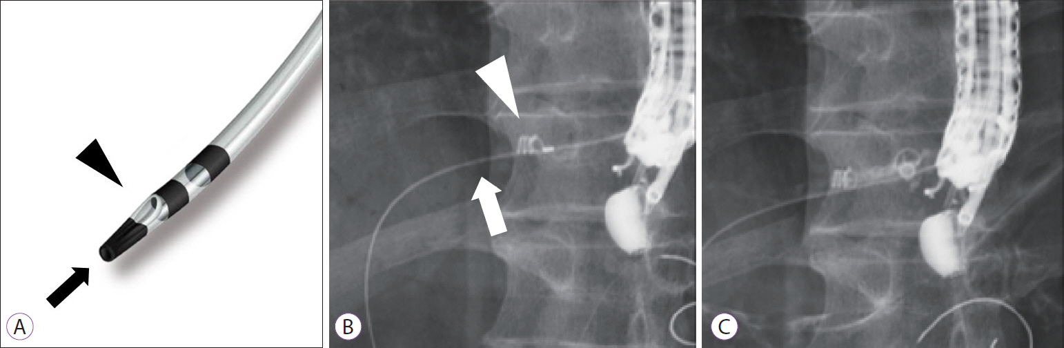
Fig. 2. (A) Image of double-lumen cannula. Main port (opening at the tip; arrow) is available for a 0.025-inch guide wire, and side port (opening at the side hole; arrowhead) is available for a 0.035-inch guide wire. (B) The guidewire (arrow) is inserted into the portal vein through the main port of the cannula and coils (arrowhead) were placed through the side port. (C) The first coil remains peripherally in the portal vein, while the second coil is placed along the puncture route.
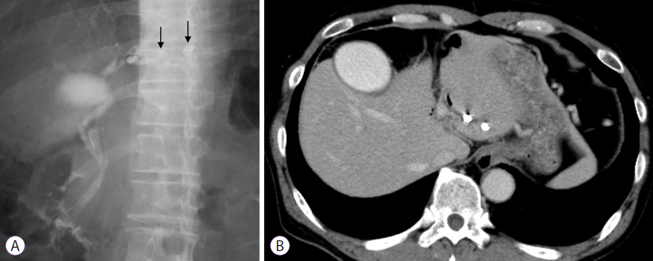
Fig. 3. (A) The final image of the procedure shows placed coils (arrows) and an inserted transpapillary stent. (B) Computed tomography the day after the procedure. The peripheral area of the intrahepatic portal vein is embolized.
Reference
-
1. Ogura T, Higuchi K. Endoscopic ultrasound-guided hepaticogastrostomy: technical review and tips to prevent adverse events. Gut Liver. 2021; 15:196–205.2. Okuno N, Hara K, Mizuno N, et al. Efficacy of the 6-mm fully covered self-expandable metal stent during endoscopic ultrasound-guided hepaticogastrostomy as a primary biliary drainage for the cases estimated difficult endoscopic retrograde cholangiopancreatography: a prospective clinical study. J Gastroenterol Hepatol. 2018; 33:1413–1421.3. Oh D, Park DH, Song TJ, et al. Optimal biliary access point and learning curve for endoscopic ultrasound-guided hepaticogastrostomy with transmural stenting. Therap Adv Gastroenterol. 2017; 10:42–53.4. Miyano A, Ogura T, Yamamoto K, et al. Clinical impact of the intra-scope channel stent release technique in preventing stent migration during EUS-guided hepaticogastrostomy. J Gastrointest Surg. 2018; 22:1312–1318.5. Kawakubo K, Isayama H, Kato H, et al. Multicenter retrospective study of endoscopic ultrasound-guided biliary drainage for malignant biliary obstruction in Japan. J Hepatobiliary Pancreat Sci. 2014; 21:328–334.6. Moryoussef F, Sportes A, Leblanc S, et al. Is EUS-guided drainage a suitable alternative technique in case of proximal biliary obstruction? Therap Adv Gastroenterol. 2017; 10:537–544.7. Levy MJ, Wong Kee Song LM, Kendrick ML, et al. EUS-guided coil embolization for refractory ectopic variceal bleeding (with videos). Gastrointest Endosc. 2008; 67:572–574.8. Robles-Medranda C, Oleas R, Valero M, et al. Endoscopic ultrasonography-guided deployment of embolization coils and cyanoacrylate injection in gastric varices versus coiling alone: a randomized trial. Endoscopy. 2020; 52:268–275.9. Chantarojanasiri T, Sirinawasatien A, Bunchorntavakul C, et al. Endoscopic ultrasound-guided vascular therapy for portoduodenal fistula. Clin Endosc. 2020; 53:750–753.10. Cho DH, Lee SS, Oh D, et al. Long-term outcomes of a newly developed hybrid metal stent for EUS-guided biliary drainage (with videos). Gastrointest Endosc. 2017; 85:1067–1075.11. Saad WEA, Davies MG, Darcy MD. Management of bleeding after percutaneous transhepatic cholangiography or transhepatic biliary drain placement. Tech Vasc Interv Radiol. 2008; 11:60–71.12. Winick AB, Waybill PN, Venbrux AC. Complications of percutaneous transhepatic biliary interventions. Tech Vasc Interv Radiol. 2001; 4:200–206.
- Full Text Links
-
- Actions
-
Cited
- CITED
-
- Close
- Share
-
- Similar articles
-
- Endoscopic Ultrasound-Guided Portal Pressure Measurement and Interventions
- Endoscopic ultrasound-guided vascular interventions: An overview of current and emerging techniques
- Endoscopic ultrasound-guided vascular intervention for portal hypertension
- Endoscopic Ultrasound-Guided Vascular Procedures: A Review
- Recent development of endoscopic ultrasound-guided biliary drainage
