Endoscopic Ultrasonography Findings for Brunner’s Gland Hamartoma in the Duodenum
- Affiliations
-
- 1Department of Internal Medicine, Yonsei University Wonju College of Medicine, Wonju, Korea
- KMID: 2527578
- DOI: http://doi.org/10.5946/ce.2020.259
Figure
-
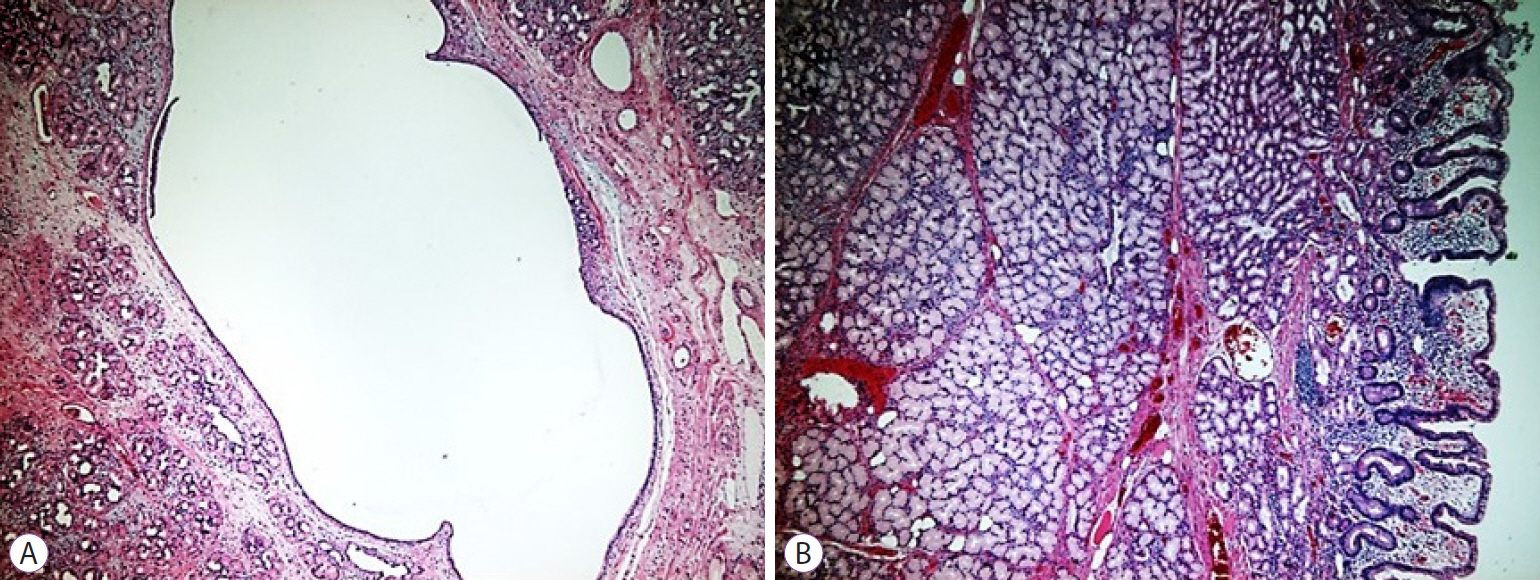
Fig. 1. Brunner's gland hamartoma or adenoma (hematoxylin & eosin stain, ×200). (A) Brunner's gland hamartoma is composed of a dilated cyst. (B) Hyperplasia of Brunner’s glands and fibrous stroma.
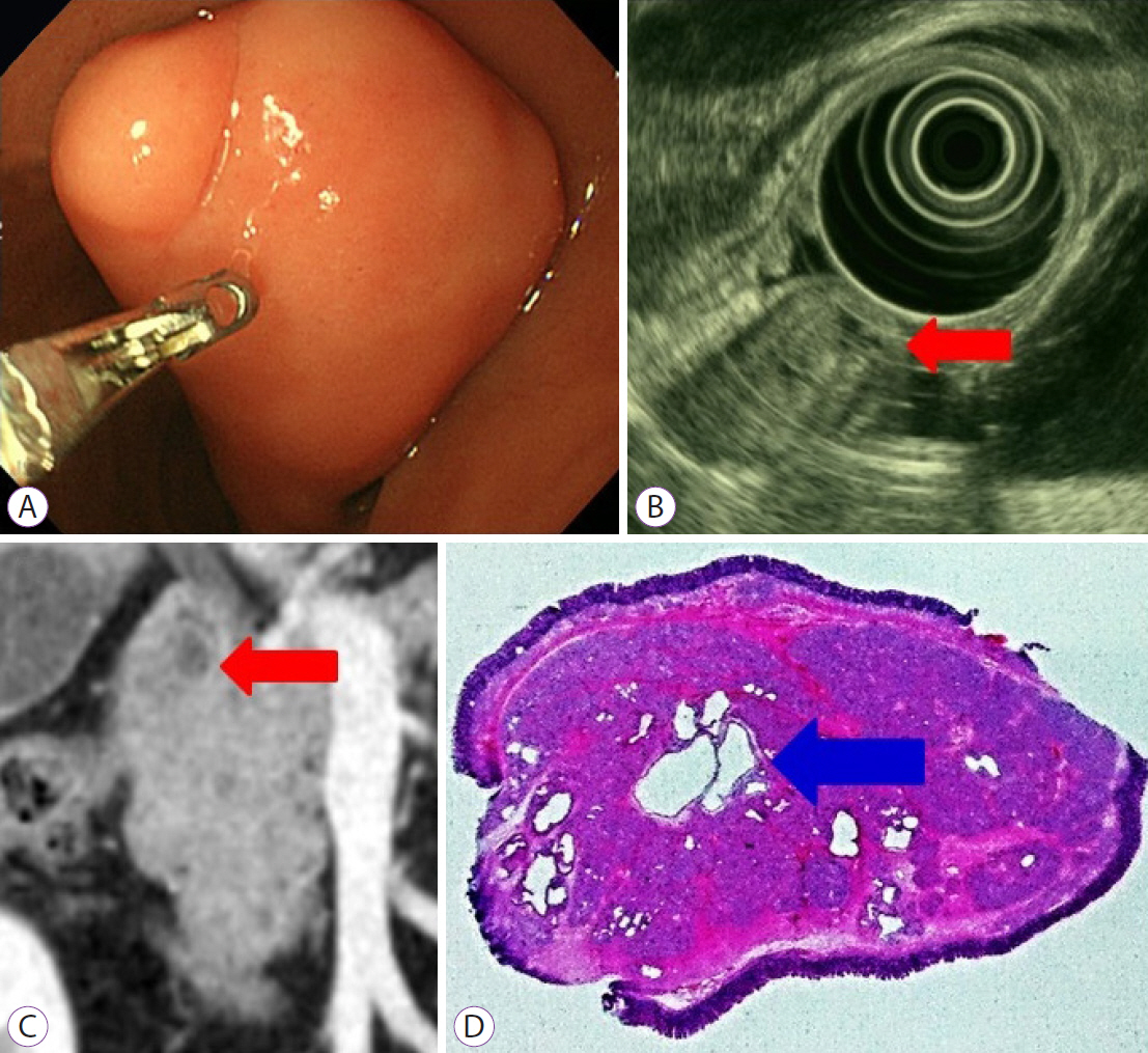
Fig. 2. (A) Endoscopy reveals a pedunculated polypoid mass in the duodenal bulb. (B) Endoscopic ultrasound reveals a heterogeneous appearance with an anechoic lesion representing cysts (red arrow). (C) On computed tomography, a round mass with a well-demarcated cystic lesion in the duodenal bulb is shown (red arrow). (D) Histological examination reveals the proliferation of Brunner's glands and fibrous stroma with several dilated cysts (blue arrow, hematoxylin & eosin stain, ×10).

Fig. 3. (A) Endoscopy reveals a round sessile polypoid mass in the second portion of the duodenum (red arrow). (B) On endoscopic ultrasond, a heterogeneous hyperechoic appearance without anechoic lesions is shown (red arrow). (C) Histological examination reveals dense Brunner's gland proliferation without definite cystic lesions (hematoxylin & eosin stain, ×10)
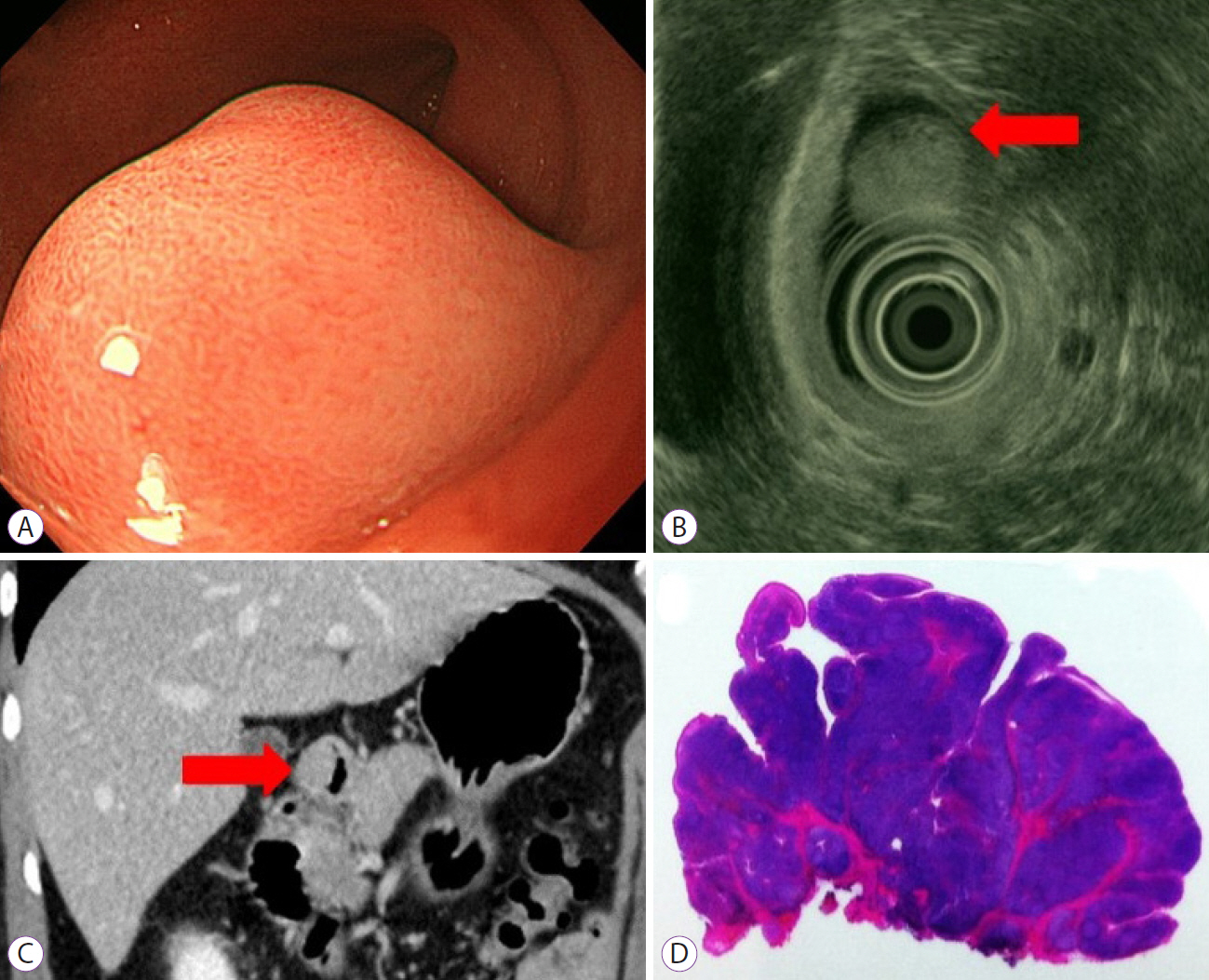
Fig. 4. (A) Endoscopy reveals a round sessile polypoid mass in the duodenal bulb. (B) Endoscopic ultrasound reveals a heterogeneous appearance, with tiny anechoic lesions representing cysts (red arrow). (C) On computed tomography, a homogeneous round mass in the duodenal bulb is shown (red arrow). (D) Histological examination reveals the proliferation of Brunner's glands and fibrous stroma without cystic lesions (hematoxylin & eosin stain, ×10).
Reference
-
1. Lu L, Li R, Zhang G, Zhao Z, Fu W, Li W. Brunner's gland adenoma of duodenum: report of two cases. Int J Clin Exp Pathol. 2015; 8:7565–7569.2. Botsford TW, Crowe P, Crocker DW. Tumors of the small intestine. A review of experience with 115 cases including a report of a rare case of malignant hemangio-endothelioma. Am J Surg. 1962; 103:358–365.3. Matsui T, Iida M, Fujischima M, Sakamoto K, Watanabe H. Brunner's gland hamartoma associated with microcarcinoids. Endoscopy. 1989; 21:37–38.4. Weisselberg B, Melzer E, Liokumovich P, Kurnik D, Koller M, Bar-Meir S. The endoscopic ultrasonographic appearance of Brunner's gland hamartoma. Gastrointest Endosc. 1997; 46:176–178.5. Matsushita M, Hajiro K, Takakuwa H, Nishio A. Characteristic EUS appearance of Brunner's gland hamartoma. Gastrointest Endosc. 1999; 49:670–672.6. Hizawa K, Iwai K, Esaki M, et al. Endosonographic features of Brunner's gland hamartomas which were subsequently resected endoscopically. Endoscopy. 2002; 34:956–958.7. Rocco A, Borriello P, Compare D, et al. Large Brunner's gland adenoma: case report and literature review. World J Gastroenterol. 2006; 12:1966–1968.8. Levine JA, Burgart LJ, Batts KP, Wang KK. Brunner's gland hamartomas: clinical presentation and pathological features of 27 cases. Am J Gastroenterol. 1995; 90:290–294.9. Walden DT, Marcon NE. Endoscopic injection and polypectomy for bleeding Brunner's gland hamartoma: case report and expanded literature review. Gastrointest Endosc. 1998; 47:403–407.10. Fujimaki E, Nakamura S, Sugai T, Takeda Y. Brunner's gland adenoma with a focus of p53-positive atypical glands. J Gastroenterol. 2000; 35:155–8.11. Changchien CS, Hsu CC, Hu TH. Endosonographic appearances of Brunner's gland hamartomas. J Clin Ultrasound. 2001; 29:243–246.
- Full Text Links
-
- Actions
-
Cited
- CITED
-
- Close
- Share
-
- Similar articles
-
- A Case of Brunner's Gland Hyperplasia with Dysplasia
- A Case of Giant Brunner's Gland Hamartoma Presenting as Gastrointestinal Hemorrhage
- A Case of a Giant Brunner's Gland Hamartoma Originating from the Pyloric Ring
- A Brunner's Gland Adenoma Removed by Endoscopic Polypectomy
- Brunner's Gland Hamartoma Causing Gastric Outlet Obstruction Treated by Endoscopic Resection
