Comparison of cone-beam computed tomography and digital panoramic radiography for detecting peri-implant alveolar bone changes using trabecular micro-structure analysis
- Affiliations
-
- 1Department of Oral and Maxillofacial Radiology, Faculty of Dentistry, Necmettin Erbakan University, Konya, Turkey
- 2Department of Periodontology, Faculty of Dentistry, Necmettin Erbakan University, Konya, Turkey
- 3Department of Oral and Maxillofacial Radiology, Faculty of Dentistry, Ankara University, Ankara, Turkey
- KMID: 2526809
- DOI: http://doi.org/10.5125/jkaoms.2022.48.1.41
Abstract
Objectives
We compared changes in fractal dimension (FD) and grayscale value (GSV) of peri-implant alveolar bone on digital panoramic radiography (DPR) and cone-beam computed tomography (CBCT) immediately after implant surgery and 12 months postoperative.
Materials and Methods
In this retrospective study, 16 patients who received posterior mandibular area dental implants with CBCT scans taken about 2 weeks after implantation and one year after implantation were analyzed. A region of interest was selected for each patient. FDs and GSVs were evaluated immediately after implant surgery and at 12-month follow-up to examine the functional loading of the implants.
Results
There were no significant differences between DPR and CBCT measurements of FD values (P>0.05). No significant differences were observed between FD values and GSVs calculated after implant surgery and at the 12-month follow-up (P>0.05). GSVs were not correlated with FD values (P>0.05).
Conclusion
The DPR and reconstructed panoramic CBCT images exhibit similar image quality for the assessment of FD. There were no changes in FD values or GSVs of the peri-implant trabecular bone structure at the 12-month postoperative evaluation of the functional loading of the implant in comparison to values immediately after implantation. GSVs representing bone mass do not align with FD values that predict bone microstructural parameters. Therefore, GSVs and FDs should be considered different parameters for assessing bone quality.
Keyword
Figure
-

Fig. 1 Region of interest selection at mesial (25×50 pixels), distal (25×50 pixels), and apical (50×25 pixels) areas on digital panoramic radiography and reformatted panoramic images of cone-beam computed tomography.
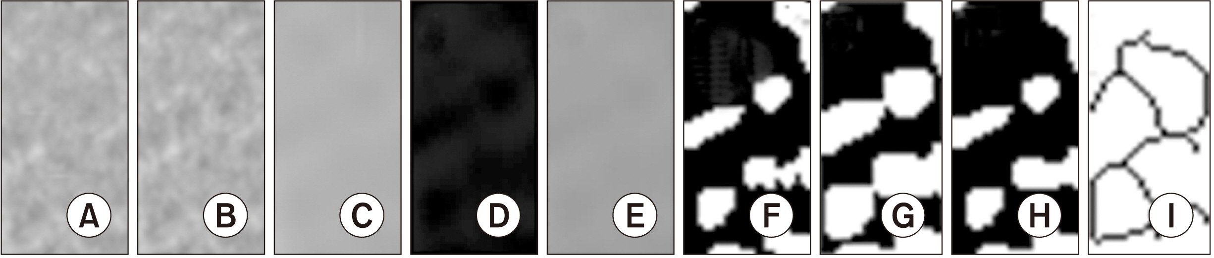
Fig. 2 A. Region of interest (ROI) on digital panoramic radiography and cone-beam volumetric tomography images were cropped and transferred to ImageJ. B, C. The cropped ROI was duplicated (B) and then blurred with a Gaussian filter (C). D, E. The blurred image was subtracted from the original image (D), and 128 was added to the result at each pixel location (E). F. The resultant image was converted to binary, to set the image into trabeculae and marrow spaces. G, H. The binary image was eroded then dilated to reduce the noise before skeletonization. I. The skeletonized image was used for fractal analysis.

Fig. 3 The selection of grayscale value (GSV) (4 mm2) at the implant apex on the cross sectional image of cone-beam computed tomography (CBCT). The mean GSV was measured automatically by the CBCT software program.
Reference
-
References
1. Donos N, Mardas N, Chadha V. 2008; Clinical outcomes of implants following lateral bone augmentation: systematic assessment of available options (barrier membranes, bone grafts, split osteotomy). J Clin Periodontol. 35(8 Suppl):173–202. https://doi.org/10.1111/j.1600-051X.2008.01269.x. DOI: 10.1111/j.1600-051X.2008.01269.x. PMID: 18724850.
Article2. Retzepi M, Donos N. 2010; Guided bone regeneration: biological principle and therapeutic applications. Clin Oral Implants Res. 21:567–76. https://doi.org/10.1111/j.1600-0501.2010.01922.x. DOI: 10.1111/j.1600-0501.2010.01922.x. PMID: 20666785.
Article3. Santing HJ, Raghoebar GM, Vissink A, den Hartog L, Meijer HJ. 2013; Performance of the Straumann Bone Level Implant system for anterior single-tooth replacements in augmented and nonaugmented sites: a prospective cohort study with 60 consecutive patients. Clin Oral Implants Res. 24:941–8. https://doi.org/10.1111/j.1600-0501.2012.02486.x. DOI: 10.1111/j.1600-0501.2012.02486.x. PMID: 22540833.
Article4. Shalabi MM, Wolke JG, Jansen JA. 2006; The effects of implant surface roughness and surgical technique on implant fixation in an in vitro model. Clin Oral Implants Res. 17:172–8. https://doi.org/10.1111/j.1600-0501.2005.01202.x. DOI: 10.1111/j.1600-0501.2005.01202.x. PMID: 16584413.
Article5. Brunski JB, Puleo DA, Nanci A. 2000; Biomaterials and biomechanics of oral and maxillofacial implants: current status and future developments. Int J Oral Maxillofac Implants. 15:15–46. PMID: 10697938.6. Stanford CM, Brand RA. 1999; Toward an understanding of implant occlusion and strain adaptive bone modeling and remodeling. J Prosthet Dent. 81:553–61. https://doi.org/10.1016/s0022-3913(99)70209-x. DOI: 10.1016/S0022-3913(99)70209-X. PMID: 10220659.
Article7. Bidez MW, Misch CE. 1992; Force transfer in implant dentistry: basic concepts and principles. J Oral Implantol. 18:264–74. PMID: 1289562.8. Drago CJ. 1992; Rates of osseointegration of dental implants with regard to anatomical location. J Prosthodont. 1:29–31. https://doi.org/10.1111/j.1532-849x.1992.tb00423.x. DOI: 10.1111/j.1532-849X.1992.tb00423.x. PMID: 1308217.
Article9. Jemt T, Lekholm U. 1993; Oral implant treatment in posterior partially edentulous jaws: a 5-year follow-up report. Int J Oral Maxillofac Implants. 8:635–40. DOI: 10.1034/j.1600-0501.1994.050304.x. PMID: 7827228.10. Lee DH, Ku Y, Rhyu IC, Hong JU, Lee CW, Heo MS, et al. 2010; A clinical study of alveolar bone quality using the fractal dimension and the implant stability quotient. J Periodontal Implant Sci. 40:19–24. https://doi.org/10.5051/jpis.2010.40.1.19. DOI: 10.5051/jpis.2010.40.1.19. PMID: 20498755. PMCID: PMC2872807.
Article11. Yasar F, Akgünlü F. 2005; Fractal dimension and lacunarity analysis of dental radiographs. Dentomaxillofac Radiol. 34:261–7. https://doi.org/10.1259/dmfr/85149245. DOI: 10.1259/dmfr/85149245. PMID: 16120874.
Article12. Huh KH, Baik JS, Yi WJ, Heo MS, Lee SS, Choi SC, et al. 2011; Fractal analysis of mandibular trabecular bone: optimal tile sizes for the tile counting method. Imaging Sci Dent. 41:71–8. https://doi.org/10.5624/isd.2011.41.2.71. DOI: 10.5624/isd.2011.41.2.71. PMID: 21977478. PMCID: PMC3174468.
Article13. Zeytinoğlu M, İlhan B, Dündar N, Boyacioğlu H. 2015; Fractal analysis for the assessment of trabecular peri-implant alveolar bone using panoramic radiographs. Clin Oral Investig. 19:519–24. https://doi.org/10.1007/s00784-014-1245-y. DOI: 10.1007/s00784-014-1245-y. PMID: 24802628.
Article14. Jolley L, Majumdar S, Kapila S. 2006; Technical factors in fractal analysis of periapical radiographs. Dentomaxillofac Radiol. 35:393–7. https://doi.org/10.1259/dmfr/30969642. DOI: 10.1259/dmfr/30969642. PMID: 17082328.
Article15. Melsen B, Lang NP. 2001; Biological reactions of alveolar bone to orthodontic loading of oral implants. Clin Oral Implants Res. 12:144–52. https://doi.org/10.1034/j.1600-0501.2001.012002144.x. DOI: 10.1034/j.1600-0501.2001.012002144.x. PMID: 11251664.
Article16. Bianchi AE, Dolci G Jr, Sberna MT, Sanfilippo S. 2005; Factors affecting bone response around loaded titanium dental implants: a literature review. J Appl Biomater Biomech. 3:135–40. PMID: 20799218.17. Jung YH. 2005; Evaluation of peri-implant bone using fractal analysis. Korean J Oral Maxillofac Radiol. 35:121–5.18. Perrotti V, Aprile G, Degidi M, Piattelli A, Iezzi G. 2011; Fractal analysis: a novel method to assess roughness organization of implant surface topography. Int J Periodontics Restorative Dent. 31:633–9. PMID: 22140665.19. Bollen AM, Taguchi A, Hujoel PP, Hollender LG. 2001; Fractal dimension on dental radiographs. Dentomaxillofac Radiol. 30:270–5. https://doi.org/10.1038/sj/dmfr/4600630. DOI: 10.1038/sj.dmfr.4600630. PMID: 11571547.
Article20. Dohan Ehrenfest DM. 2011; Fractal patterns applied to implant surface: definitions and perspectives. J Oral Implantol. 37:506–9. https://doi.org/10.1563/AAID-JOI-D-11-00081. DOI: 10.1563/AAID-JOI-D-11-00081. PMID: 21668354.
Article21. González-García R, Monje F. 2013; Is micro-computed tomography reliable to determine the microstructure of the maxillary alveolar bone? Clin Oral Implants Res. 24:730–7. https://doi.org/10.1111/j.1600-0501.2012.02478.x. DOI: 10.1111/j.1600-0501.2012.02478.x. PMID: 22540518.
Article22. Müller R, Van Campenhout H, Van Damme B, Van Der Perre G, Dequeker J, Hildebrand T, et al. 1998; Morphometric analysis of human bone biopsies: a quantitative structural comparison of histological sections and micro-computed tomography. Bone. 23:59–66. https://doi.org/10.1016/s8756-3282(98)00068-4. DOI: 10.1016/S8756-3282(98)00068-4. PMID: 9662131.
Article23. Swain MV, Xue J. 2009; State of the art of Micro-CT applications in dental research. Int J Oral Sci. 1:177–88. https://doi.org/10.4248/IJOS09031. DOI: 10.4248/IJOS09031. PMID: 20690421. PMCID: PMC3470105.
Article24. Aranyarachkul P, Caruso J, Gantes B, Schulz E, Riggs M, Dus I, et al. 2005; Bone density assessments of dental implant sites: 2. Quantitative cone-beam computerized tomography. Int J Oral Maxillofac Implants. 20:416–24. PMID: 15973953.25. Turkyilmaz I, Sennerby L, McGlumphy EA, Tözüm TF. 2009; Biomechanical aspects of primary implant stability: a human cadaver study. Clin Implant Dent Relat Res. 11:113–9. https://doi.org/10.1111/j.1708-8208.2008.00097.x. DOI: 10.1111/j.1708-8208.2008.00097.x. PMID: 18422713.
Article26. Naitoh M, Hirukawa A, Katsumata A, Ariji E. 2009; Evaluation of voxel values in mandibular cancellous bone: relationship between cone-beam computed tomography and multislice helical computed tomography. Clin Oral Implants Res. 20:503–6. https://doi.org/10.1111/j.1600-0501.2008.01672.x. DOI: 10.1111/j.1600-0501.2008.01672.x. PMID: 19250241.
Article27. Pauwels R, Nackaerts O, Bellaiche N, Stamatakis H, Tsiklakis K, Walker A, et al. 2013; SEDENTEXCT Project Consortium. Variability of dental cone beam CT grey values for density estimations. Br J Radiol. 86:20120135. https://doi.org/10.1259/bjr.20120135. DOI: 10.1259/bjr.20120135. PMID: 23255537. PMCID: PMC4651064.
Article28. Vandenberghe B, Jacobs R, Bosmans H. 2010; Modern dental imaging: a review of the current technology and clinical applications in dental practice. Eur Radiol. 20:2637–55. https://doi.org/10.1007/s00330-010-1836-1. DOI: 10.1007/s00330-010-1836-1. PMID: 20544352.
Article29. Tyndall DA, Price JB, Tetradis S, Ganz SD, Hildebolt C. Scarfe WC; American Academy of Oral and Maxillofacial Radiology. 2012; Position statement of the American Academy of Oral and Maxillofacial Radiology on selection criteria for the use of radiology in dental implantology with emphasis on cone beam computed tomography. Oral Surg Oral Med Oral Pathol Oral Radiol. 113:817–26. https://doi.org/10.1016/j.oooo.2012.03.005. DOI: 10.1016/j.oooo.2012.03.005. PMID: 22668710.
Article30. White SC, Cohen JM, Mourshed FA. 2000; Digital analysis of trabecular pattern in jaws of patients with sickle cell anemia. Dentomaxillofac Radiol. 29:119–24. https://doi.org/10.1038/sj/dmfr/4600516. DOI: 10.1038/sj.dmfr.4600516. PMID: 10808227.
Article31. Koh KJ, Park HN, Kim KA. 2012; Prediction of age-related osteoporosis using fractal analysis on panoramic radiographs. Imaging Sci Dent. 42:231–5. https://doi.org/10.5624/isd.2012.42.4.231. DOI: 10.5624/isd.2012.42.4.231. PMID: 23301209. PMCID: PMC3534177.
Article32. Brånemark PI, Hansson BO, Adell R, Breine U, Lindström J, Hallén O, et al. 1977; Osseointegrated implants in the treatment of the edentulous jaw. Experience from a 10-year period. Scand J Plast Reconstr Surg Suppl. 16:1–132. PMID: 356184.33. Haire TJ, Hodgskinson R, Ganney PS, Langton CM. 1998; A comparison of porosity, fabric and fractal dimension as predictors of the Young's modulus of equine cancellous bone. Med Eng Phys. 20:588–93. https://doi.org/10.1016/s1350-4533(98)00063-0. DOI: 10.1016/S1350-4533(98)00063-0.
Article34. Wilding RJ, Slabbert JC, Kathree H, Owen CP, Crombie K, Delport P. 1995; The use of fractal analysis to reveal remodelling in human alveolar bone following the placement of dental implants. Arch Oral Biol. 40:61–72. https://doi.org/10.1016/0003-9969(94)00138-2. DOI: 10.1016/0003-9969(94)00138-2. PMID: 7748114.
Article35. Southard TE, Southard KA, Jakobsen JR, Hillis SL, Najim CA. 1996; Fractal dimension in radiographic analysis of alveolar process bone. Oral Surg Oral Med Oral Pathol Oral Radiol Endod. 82:569–76. https://doi.org/10.1016/s1079-2104(96)80205-8. DOI: 10.1016/S1079-2104(96)80205-8. PMID: 8936523.
Article36. Geraets WG, van der Stelt PF. 2000; Fractal properties of bone. Dentomaxillofac Radiol. 29:144–53. https://doi.org/10.1038/sj/dmfr/4600524. DOI: 10.1038/sj.dmfr.4600524. PMID: 10849540.
Article37. Pawelzik J, Cohnen M, Willers R, Becker J. 2002; A comparison of conventional panoramic radiographs with volumetric computed tomography images in the preoperative assessment of impacted mandibular third molars. J Oral Maxillofac Surg. 60:979–84. https://doi.org/10.1053/joms.2002.34399. DOI: 10.1053/joms.2002.34399. PMID: 12215976.
Article38. Mischkowski RA, Ritter L, Neugebauer J, Dreiseidler T, Keeve E, Zöller JE. 2007; Diagnostic quality of panoramic views obtained by a newly developed digital volume tomography device for maxillofacial imaging. Quintessence Int. 38:763–72. PMID: 17873983.39. Pittayapat P, Galiti D, Huang Y, Dreesen K, Schreurs M, Souza PC, et al. 2013; An in vitro comparison of subjective image quality of panoramic views acquired via 2D or 3D imaging. Clin Oral Investig. 17:293–300. https://doi.org/10.1007/s00784-012-0698-0. DOI: 10.1007/s00784-012-0698-0. PMID: 22382448.
Article40. dos Santos Corpas L, Jacobs R, Quirynen M, Huang Y, Naert I, Duyck J. 2011; Peri-implant bone tissue assessment by comparing the outcome of intra-oral radiograph and cone beam computed tomography analyses to the histological standard. Clin Oral Implants Res. 22:492–9. https://doi.org/10.1111/j.1600-0501.2010.02029.x. DOI: 10.1111/j.1600-0501.2010.02029.x. PMID: 21143531.
Article41. Sansare K, Singh D, Karjodkar F. 2012; Changes in the fractal dimension on pre- and post-implant panoramic radiographs. Oral Radiol. 28:15–23. https://doi.org/10.1007/s11282-011-0075-8. DOI: 10.1007/s11282-011-0075-8.
Article42. Ilhan B, Güneri P, Saraçoglu A, Koca H, Boyacioglu H. 2015; A comparison of fractal dimension values of peri-implant bone and healthy contralateral side using panoramic radiographs. J Oral Maxillofac Radiol. 3:1–6. https://doi.org/10.4103/2321-3841.151636. DOI: 10.4103/2321-3841.151636.
Article43. Hua Y, Nackaerts O, Duyck J, Maes F, Jacobs R. 2009; Bone quality assessment based on cone beam computed tomography imaging. Clin Oral Implants Res. 20:767–71. https://doi.org/10.1111/j.1600-0501.2008.01677.x. DOI: 10.1111/j.1600-0501.2008.01677.x. PMID: 19489931.
Article44. González-Martín O, Lee EA, Veltri M. 2012; CBCT fractal dimension changes at the apex of immediate implants placed using undersized drilling. Clin Oral Implants Res. 23:954–7. https://doi.org/10.1111/j.1600-0501.2011.02246.x. DOI: 10.1111/j.1600-0501.2011.02246.x. PMID: 21806684.
Article45. Huang Y, Van Dessel J, Liang X, Depypere M, Zhong W, Ma G, et al. 2014; Effects of immediate and delayed loading on peri-implant trabecular structures: a cone beam CT evaluation. Clin Implant Dent Relat Res. 16:873–83. https://doi.org/10.1111/cid.12063. DOI: 10.1111/cid.12063. PMID: 23551564.
Article46. Appleton RS, Nummikoski PV, Pigno MA, Cronin RJ, Chung KH. 2005; A radiographic assessment of progressive loading on bone around single osseointegrated implants in the posterior maxilla. Clin Oral Implants Res. 16:161–7. https://doi.org/10.1111/j.1600-0501.2004.01089.x. DOI: 10.1111/j.1600-0501.2004.01089.x. PMID: 15777325.
Article47. Barone A, Covani U, Cornelini R, Gherlone E. 2003; Radiographic bone density around immediately loaded oral implants. Clin Oral Implants Res. 14:610–5. https://doi.org/10.1034/j.1600-0501.2003.00878.x. DOI: 10.1034/j.1600-0501.2003.00878.x. PMID: 12969365.
Article48. Goodsitt MM, Chan HP, Way TW, Larson SC, Christodoulou EG, Kim J. 2006; Accuracy of the CT numbers of simulated lung nodules imaged with multi-detector CT scanners. Med Phys. 33:3006–17. https://doi.org/10.1118/1.2219332. DOI: 10.1118/1.2219332. PMID: 16964879. PMCID: PMC2742214.
Article49. Mori S, Endo M, Tsunoo T, Kandatsu S, Tanada S, Aradate H, et al. 2004; Physical performance evaluation of a 256-slice CT-scanner for four-dimensional imaging. Med Phys. 31:1348–56. https://doi.org/10.1118/1.1747758. DOI: 10.1118/1.1747758. PMID: 15259638.
Article50. Yi WJ, Heo MS, Lee SS, Choi SC, Huh KH, Lee SP. 2007; Direct measurement of trabecular bone anisotropy using directional fractal dimension and principal axes of inertia. Oral Surg Oral Med Oral Pathol Oral Radiol Endod. 104:110–6. https://doi.org/10.1016/j.tripleo.2006.11.005. DOI: 10.1016/j.tripleo.2006.11.005. PMID: 17368056.
Article
- Full Text Links
-
- Actions
-
Cited
- CITED
-
- Close
- Share
-
- Similar articles
-
- Commentary on "Reliability of two different presurgical preparation methods for implant dentistry based on panoramic radiography and cone-beam computed tomography in cadavers"
- Evaluation of peri-implant bone defects on cone-beam computed tomography and the diagnostic accuracy of detecting these defects on panoramic images
- Reply on "Reliability of two different presurgical preparation methods for implant dentistry based on panoramic radiography and cone-beam computed tomography in cadavers"
- Comparing the precision of panoramic radiography and cone-beam computed tomography in avoiding anatomical structures critical to dental implant surgery: A retrospective study
- Comparison of panoramic radiography and cone beam computed tomography for assessing the relationship between the maxillary sinus floor and maxillary molars
