Segmental schwannomatosis in the upper extremity: a case report and review of the literature
- Affiliations
-
- 1Department of Orthopaedic Surgery, CHA Bundang Medical Center, CHA University School of Medicine, Seongnam, Korea
- 2Department of Pathology, Seoul Paik Hospital, Inje University College of Medicine, Seoul, Korea
- 3Department of Orthopaedic Surgery, National Health Insurance Service Ilsan Hospital, Goyang, Korea
- KMID: 2526656
- DOI: http://doi.org/10.12790/ahm.21.0103
Abstract
- Schwannomas, the most frequently occurring benign tumors of the peripheral nerve sheath, generally remain as painless swellings for several years before diagnosis. Multiple schwannomas involving different nerves within the same extremity are rare. We report a rare case of a 61-year-old female patient who presented with multiple schwannomas in the palmar common and proper digital nerves, 15 years after the resection of a median nerve schwannoma within the same upper extremity. Using pre-established diagnostic criteria, she was diagnosed with segmental schwannomatosis. After careful surgical resection, biopsy confirmed the diagnosis, and she recovered without neurological symptoms or limitations in the range of motion. A literature review revealed only four case series on segmental schwannomatosis, indicating its rarity. Postoperative sensory deficits are more likely in cases with multiple schwannomas in the common and proper digital nerves. We demonstrate that such complications can be avoided by meticulous dissection and separation of the tumors from the nerve fibers.
Keyword
Figure
-

Fig. 1. Magnetic resonance imaging scans (taken 15 years ago) reveal an 8×5-cm schwannoma encapsulating the median nerve in the right wrist. T2-weighted coronal (A) and axial (B) images show bright enhancement mainly in the tumor’s edge, while the remaining region appears to emit an amorphous low signal. On T2-weighted gadolinium enhancement (C), the low-signal areas show inhomogeneous enhancement.

Fig. 2. Preoperative magnetic resonance imaging reveals multiple masses encapsulated by the common and palmar digital nerves in the palmar aspect of the right hand. A T2-weighted short tau inversion recovery coronal image (A) reveals heterogeneous signal intensities for multiple masses. The T1-weighted axial images (B and C) reveal masses with low-signal intensities along with the digital nerve routes.

Fig. 3. Intraoperative image taken during excisional biopsy of the mass. (A) A V-shaped incision is made along with the mass. (B) The mass is observed to be encapsulated by the palmar common digital nerves. (C) After careful excision, the nerve fiber maintains continuity. (D) A total of four masses are excised.

Fig. 4. A low-power microscopic view of multiple resected tumors shows a multinodular growth pattern consistent with plexiform schwannomas (H&E, ×40) (A). (B) A high-power microscopic view shows spindle-shaped tumor cells forming a Verocay body made of typical nuclear palisading regions and an anuclear zone (H&E, ×200).
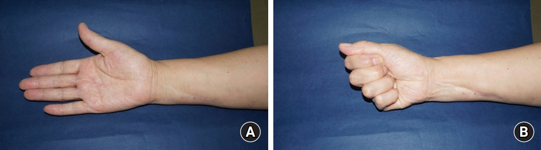
Fig. 5. (A, B) Postoperative images reveal neither a limited range of motion nor any neurological deficits.
Reference
-
References
1. Jiang S, Shen H, Lu H. Multiple schwannomas of the digital nerves and common palmar digital nerves: an unusual case report of multiple schwannomas in one hand. Medicine (Baltimore). 2019; 98:e14605.2. Nicolescu R, Agrawal NA, Pettit RW, Netscher DT. Recurrent schwannomatosis of the hand. Hand (N Y). 2020; 15:732–38.
Article3. Lans J, Yue KC, Castelein RM, et al. Benign hand tumors (part ii): soft tissue tumors. Hand (N Y). 2020; Jul. 15. [Epub]. https://doi.org/10.1177/1558944720928499.
Article4. Athanasian EA. Bone and soft tissue tumors. In : Green DP, Wolfe SW, Cohen MS, Hotchkiss RN, Kozin SH, Pederson WC, editors. Green’s operative hand surgery. 7th ed. Philadelphia: Elsevier;2017. p. 1987–2035.5. Baser ME, Friedman JM, Evans DG. Increasing the specificity of diagnostic criteria for schwannomatosis. Neurology. 2006; 66:730–2.
Article6. Kütahya H, Güleç A, Güzel Y, Kacira B, Toker S. Schwannoma of the median nerve at the wrist and palmar regions of the hand: a rare case report. Case Rep Orthop. 2013; 2013:950106.
Article7. Wang ZX, Chen SL, Yi CJ, Li C, Rong YB, Tian GL. Beijing Da Xue Xue Bao Yi Xue Ban. 2013; 45:698–703.8. Chen SL, Liu C, Liu B, et al. Schwannomatosis: a new member of neurofibromatosis family. Chin Med J (Engl). 2013; 126:2656–60.9. Gosk J, Gutkowska O, Kulinski S, Urban M, Halon A. Multiple schwannomas of the digital nerves and superficial radial nerve: two unusual cases of segmental schwannomatosis. Folia Neuropathol. 2015; 53:158–67.10. Alaidarous A, Parfait B, Ferkal S, Cohen J, Wolkenstein P, Mazereeuw-Hautier J. Segmental schwannomatosis: characteristics in 12 patients. Orphanet J Rare Dis. 2019; 14:207.
Article
- Full Text Links
-
- Actions
-
Cited
- CITED
-
- Close
- Share
-
- Similar articles
-
- Multiple Lower Extremity Mononeuropathies by Segmental Schwannomatosis: A Case Report
- Recurred Segmental Schwannomatosis Without Neurofibromatosis Type 2
- Trigeminal Neuralgia with Persistent Trigeminal Artery Variant and Schwannomatosis of the Abducens and Lower Cranial Nerves: A Case Report
- Multiple Schwannomas of the Spine: Review of the Schwannomatosis or Congenital Neurilemmomatosis: A Case Report
- Upper Extremity Deep Vein Thrombosis after Clavicle Fracture and Immobilization
