Lab Med Online.
2021 Jul;11(3):155-161. 10.47429/lmo.2021.11.3.155.
A Study for Accurate Reporting of Bacteria in Urine by Manual Microscopic Examination
- Affiliations
-
- 1Department of Laboratory Medicine, Green Cross Laboratories, Yongin, Korea
- 2Department of Laboratory Medicine, School of Medicine, Kyung Hee University, Seoul, Korea
- KMID: 2526060
- DOI: http://doi.org/10.47429/lmo.2021.11.3.155
Abstract
- Background
Since there is no standardized criterion for the semi quantitation of bacteria in manual microscopic examination, activities for reducing the subjectiveness of manual microscopic examination for detecting urinary bacteria are required.
Methods
This study was performed on specimens with result of WBC 0-1/a few bacteria in an automated urine sediment analyzer (Roche Diagnostic International, Switzerland). To establish the criterion for semi quantitation of bacterial counting, 43 specimens were examined by five technologists using manual microscopy and compared with the results of Gram staining. After application of the criterion, 71 specimens were examined by manual microscopy, following which, Gram staining and a urine culture were also performed.
Results
The newly established criterion was as follows: negative ( < 20/high-power field, HPF), a few (20-30/HPF), moderate (31-49/HPF), and many ( ≥ 50/HPF). The analytical sensitivity of the instrument was adjusted (from 18.18/field to 30/field) to decrease false positivity. After establishment of the criterion and education, the agreement rate was increased from 52.8% to 95.8%, and the specificity increased from 32.5% to 87.7% with the same sensitivity.
Conclusions
It will be necessary to ensure that all technologists apply the same criterion in the laboratory and clinical settings, assess the analytical sensitivity of an automated analyzer, and educate on the correct interpretation of urine microscopic examination.
Keyword
Figure
-

Fig. 1 An outline of the study. Abbreviation: WBC, white blood cell.
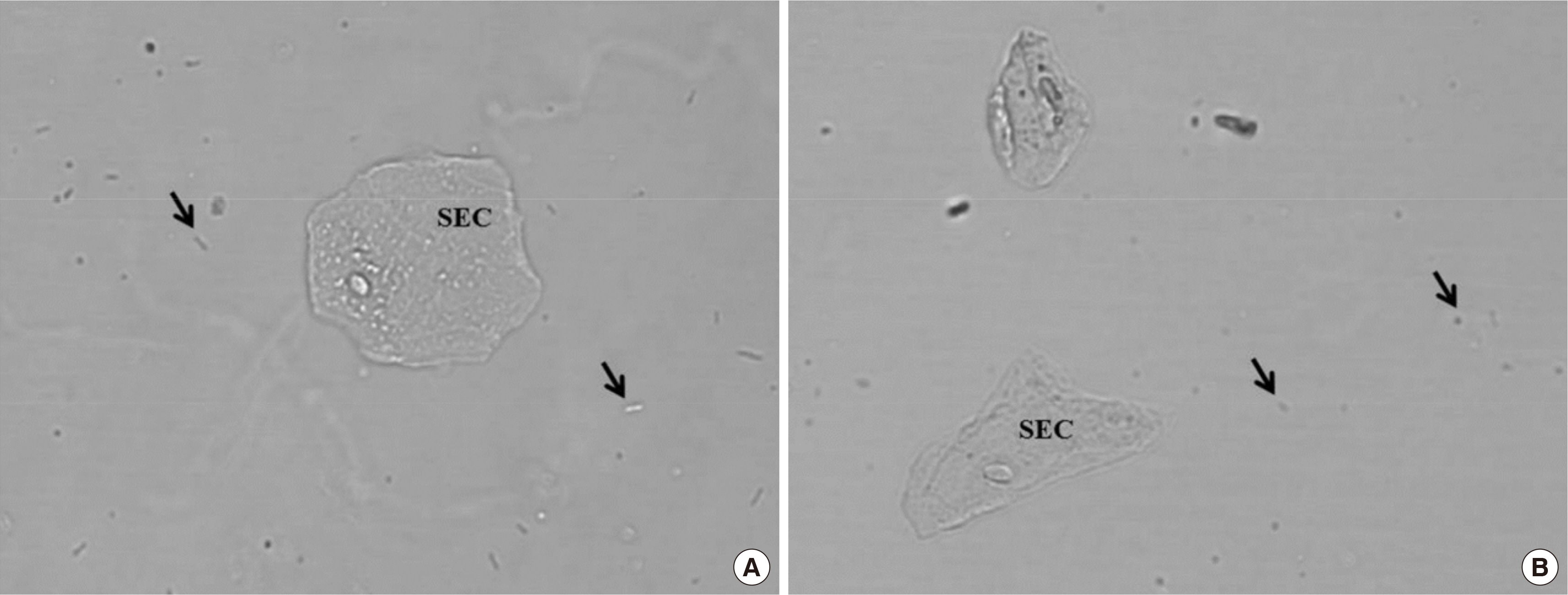
Fig. 2 Digital images from Cobas u 701. (A) Arrows indicate rod bacteria. (B) Arrows indicated cocci-like features, but were confirmed as artifacts by manual microscopic examination. Abbreviation: SEC, squamous epithelial cell.
Reference
-
1. dos Santos JC, Weber LP, Perez LR. 2007; Evaluation of urinalysis parameters to predict urinary-tract infection. Braz J Infect Dis. 11:479–81. DOI: 10.1590/S1413-86702007000500008. PMID: 17962874.
Article2. Broeren MA, Bahçeci S, Vader HL, Arents NL. 2011; Screening for urinary tract infection with the Sysmex UF-1000i urine flow cytometer. J Clin Microbiol. 49:1025–9. DOI: 10.1128/JCM.01669-10. PMID: 21248088. PMCID: PMC3067737.
Article3. Hsiao CY, Yang HY, Chang CH, Lin HL, Wu CY, Hsiao MC, et al. 2015; Risk factors for development of septic shock in patients with urinary tract infection. Biomed Res Int. 2015:717094. DOI: 10.1155/2015/717094. PMID: 26380292. PMCID: PMC4561874.
Article4. Mundt LA, Shanahan K, editors. 2011. Graff’s textbook of routine urinalysis and body fluids. 2nd ed. Wolters Kluwer Health/Lippincott Williams & Wilkins Health;PA: p. 36.5. Zaman Z, Fogazzi GB, Garigali G, Croci MD, Bayer G, Kránicz T. 2010; Urine sediment analysis: Analytical and diagnostic performance of sediMAX - a new automated microscopy image-based urine sediment analyser. Clin Chim Acta. 411:147–54. DOI: 10.1016/j.cca.2009.10.018. PMID: 19861122.
Article6. Ko DH, Ji M, Kim S, Cho EJ, Lee W, Yun YM, et al. 2016; An approach to standardization of urine sediment analysis via suggestion of a common manual protocol. Scand J Clin Lab Invest. 76:256–63. DOI: 10.3109/00365513.2016.1144141. PMID: 26963924.
Article7. Winkel P, Statland BE, Jorgensen K. 1974; Urine microscopy, an ill-defined method, examined by a multifactorial technique. Clin Chem. 20:436–9. DOI: 10.1093/clinchem/20.4.436. PMID: 4818195.8. Lee W, Ha JS, Ryoo NH. 2016; Comparison of the automated cobas u 701 urine microscopy and UF-1000i flow cytometry systems and manual microscopy in the examination of urine sediments. J Clin Lab Anal. 30:663–71. DOI: 10.1002/jcla.21919. PMID: 26842372. PMCID: PMC6807231.
Article9. Yüksel H, Kiliç E, Ekinci A, Evliyaoğlu O. 2013; Comparison of fully automated urine sediment analyzers H800-FUS100 and LabUMat-UriSed with manual microscopy. J Clin Lab Anal. 27:312–6. DOI: 10.1002/jcla.21604. PMID: 23852791. PMCID: PMC6807495.
Article10. İnce FD, Ellidağ HY, Koseoğlu M, Şimşek N, Yalçın H, Zengin MO. 2016; The comparison of automated urine analyzers with manual microscopic examination for urinalysis automated urine analyzers and manual urinalysis. Pract Lab Med. 5:14–20. DOI: 10.1016/j.plabm.2016.03.002. PMID: 28856199. PMCID: PMC5574505.
Article11. Kim SH, Song SA, Urm SH, Kook JK, Kim HR, Yong D, et al. 2017; Evaluation of the Cobas u 701 microscopy analyser compared with urine culture in screening for urinary tract infection. J Med Microbiol. 66:1110–3. DOI: 10.1099/jmm.0.000553. PMID: 28771134.
Article12. Pfaller MA, Baum CA, Niles AC, Murray PR. 1983; Clinical laboratory evaluation of a urine screening device. J Clin Microbiol. 18:674–9. DOI: 10.1128/jcm.18.3.674-679.1983. PMID: 6195180. PMCID: PMC270873.
Article13. European Confederation of Laboratory Medicine. 2000; European urinalysis guidelines. Scand J Clin Lab Invest Suppl. 231:1–86. DOI: 10.1080/00365513.2000.12056993.14. Kunin CM. 1997. Urinary tract infections: detection, prevention, and management. 5th ed. Williams & Wilkins;Baltimore, MD: p. 59.15. Deindoerfer FH, Gangwer JR, Laird CW, Ringold RR. 1985; "The Yellow IRIS" urinalysis workstation--the first commercial application of "automated intelligent microscopy". Clin Chem. 31:1491–9. DOI: 10.1093/clinchem/31.9.1491. PMID: 4028398.
Article16. Budak YU, Huysal K. 2011; Comparison of three automated systems for urine chemistry and sediment analysis in routine laboratory practice. Clin Lab. 57:47–52.17. Lamchiagdhase P, Preechaborisutkul K, Lomsomboon P, Srisuchart P, Tantiniti P, Khan-u-Ra N, et al. 2005; Urine sediment examination: a comparison between the manual method and the iQ200 automated urine microscopy analyzer. Clin Chim Acta. 358:167–74. DOI: 10.1016/j.cccn.2005.02.021. PMID: 16018883.
Article18. Alves L, Ballester F, Camps J, Joven J. 2005; Preliminary evaluation of the Iris IQ 200 automated urine analyser. Clin Chem Lab Med. 43:967–70. DOI: 10.1515/CCLM.2005.166. PMID: 16176179.
Article19. Littlewood JM, Jacobs SI, Ramsden CH. 1977; Comparison between microscopical examination of unstained deposits of urine and quantitative culture. Arch Dis Child. 52:894–6. DOI: 10.1136/adc.52.11.894. PMID: 339849. PMCID: PMC1544817.
Article20. Pryles CV, Eliot CR. 1965; Pyuria and bacteriuria in infants and children. The value of pyuria as a diagnostic criterion of urinary tract infections. Am J Dis Child. 110:628–35. DOI: 10.1001/archpedi.1965.02090030656007. PMID: 5320881.21. Jenkins RD, Fenn JP, Matsen JM. 1986; Review of urine microscopy for bacteriuria. JAMA. 255:3397–403. DOI: 10.1001/jama.1986.03370240067039. PMID: 2423720.
Article
- Full Text Links
-
- Actions
-
Cited
- CITED
-
- Close
- Share
-
- Similar articles
-
- Microscopic Examination and Bacterial Culture of the Prostatic Secretion of Chronic Prostatitis: Its Diagnostic Significance
- Evaluation of iQ200 Automated Urine Microscopy Analyzer
- The Comparison of Microscopic Urine Sediment, Nitrite, and Leukocyte Esterase Tests for Bacteriuria
- Comparison of Analytical Performance between the Sysmex UF-100 flow cytometer and the Iris iQ200 Urine Microscopy System
- Diagnostic value of dipstick urinalysis as a screening test for urinary tract infection
