Primary Hepatic Actinomycosis Mimicking Hepatic Malignancy
- Affiliations
-
- 1Department of Surgery, Inje University Haeundae Paik Hospital, Inje University College of Medicine, Busan, Korea
- KMID: 2525523
- DOI: http://doi.org/10.4166/kjg.2021.165
Figure
-
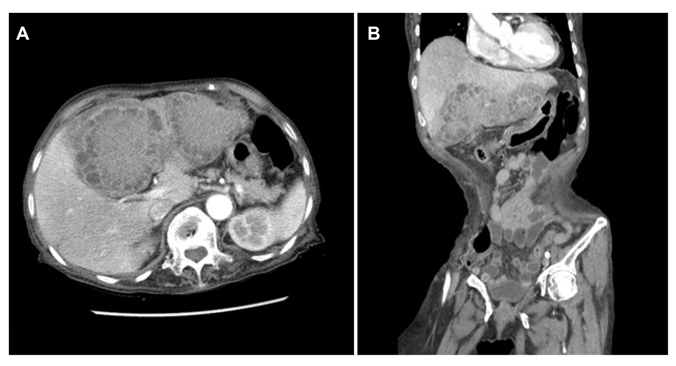
Fig. 1 (A, B) Abdominal computed tomography demonstrated two large masses on left lobe of the liver, about 7 cm, 7.5 cm showing peripherally necrotic portions with central delayed enhancing solid portions with perihepatic fat infiltrations and fat plane loss between the stomach, duodenum, suggesting invasion.

Fig. 2 (A) Increased size of peripheral low density portions of previous lobulating hepatic masses on left lobe with (B) rapid necrotic volume change of S4 lesion extending superiorly to S8 subcapsular area, with suspected parenchymal rupture, capsular defect.
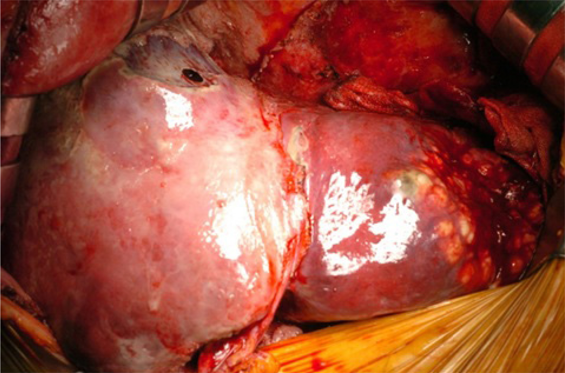
Fig. 3 Partial ruptured mass on S4 and multilobulating mass on left lateral segment.

Fig. 4 The specimen shows a whitish capsular retraction with rupture and hemorrhagic area which had an ill-defined tan/yellow mass-like lesion, measuring 18.0×9.5×6.0 cm. (A) The lesion is composed of solid area and many cystic spaces containing inflammatory exudate. (B) Hematoxylin and eosin stain, ×200; (C) GMS, ×200 and (D) Gram stain, ×200 revealed positive in filamentous bacilli.
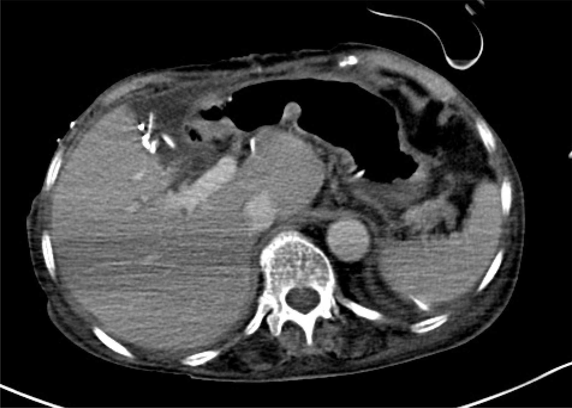
Fig. 5 Postoperative computed tomography scan at postoperative 14th day. There was no abnormal findings except small amount of fluid collection on resection margin.
Reference
-
1. Hayashi M, Asakuma M, Tsunemi S, et al. 2010; Surgical treatment for abdominal actinomycosis: a report of two cases. World J Gastrointest Surg. 2:405–408. DOI: 10.4240/wjgs.v2.i12.405. PMID: 21206723. PMCID: PMC3014523.
Article2. Brook I. 2008; Actinomycosis: diagnosis and management. South Med J. 101:1019–1023. DOI: 10.1097/SMJ.0b013e3181864c1f. PMID: 18791528.
Article3. Yang XX, Lin JM, Xu KJ, et al. 2014; Hepatic actinomycosis: report of one case and analysis of 32 previously reported cases. World J Gastroenterol. 20:16372–16376. DOI: 10.3748/wjg.v20.i43.16372. PMID: 25473199. PMCID: PMC4239533.
Article4. Kanellopoulou T, Alexopoulou A, Tiniakos D, Koskinas J, Archimandritis AJ. 2010; Primary hepatic actinomycosis mimicking metastatic liver tumor. J Clin Gastroenterol. 44:458–459. DOI: 10.1097/MCG.0b013e3181d2ef30. PMID: 20195165.
Article5. Wong JJ, Kinney TB, Miller FJ, Rivera-Sanfeliz G. 2006; Hepatic actinomycotic abscesses: diagnosis and management. AJR Am J Roentgenol. 186:174–176. DOI: 10.2214/AJR.04.1691. PMID: 16357398.
Article6. Ha YJ, An JH, Shim JH, et al. 2015; A case of primary hepatic actinomycosis: an enigmatic inflammatory lesion of the liver. Clin Mol Hepatol. 21:80–84. DOI: 10.3350/cmh.2015.21.1.80. PMID: 25834805. PMCID: PMC4379201.
Article7. Miyamoto MI, Fang FC. 1993; Pyogenic liver abscess involving actinomyces: case report and review. Clin Infect Dis. 16:303–309. DOI: 10.1093/clind/16.2.303. PMID: 8443315.
Article8. Wayne MG, Narang R, Chauhdry A, Steele J. 2011; Hepatic actinomycosis mimicking an isolated tumor recurrence. World J Surg Oncol. 9:70. DOI: 10.1186/1477-7819-9-70. PMID: 21745394. PMCID: PMC3160369.
Article9. Ávila F, Santos V, Massinha P, et al. 2015; Hepatic actinomycosis. GE Port J Gastroenterol. 22:19–23. DOI: 10.1016/j.jpge.2014.08.002. PMID: 28868364. PMCID: PMC5580170.
Article10. Felekouras E, Menenakos C, Griniatsos J, et al. 2004; Liver resection in cases of isolated hepatic actinomycosis: case report and review of the literature. Scand J Infect Dis. 36:535–538. DOI: 10.1080/00365540410020866-1. PMID: 15307597.
Article11. Sharma M, Briski LE, Khatib R. 2002; Hepatic actinomycosis: an overview of salient features and outcome of therapy. Scand J Infect Dis. 34:386–391. DOI: 10.1080/00365540110080304. PMID: 12069027.
Article12. Lai AT, Lam CM, Ng KK, et al. 2004; Hepatic actinomycosis presenting as a liver tumour: case report and literature review. Asian J Surg. 27:345–347. DOI: 10.1016/S1015-9584(09)60066-X. PMID: 15564194.
Article13. Kanellopoulou T, Alexopoulou A, Tanouli MI, et al. 2010; Primary hepatic actinomycosis. Am J Med Sci. 339:362–365. DOI: 10.1097/MAJ.0b013e3181cbf47c. PMID: 20195148.
Article14. Murphy P, Mar WA, Allison D, Cornejo GA, Setty S, Giulianotti PC. 2019; Hepatic actinomycosis - a potential mimicker of malignancy. Radiol Case Rep. 15:105–109. DOI: 10.1016/j.radcr.2019.10.014. PMID: 31762867. PMCID: PMC6864297.
Article15. Chou HH, Huang YT, Yang CJ. 2016; Actinomycosis resembling liver tumor with multiple metastasis. Int J Infect Dis. 45:98–99. DOI: 10.1016/j.ijid.2016.02.023. PMID: 26948481.
Article16. Yang SS, Im YC. 2018; Severe abdominopelvic actinomycosis with colon perforation and hepatic involvement mimicking advanced sigmoid colon cancer with hepatic metastasis: a case study. BMC Surg. 18:51. DOI: 10.1186/s12893-018-0386-3. PMID: 30068330. PMCID: PMC6090905.
Article17. Soardo G, Basan L, Intini S, Avellini C, Sechi LA. 2005; Elevated serum CA 19-9 in hepatic actinomycosis. Scand J Gastroenterol. 40:1372–1373. DOI: 10.1080/00365520510024232. PMID: 16334448.
Article18. Hansen JM, Fjeldsøe-Nielsen H, Sulim S, Kemp M, Christensen JJ. 2009; Actinomyces species: a Danish survey on human infections and microbiological characteristics. Open Microbiol J. 3:113–120. DOI: 10.2174/1874285800903010113. PMID: 19657460. PMCID: PMC2720514.
Article19. Filipović B, Milinić N, Nikolić G, Ranthelović T. 2005; Primary actinomycosis of the anterior abdominal wall: case report and review of the literature. J Gastroenterol Hepatol. 20:517–520. DOI: 10.1111/j.1440-1746.2004.03564.x. PMID: 15836698.
Article20. Zeng QQ, Zheng XW, Wang QJ, Yu ZP, Zhang QY. 2018; Primary hepatic actinomycosis mimicking liver tumour. ANZ J Surg. 88:E629–E630. DOI: 10.1111/ans.13586. PMID: 27080813.
Article
- Full Text Links
-
- Actions
-
Cited
- CITED
-
- Close
- Share
-
- Similar articles
-
- Hepatic Actinomycosis Mimicking a Malignant Tumor: Three Case Reports
- Primary Hepatic Actinomycosis Mimicking Hepatic Malignancy with Metastatic Lymph Nodes by F-18 FDG PET/CT
- MR Findings of Hepatic Actinomycosis: Case Report
- Unique Imaging Features in Hepatic Actinomycosis Accompanied by an IgG4-Related Inflammatory Pseudotumor: A Case Report
- A case of primary hepatic actinomycosis coinfected with alpha-streptococcus
