Clinical Manifestations and Genetic Analysis of 5 Korean Choroideremia Patients Initially Diagnosed With Retinitis Pigmentosa
- Affiliations
-
- 1Department of Ophthalmology, Soonchunhyang University Hospital Bucheon, Bucheon, Korea
- 2Hyangseol Medical Research Center, Soonchunhyang University Bucheon Hospital, Bucheon, Korea
- 3Department of Interdisciplinary Program in Biomedical Science, Soonchunhyang University, Graduate School, Bucheon Hospital, Bucheon, Korea
- 4Department of Laboratory Medicine and Genetics, Soonchunhyang University Bucheon Hospital, Soonchunhyang University College of Medicine, Bucheon, Korea
- 5Oneomics Institute, Soonchunhyang Mirai Medical Center, Bucheon, Korea
- 6Korean Foundation for Fighting Blindness, Seoul, Korea
- 7Department of Anatomy and BK21 FOUR Project, College of Medicine, Soonchunhyang University, Cheonan, Korea
- 8Laboratory for Translational Research on Retinal and Macular Degeneration, Soonchunhyang University Hospital Bucheon, Bucheon, Korea
- 9Department of Ophthalmology, College of Medicine, Soonchunhyang University, Cheonan, Korea
- 10Ex Lumina Therapeutics and Technologies, Bucheon, Korea
- KMID: 2524894
- DOI: http://doi.org/10.3346/jkms.2022.37.e5
Abstract
- Background
To investigate the clinical findings of choroideremia patients and perform genetic analysis by whole-exome sequencing (WES).
Methods
A total of 94 patients initially diagnosed with retinitis pigmentosa (RP) at another hospital, and who visited our hospital for genetic analysis by WES, were included in the study, along with 64 family members. All subjects underwent comprehensive ophthalmic evaluation, including best-corrected visual acuity, slit lamp examination, fundus photography, fundus autofluorescence (FAF), fluorescein angiography (FAG), visual field (VF), electroretinogram (ERG), and optical coherence tomography (OCT).
Results
In six male patients with suspected choroideremia, extensive retinal pigment epithelium (RPE) and severe loss of choroid were observed in the fundus, but not in the macula. CHM gene mutation was confirmed in five patients. A novel single nucleotide variant at a splice site was observed in one patient. OCT showed marked thinning of the outernuclear layer and choroid, except in the macula. FAF showed a small area of hyperfluorescence in the posterior pole. In addition, characteristic interlaminar bridges were observed in four patients. On FAG, hypofluorescence was seen up to the far-peripheral retina in five patients.
Conclusion
Of the 94 patients initially diagnosed with RP, CHM mutation was identified in five (5.3%) by WES. Choroideremia should be considered as a differential diagnosis of RP. WES would be useful for identifying the causes of hereditary retinal disease.
Figure
-

Fig. 1 Wide field color fundus photography of the right and left eyes of a patient with advanced choroideremia.Wide field color fundus photographs show granular and lumpy material, chorioretinal atrophy, and a yellow-whitish sclera in peripheral and peripapillary regions. RPE and choroid remain in only a small area of the macula.RPE = retinal pigment epithelium.
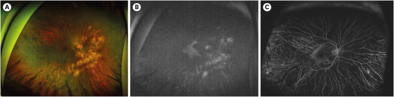
Fig. 2 Multimodal imaging of a male choroideremia patient. (A) Color fundus photography shows granular material in the mid-peripheral fundus, chorioretinal atrophy, and preservation of the posterior pole. (B) FAF showed a small area of hyperfluorescence in the posterior pole, in line with the results of color fundus photography shown in (A). (C) FAG revealed hypofluorescence up to the far-peripheral retina, with prominent choroidal vessels.FAF = fundus autofluorescence, FAG = fluorescein angiography.

Fig. 3 Representative OCT images of choroideremia patients. OCT shows marked atrophy of the RPE, except in the macula, with intraretinal cysts, ORT (arrowhead), and ILB (arrow). Marked thinning of the ONLs was observed and the choroid was virtually absent, both of which are hallmarks of choroideremia.OCT = optical coherence tomography, RPE = retinal pigment epithelium, ORT = outer retinal tubulation, ILB = interlaminar bridge, ONL = outer nuclear layer.
Reference
-
1. Dimopoulos IS, Radziwon A, St Laurent CD, MacDonald IM. Choroideremia. Curr Opin Ophthalmol. 2017; 28(5):410–415. PMID: 28520608.
Article2. Coussa RG, Traboulsi EI. Choroideremia: a review of general findings and pathogenesis. Ophthalmic Genet. 2012; 33(2):57–65. PMID: 22017263.
Article3. MacDonald IM, Binczyk N, Radziwon A, Dimopoulos I. Choroideremia. Cheung G, editor. Hereditary Chorioretinal Disorders. Singapore: Springer Nature Singapore Pte Ltd;2020. p. 99–106.4. Jacobson SG, Cideciyan AV, Sumaroka A, Aleman TS, Schwartz SB, Windsor EA, et al. Remodeling of the human retina in choroideremia: Rab escort protein 1 (REP-1) mutations. Invest Ophthalmol Vis Sci. 2006; 47(9):4113–4120. PMID: 16936131.5. MacDonald IM, Sereda C, McTaggart K, Mah D. Choroideremia gene testing. Expert Rev Mol Diagn. 2004; 4(4):478–484. PMID: 15225095.
Article6. Goody RS, Rak A, Alexandrov K. The structural and mechanistic basis for recycling of Rab proteins between membrane compartments. Cell Mol Life Sci. 2005; 62(15):1657–1670. PMID: 15924270.
Article7. Pereira-Leal JB, Hume AN, Seabra MC. Prenylation of Rab GTPases: molecular mechanisms and involvement in genetic disease. FEBS Lett. 2001; 498(2-3):197–200. PMID: 11412856.
Article8. Battu R, Jeyabalan N, Murthy P, Reddy KS, Schouten JS, Webers CA. Genetic analysis and clinical phenotype of two Indian families with X-linked choroideremia. Indian J Ophthalmol. 2016; 64(12):924–929. PMID: 28112135.
Article9. Wu AL, Wang JP, Tseng YJ, Liu L, Kang YC, Chen KJ, et al. Multimodal imaging of mosaic retinopathy in carriers of hereditary X-linked recessive diseases. Retina. 2018; 38(5):1047–1057. PMID: 28376043.
Article10. Rodrigues MM, Ballintine EJ, Wiggert BN, Lee L, Fletcher RT, Chader GJ. Choroideremia: a clinical, electron microscopic, and biochemical report. Ophthalmology. 1984; 91(7):873–883. PMID: 6089068.11. Mitsios A, Dubis AM, Moosajee M. Choroideremia: from genetic and clinical phenotyping to gene therapy and future treatments. Ther Adv Ophthalmol. 2018; 10:2515841418817490. PMID: 30627697.
Article12. Krill AE, Archer D. Classification of the choroidal atrophies. Am J Ophthalmol. 1971; 72(3):562–585. PMID: 5315093.
Article13. Lee TK, McTaggart KE, Sieving PA, Heckenlively JR, Levin AV, Greenberg J, et al. Clinical diagnoses that overlap with choroideremia. Can J Ophthalmol. 2003; 38(5):364–372. PMID: 12956277.
Article14. Gao FJ, Tian GH, Hu FY, Wang DD, Li JK, Chang Q, et al. Next-generation sequencing-based clinical diagnosis of choroideremia and comprehensive mutational and clinical analyses. BMC Ophthalmol. 2020; 20(1):212. PMID: 32487042.
Article15. Kim MS, Joo K, Seong MW, Kim MJ, Park KH, Park SS, et al. Genetic mutation profiles in Korean patients with inherited retinal diseases. J Korean Med Sci. 2019; 34(21):e161. PMID: 31144483.
Article16. Ma DJ, Lee HS, Kim K, Choi S, Jang I, Cho SH, et al. Whole-exome sequencing in 168 Korean patients with inherited retinal degeneration. BMC Med Genomics. 2021; 14(1):74. PMID: 33691693.
Article17. Guo H, Li J, Gao F, Li J, Wu X, Liu Q. Whole-exome sequencing reveals a novel CHM gene mutation in a family with choroideremia initially diagnosed as retinitis pigmentosa. BMC Ophthalmol. 2015; 15(1):85. PMID: 26216097.
Article18. MacDonald IM, Russell L, Chan CC. Choroideremia: new findings from ocular pathology and review of recent literature. Surv Ophthalmol. 2009; 54(3):401–407. PMID: 19422966.
Article19. MacDonald IM, Mah DY, Ho YK, Lewis RA, Seabra MC. A practical diagnostic test for choroideremia. Ophthalmology. 1998; 105(9):1637–1640. PMID: 9754170.
Article20. Di Iorio V, Esposito G, De Falco F, Boccia R, Fioretti T, Colucci R, et al. CHM/REP1 transcript expression and loss of visual function in patients affected by choroideremia. Invest Ophthalmol Vis Sci. 2019; 60(5):1547–1555. PMID: 30995293.21. Li S, Guan L, Fang S, Jiang H, Xiao X, Yang J, et al. Exome sequencing reveals CHM mutations in six families with atypical choroideremia initially diagnosed as retinitis pigmentosa. Int J Mol Med. 2014; 34(2):573–577. PMID: 24913019.22. Bae K, Song JS, Lee C, Kim NK, Park WY, Kim BJ, et al. Identification of pathogenic variants in the CHM gene in two Korean patients with choroideremia. Ann Lab Med. 2017; 37(5):438–442. PMID: 28643494.23. Khan KN, Islam F, Moore AT, Michaelides M. Clinical and genetic features of choroideremia in childhood. Ophthalmology. 2016; 123(10):2158–2165. PMID: 27506488.
Article24. Xue K, Oldani M, Jolly JK, Edwards TL, Groppe M, Downes SM, et al. Correlation of optical coherence tomography and autofluorescence in the outer retina and choroid of patients with choroideremia. Invest Ophthalmol Vis Sci. 2016; 57(8):3674–3684. PMID: 27403996.
Article25. Heon E, Alabduljalil T, McGuigan DB 3rd, Cideciyan AV, Li S, Chen S, et al. Visual function and central retinal structure in choroideremia. Invest Ophthalmol Vis Sci. 2016; 57(9):OCT377–OCT387. PMID: 27409497.
Article26. Lazow MA, Hood DC, Ramachandran R, Burke TR, Wang YZ, Greenstein VC, et al. Transition zones between healthy and diseased retina in choroideremia (CHM) and Stargardt disease (STGD) as compared to retinitis pigmentosa (RP). Invest Ophthalmol Vis Sci. 2011; 52(13):9581–9590. PMID: 22076985.
Article27. Sun LW, Johnson RD, Williams V, Summerfelt P, Dubra A, Weinberg DV, et al. Multimodal imaging of photoreceptor structure in choroideremia. PLoS One. 2016; 11(12):e0167526. PMID: 27936069.
Article28. Syed R, Sundquist SM, Ratnam K, Zayit-Soudry S, Zhang Y, Crawford JB, et al. High-resolution images of retinal structure in patients with choroideremia. Invest Ophthalmol Vis Sci. 2013; 54(2):950–961. PMID: 23299470.
Article29. Popović P, Jarc-Vidmar M, Hawlina M. Abnormal fundus autofluorescence in relation to retinal function in patients with retinitis pigmentosa. Graefes Arch Clin Exp Ophthalmol. 2005; 243(10):1018–1027. PMID: 15906064.
Article30. McTaggart KE, Tran M, Mah DY, Lai SW, Nesslinger NJ, MacDonald IM. Mutational analysis of patients with the diagnosis of choroideremia. Hum Mutat. 2002; 20(3):189–196. PMID: 12203991.
Article31. Freund PR, Sergeev YV, MacDonald IM. Analysis of a large choroideremia dataset does not suggest a preference for inclusion of certain genotypes in future trials of gene therapy. Mol Genet Genomic Med. 2016; 4(3):344–358. PMID: 27247961.
Article32. van den Hurk JA, van de Pol DJ, Wissinger B, van Driel MA, Hoefsloot LH, de Wijs IJ, et al. Novel types of mutation in the choroideremia (CHM) gene: a full-length L1 insertion and an intronic mutation activating a cryptic exon. Hum Genet. 2003; 113(3):268–275. PMID: 12827496.
- Full Text Links
-
- Actions
-
Cited
- CITED
-
- Close
- Share
-
- Similar articles
-
- A Case of Retinitis Pigmentosa without Pigment
- A Case of Unilateral Retinitis Pigmentosa
- Strategies for Mutation Discovery in Retinitis Pigmentosa: Transition to the Next Generation
- Visual Function and Functional Vision of Retinitis Pigmentosa
- Pars Plana Vitrectomy for Vitreous Hemorrhage in Coats-Type Retinitis Pigmentosa
