Pars Plana Vitrectomy for Vitreous Hemorrhage in Coats-Type Retinitis Pigmentosa
- Affiliations
-
- 1Department of Ophthalmology and Visual Science, Uijeongbu St. Mary's Hospital, College of Medicine, The Catholic University of Korea, Uijeongbu, Korea. deenie@daum.net
- KMID: 2212806
- DOI: http://doi.org/10.3341/jkos.2016.57.4.677
Abstract
- PURPOSE
For vitreous hemorrhage induced by coats-types retinitis pigmentosa, we report a case treated with pars plana vitrectomy and endolaser photocoagulation.
CASE SUMMARY
A 24-year-old male who was diagnosed with retinitis pigmentosa in both eyes 6 years earlier presented with decreased visual acuity in his left eye for the last 7 months. Corrected visual acuity was measured at 0.06 in the left eye and fundus examination revealed a vitreous hemorrhage in the left eye as well as an exudative lesion in the right eye's peripheral retina, which suggested Coats-type retinitis pigmentosa. The left eye was treated with pars plana vitrectomy. After removal of the vitreous hemorrhage, endolaser photocoagulation was performed around the peripheral exudative lesion that caused the vitreous hemorrhage. One month later, the best-corrected visual acuity increased to 0.20 in the left eye, and there was an improvement in the vitreous hemorrhage and the exudative lesion.
CONCLUSIONS
Pars plana vitrectomy and endolaser can be helpful in vitreous hemorrhage induced by coats-type retinitis pigmentosa.
Keyword
MeSH Terms
Figure
-
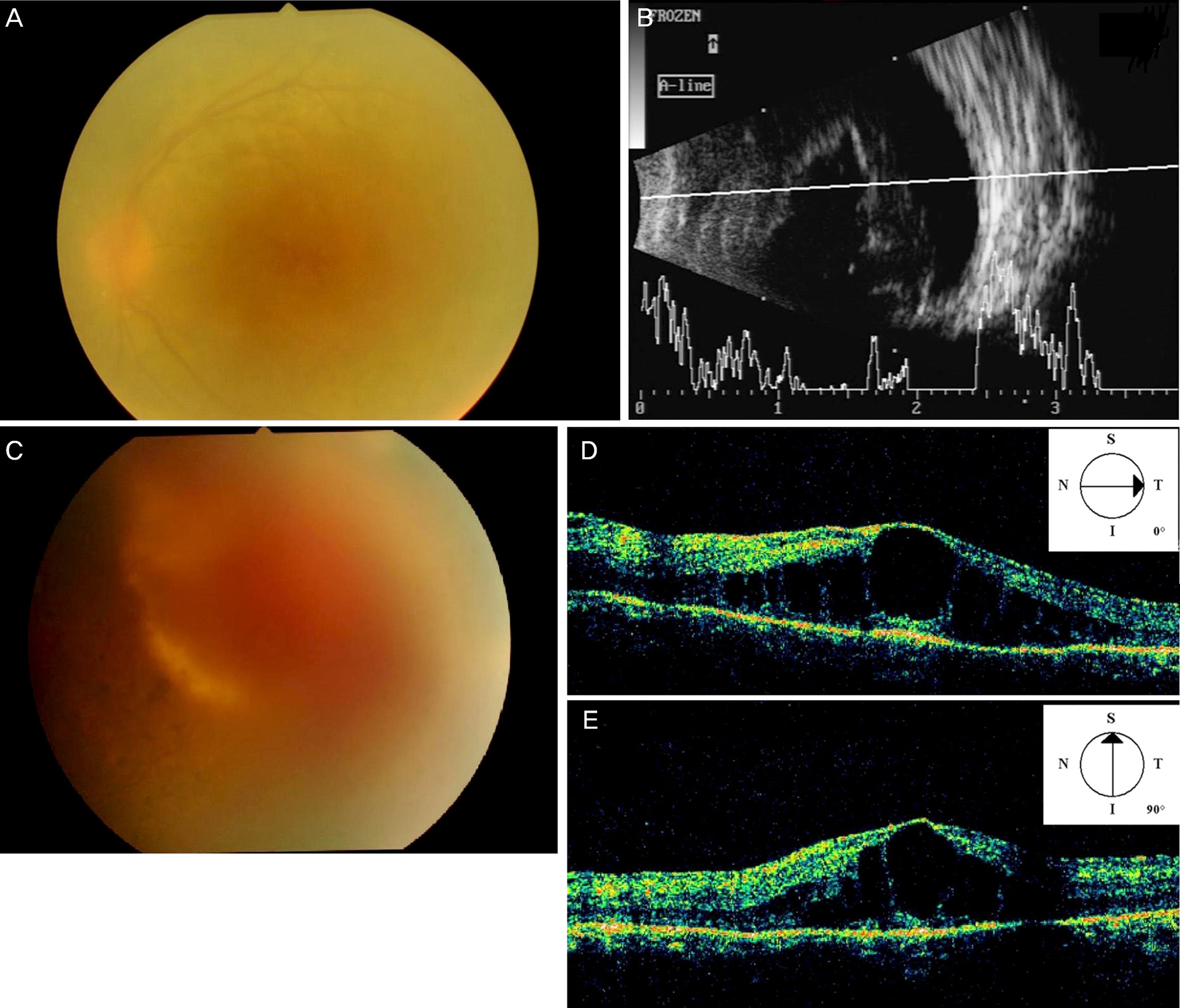
Figure 1. Preoperative imagings of the left eye. (A, B) Fundus photograph and ultrasound B scan of the left eye showing a vitreous hemorrhage. (C) Fundus photograph of the left eye showing putative exudative lesions; the image is blurry because of the vitreous hemorrhage. (D, E) Optical coherence tomography scan of the left eye showing cystoid macular oedema. S = superior; N = nasal; I = inferior; T = temporal.

Figure 2. Retinal photograph of the right eye showing a bone spicule pattern of pigmentation and peripheral exudates in the temporal retina accompanied by telangiectatic vessels.
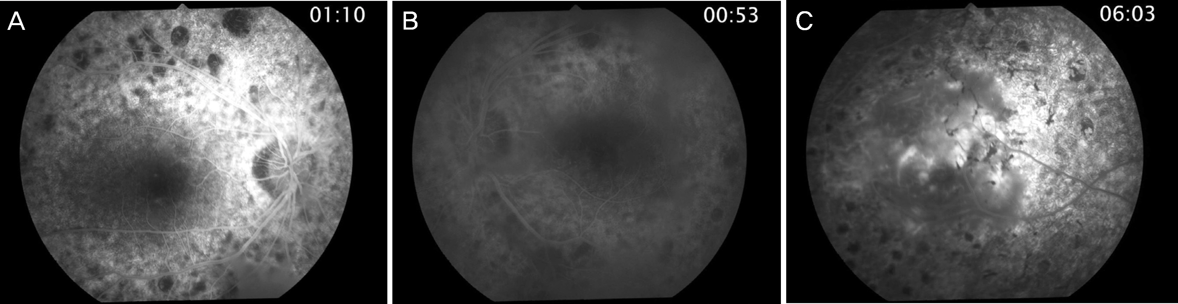
Figure 3. Initial fluorescein angiography of patient's both eye. (A, B) Fluorescein angiography of both eyes reveals hyper-fluorescence due to atrophy of the retinal pigment epithelium. (C) Late phase fluorescein angiography of left eye shows blocked fluorescence due to exudation accompanied by telangiectatic retinal vessels with leakage of the dye.
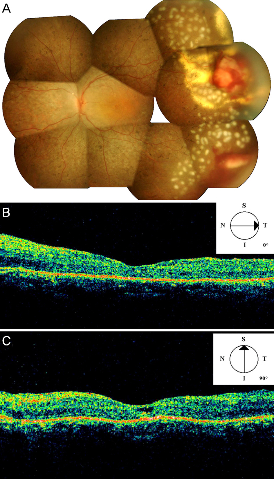
Figure 4. Postoperative imagings of the left eye. (A) Retinal photograph of the left eye three days after surgery shows a vitreous hemorrhage-free, well-attached retina. The localized subretinal exudative lesion was surrounded by photocoagulation scarring. (B, C) Optical coherence tomography scan of the left eye demonstrates resolved cystoid macular oedema. S = superior; N = nasal; I = inferior; T = temporal.
Cited by 1 articles
-
Surgical Repair of a Full-thickness Macular Hole in Retinitis Pigmentosa: a Case Report
Seungmo Kim, Joon Hyung Yeo, June-Gone Kim
J Korean Ophthalmol Soc. 2019;60(3):287-291. doi: 10.3341/jkos.2019.60.3.287.
Reference
-
References
1. Khan JA, Ide CH, Strickland MP. Coats’-type retinitis pigmentosa. Surv Ophthalmol. 1988; 32:317–32.
Article2. Kan E, Yilmaz T, Aydemir O, et al. Coats-like retinitis pigmentosa: Reports of three cases. Clin Ophthalmol. 2007; 1:193–8.3. Sarao V, Veritti D, Prosperi R, et al. A case of CRB1-negative Coats-like retinitis pigmentosa. J AAPOS. 2013; 17:414–6.
Article4. Bansal S, Saha N, Woon WH. The management of “coats’ response” in a patient with x-linked retinitis pigmentosa-a case report. ISRN Surg. 2011; 2011. 970361.
Article5. De Salvo G, Gemenetzi M, Luff AJ, Lotery AJ. Cystoid macular oedema successfully treated by cryotherapy in retinitis pigmentosa with Coats’-like retinal exudation. Eye (Lond). 2011; 25:821–2.
Article6. Artunay O, Yuzbasioglu E, Rasier R, et al. Intravitreal ranibizumab in the treatment of cystoid macular edema associated with retinitis pigmentosa. J Ocul Pharmacol Ther. 2009; 25:545–50.
Article
- Full Text Links
-
- Actions
-
Cited
- CITED
-
- Close
- Share
-
- Similar articles
-
- Retinitis Pigmentosa Complicated by Vitreous Hemorrhage in a Young Patient: A Case Report
- Pars Plana Vitrectomy for Cystoid Macular Edema in a Retinitis Pigmentosa Patient
- Vitrectomy for Vitreous Opacity
- Gas Injection and Simple Pars Plana Vitrecomy in Submacular hemorrhage without using t-PA
- Clinical evaluation of severe penetrating injury involving vitreous gel treated with pars plana vitrectomy
