Child Kidney Dis.
2021 Dec;25(2):117-121. 10.3339/jkspn.2021.25.2.117.
Extraskeletal Calcifications in Children with Maintenance Peritoneal Dialysis
- Affiliations
-
- 1Department of Pediatrics, Seoul National University Children's Hospital, Seoul, Republic of Korea
- 2Department of Pediatrics, Uijeongbu Eulji Medical Center, Uijeongbu-si, Republic of Korea
- 3Department of Pediatrics, Seoul National University Bundang Hospital, Seongnam, Republic of Korea
- 4Department of Pediatrics, Seoul National University College of Medicine, Seoul, Republic of Korea
- 5Kidney Research Institute, Seoul National University College of Medicine, Seoul, Republic of Korea
- 6Wide River Institute of Immunology, Seoul National University, Hongcheon, Republic of Korea
- KMID: 2524513
- DOI: http://doi.org/10.3339/jkspn.2021.25.2.117
Abstract
- Chronic kidney disease (CKD)-mineral and bone disorder (CKD-MBD) is a common complication of CKD, often accompanied by extra-skeletal calcification in adult patients. As increased vascular calcification is predicted to increase cardiovascular mortality and morbidity, the revised Kidney Disease: Improving Global Outcomes guidelines recommend avoiding calcium-containing phosphate chelators. However, extra-skeletal calcification is less commonly noticed in pediatric patients. Here, we report our experience of such a complication in pediatric patients receiving maintenance peritoneal dialysis. Extra-skeletal calcification was noticed at the corneas, pelvic cavity, and soft tissues of the lower leg in 4 out of 32 patients on maintenance peritoneal dialysis. These patients experienced the aggravation of extra-skeletal calcifications during peritoneal dialysis, and 2 of them underwent excisional operations. It is required to monitor extra-skeletal calcifications in children on kidney replacement therapy.
Keyword
Figure
-
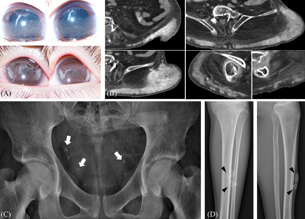
Fig. 1. Calcifications shown in patients. (A) Band keratopathy in the bilateral corneas (case 1) (B) Computerized tomography image of a palpable mass at the left flank and both hips (white arrows, case 2) (C) Incidentally found calcifications of the pelvic cavity (white arrows, case 3) (D) Palpable and slowly growing mass of the anterior tibial area (case 4).
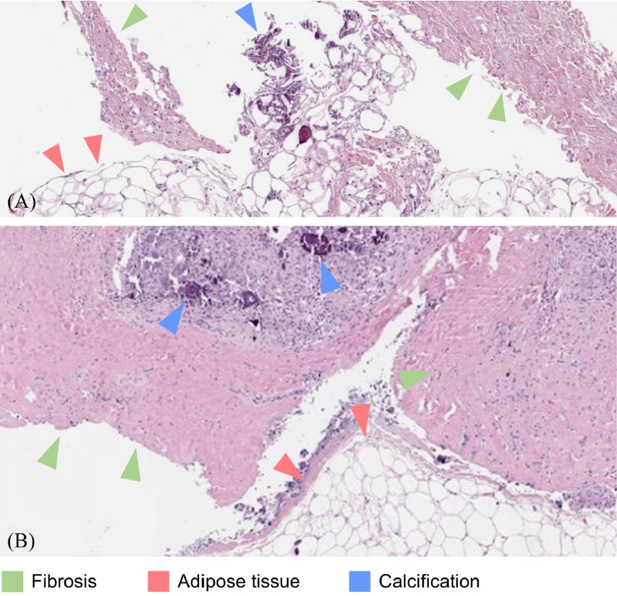
Fig. 2. Pathological slide of the palpable mass. Both slides show fibroadipose tissues with calcifications. (green arrowhead, fibrosis; red arrowhead, adipose tissue; blue arrowhead, calcification) (A) Specimen obtained by needle biopsy (case 2) (B) Specimen obtained by excision (case 4).
Reference
-
References
1. Suh JS. Diagnosis and Management of Chronic Kidney DiseaseMineral Bone Disease in Children. Childhood Kidney Diseases. 2020; 24:14–8.
Article2. Heaf JG. Chronic Kidney Disease-Mineral Bone Disorder in the Elderly Peritoneal Dialysis Patient. Perit Dial Int. 2015; 35:640–4.
Article3. Goel SK, Bellovich K, McCullough PA. Treatment of severe metastatic calcification and calciphylaxis in dialysis patients. Int J Nephrol. 2011; 2011:70160.
Article4. Drüeke TB. A clinical approach to the uraemic patient with extraskeletal calcifications. Nephrology Dialysis Transplantation. 1996; 11:37–42.
Article5. Milliner DS, Zinsmeister AR, Lieberman E, Landing B. Soft tissue calcification in pediatric patients with end-stage renal disease. Kidney Int. 1990; 38:931–6.
Article6. Sheth RD, Perez MD, Goldstein SL. Cardiovascular calcifications in pediatric patients receiving maintenance dialysis. Pediatr Nephrol. 2003; 18:810–3.
Article7. Kidney Disease: Improving Global Outcomes (KDIGO) CKD-MBD Update Work Group. KDIGO 2017 Clinical Practice Guideline Update for the Diagnosis, Evaluation, Prevention, and Treatment of Chronic Kidney Disease–Mineral and Bone Disorder (CKD-MBD). Kidney Int Suppl. 2017; 7:1–59.8. Wesseling K, Bakkaloglu S, Salusky I. Chronic kidney disease mineral and bone disorder in children. Pediatr Nephrol. 2008; 23:195–207.
Article9. Palit S, Kendrick J. Vascular calcification in chronic kidney disease: role of disordered mineral metabolism. Curr Pharm Des. 2014; 20:5829–33.
Article10. Kim C, Cheong HI, Kim JH, Yu YS, Kwon JW. Presumed atypical HDR syndrome associated with Band Keratopathy and pigmentary retinopathy. J Pediatr Ophthalmol Strabismus. 2011; 48 Online:e1–3.
Article11. Khouzam N, Wesseling-Perry K. Pathophysiology and treatment of cardiovascular disease in pediatric chronic kidney disease. Pediatr Nephrol. 2019; 34:1–10.
Article12. Fathallah-Shaykh S, Drozdz D, Flynn J, Jenkins R, Wesseling-Perry K, Swartz SJ, et al. Efficacy and safety of sevelamer carbonate in hyperphosphatemic pediatric patients with chronic kidney disease. Pediatr Nephrol. 2018; 33:325–33.
Article13. Toussaint N, Cooney P, Kerr PG. Review of dialysate calcium concentration in hemodialysis. Hemodial Int. 2006; 10:326–37.
Article14. Warady BA, Iles JN, Ariceta G, Dehmel B, Hidalgo G, Jiang X, et al. A randomized, double-blind, placebo-controlled study to assess the efficacy and safety of cinacalcet in pediatric patients with chronic kidney disease and secondary hyperparathyroidism receiving dialysis. Pediatr Nephrol. 2019; 34:475–86.
Article
- Full Text Links
-
- Actions
-
Cited
- CITED
-
- Close
- Share
-
- Similar articles
-
- Peritoneal dialysis in children and adolescents
- Peritoneal-pleural leak improved by switching from continuous ambulatory peritoneal dialysis to automated peritoneal dialysis
- Extensive Peritoneal Calcifications Associated with Continuous Ambulatory Peritoneal Dialysis
- A clinical study of continuous ambulatory peritoneal dialysis in childhood
- A Case of Conjunctival and Corneal Calcification in a Child on Peritoneal Dialysis
