Korean Circ J.
2022 Jan;52(1):87-88. 10.4070/kcj.2021.0341.
The Use of New Emerging Technology in Echocardiography-Glass View
- Affiliations
-
- 1Department of Internal Medicine, University of Cincinnati College of Medicine, Cincinnati, OH, USA
- 2Department of Cardiovascular Diseases, Mayo Clinic School of Medicine, Scottsdale, AZ, USA
- KMID: 2524174
- DOI: http://doi.org/10.4070/kcj.2021.0341
Abstract
- no abstract available.
Figure
-
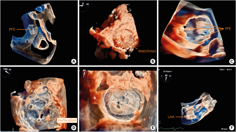
Figure 1 Examples of glass-view images. (A) Glass view showing a PFO. (B) Glass view showing a LAA occlusion device (Watchman). (C) Glass view showing multiple PFE on the aortic valve. (D) Glass view showing a small perforation of a tissue mitral valve prosthesis cusp. (E) Glass view showing a Barlow-type mitral valve prolapse with a flail P2 scallop. (F) Glass view of the side view showing a LAA anatomy after increasing transparency.LAA = left atrial appendage; PFE = papillary fibroelastomas; PFO = patent foramen ovale.
- Full Text Links
-
- Actions
-
Cited
- CITED
-
- Close
- Share
-
- Similar articles
-
- Stress-Induced Cardiomyopathy: The Role of Echocardiography
- Two Cases of Double-Orifice Mitral Valve Detected by Echocardiography
- Right Ventricular Area, Dimension, and Volume Measured by Two-dimensional Echocardiography in Normal Children
- Direct Visualization of Coronary Artery and Flow using Transthoracic Doppler Echocardiography
- Screening Fetal Echocardiography Made Easy
