Measurements of Neutron Activation and Dose Rate Induced by High-Energy Medical Linear Accelerator
- Affiliations
-
- 1Department of Radiation Oncology, Yonsei Cancer Center, Yonsei University College of Medicine, Seoul, Korea
- 2Department of Accelerator Science, Korea University, Sejong, Korea
- 3National Radiation Emergency Medical Center, Korea Institute of Radiological and Medical Sciences, Seoul, Korea
- 4Research Team of Radiological Physics & Engineering, Korea Institute of Radiological & Medical Sciences, Seoul, Korea
- KMID: 2524161
- DOI: http://doi.org/10.14316/pmp.2021.32.4.145
Abstract
- Purpose
During the treatments of cancer patients with a linear accelerator (LINAC) using photon beams with energies ≥8 MV, the components inside the LINAC head get activated through the interaction of photonuclear reaction (γ, n) and neutron capture (n, γ). We used spectroscopy and measured the dose rate for the LINAC in operation after the treatment ended.
Methods
We performed spectroscopy and dose rate measurements for three units of LINACs with a portable high-purity Germanium (HPGe) detector and a survey meter. The spectra were obtained after the beams were turned off. Spectroscopy was conducted for 3,600 seconds, and the dose rate was measured three times. We identified the radionuclides for each LINAC.
Results
According to gamma spectroscopy results, most of the nuclides were short-lived radionuclides with half-lives of 100 days, except for 60 Co, 65 Zn, and 181 W nuclides. The dose rate for three LINACs obtained immediately in front of the crosshair was in the range of 0.113 to 0.129 µSv/h. The maximum and minimum dose rates measured on weekends were 0.097 µSv/h and 0.092 µSv/h, respectively. Compared with the differences in weekday data, there was no significant difference between the data measured on Saturday and Sunday.
Conclusions
Most of the detected radionuclides had half-lives <100 days, and the dose rate decreased rapidly. For equipment that primarily used energies ≤10 MV, when the equipment was transferred after at least 10 minutes after shutting it down, it is expected that there will be little effect on the workers’ exposure.
Keyword
Figure
-
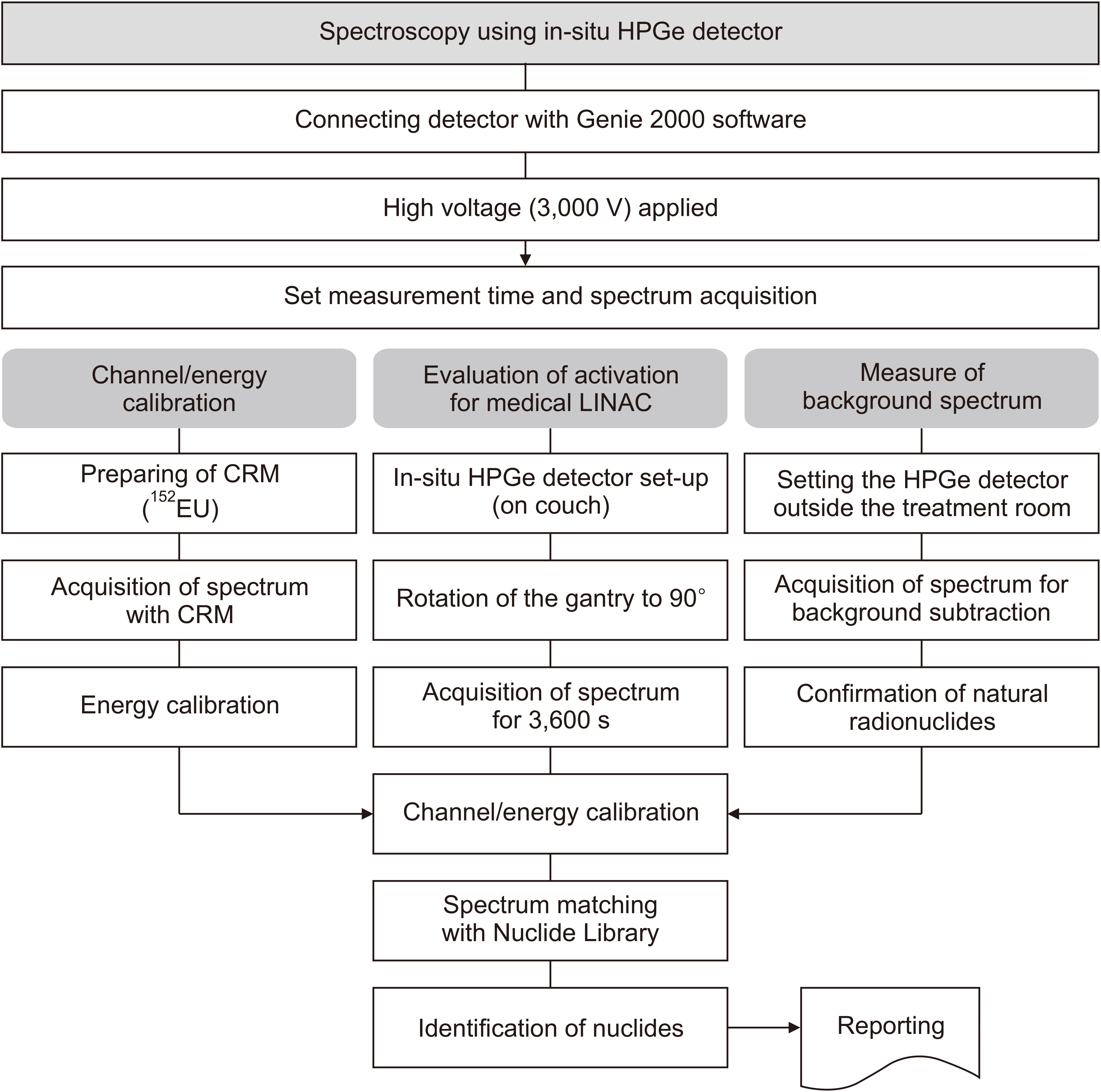
Fig. 1 Diagram showing the measurement procedure and evaluation of activation of medical linear accelerators (LINACs). HPGe, high-purity Germanium; CRM, certified reference material.
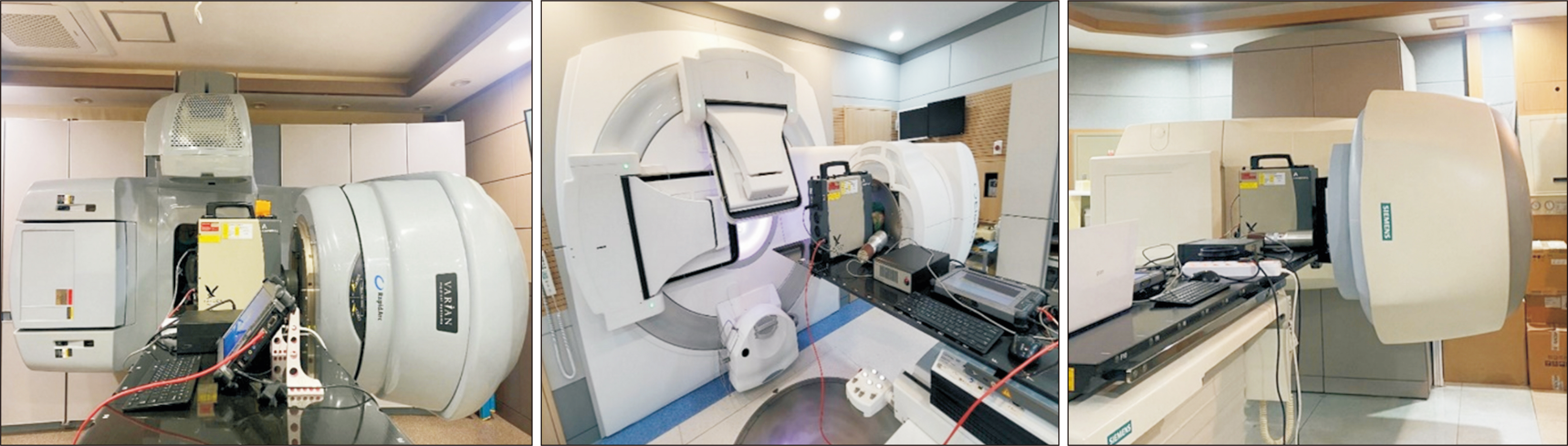
Fig. 2 Setup of the in-situ high-purity Germanium detector in front of the linear accelerator crosshair and relevant measurements.
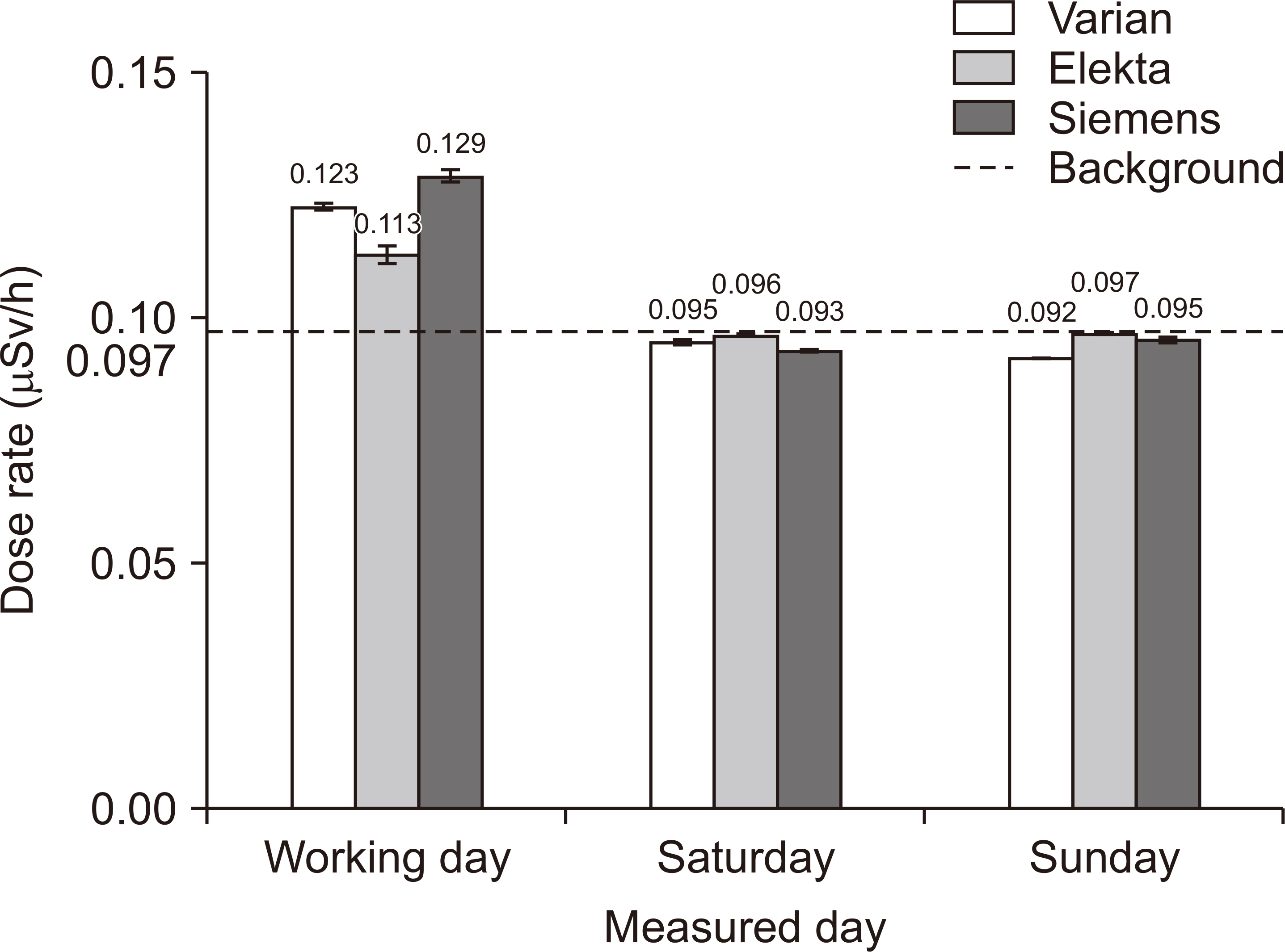
Fig. 3 LINAC dose rate measurement outcomes. LINAC, linear accelerator.
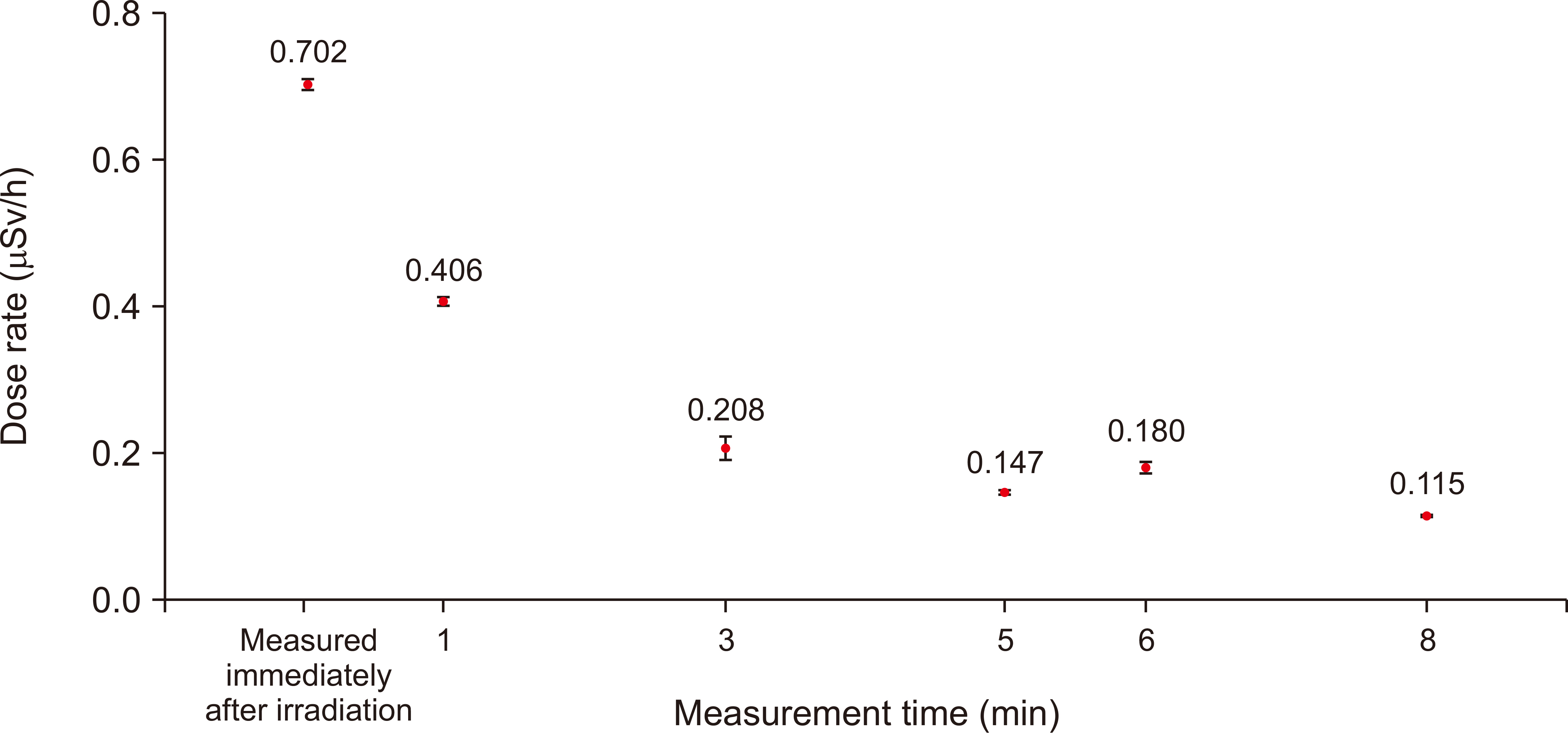
Fig. 4 Dose rate changes after beam irradiation with the use of a 10 MV energy beam.
Cited by 1 articles
-
Dead Layer Thickness and Geometry Optimization of HPGe Detector Based on Monte Carlo Simulation
Suah Yu, Na Hye Kwon, Young Jae Jang, Byungchae Lee, Jihyun Yu, Dong-Wook Kim, Gyu-Seok Cho, Kum-Bae Kim, Geun Beom Kim, Cheol Ha Baek, Sang Hyoun Choi
Prog Med Phys. 2022;33(4):129-135. doi: 10.14316/pmp.2022.33.4.129.
Reference
-
References
1. Korea Institute of Nuclear Safety. 2020. Current status of medical radiation devices. Korea Institute of Nuclear Safety;Daejeon:2. Kwon NH, Jang YJ, Kim DW, Shin DO, Kim KB, Kim JS, et al. 2020; Trend analysis on Korean and international management for activated material waste from medical linear accelerator. Prog Med Phys. 31:194–204. DOI: 10.14316/pmp.2020.31.4.194.
Article3. Çeçen Y, Gülümser T, Yazgan Ç, Dapo H, Üstün M, Boztosun I. 2017; Photoneutron flux measurement via neutron activation analysis in a radiotherapy bunker with an 18 MV linear accelerator. EPJ Web Conf. 153:07006. DOI: 10.1051/epjconf/201715307006.
Article4. Vichi S, Dean D, Ricci S, Zagni F, Berardi P, Mostacci D. 2020; Activation study of a 15MeV LINAC via Monte Carlo simulations. Radiat Phys Chem. 172:108758. DOI: 10.1016/j.radphyschem.2020.108758.
Article5. Lee DY, Park ET, Kim JH. 2017; Evaluate the activation of linear accelerator components and shielding wall through simulation. J Korea Contents Assoc. 17:69–76.6. Patil B, Chavan S, Pethe SN, Krishnan R, Bhoraskar V, Dhole S. 2011; Estimation of neutron production from accelerator head assembly of 15 MV medical LINAC using FLUKA simulations. Nucl Instrum Methods Phys. 269:3261–3265. DOI: 10.1016/j.nimb.2011.04.013.7. Morató S, Juste B, Miró R, Verdú G, Diez S. 2016. Aug. 16-20. Experimental validation of neutron activation simulation of a varian medical linear accelerator. Paper presented at: 2016 38th Annual International Conference of the IEEE Engineering in Medicine and Biology Society (EMBC). Orlando, USA: p. 5656–5659. DOI: 10.1109/EMBC.2016.7592010. PMID: 28269538.
Article8. Lee DY, Kim JH, Park ET. 2017; Assessment of human exposure doses received by activation of medical linear accelerator components. J Instrum. 12:P08022. DOI: 10.1088/1748-0221/12/08/P08022.
Article9. McMorrow A. 2019; Radiation shielding and bunker design. Biomed J Sci Tech Res. 18:13320–13330. DOI: 10.26717/BJSTR.2019.18.003106.
Article10. Lonski P, Taylor ML, Franich RD, Harty P, Kron T. 2012; Assessment of leakage doses around the treatment heads of different linear accelerators. Radiat Prot Dosimetry. 152:304–312. DOI: 10.1093/rpd/ncs049. PMID: 22511732.
Article11. Varian Medical Systems. Confidential data package. Monte Carlo data package high energy accelerator. Varian Medical Systems;Crowley:12. Elekta. High energy accelerator Monte Carlo. Confidential data package. Elekta;Crowley:13. Joint working group of medical societies related to clearance and radiotherapy in Japan. 2016. Society standard for radiotherapy management in radiotherapy equipment. Japanese Society for Radiation Oncology;Chuo:14. Siemens. Monte Carlo simulation for teletherapy equipment. Confidential data. Siemens Healthineers;Crowley:15. Terashima S, Kimura N, Seino M, Kubota M, Saito H, Hosokawa Y. 2015; Analysis using an NaI(Tl) detector for radioactive materials when dismantling a linear accelerator. Radiat Emerg Med. 4:47–52.16. Konefał A, Orlef A, Bieniasiewicz M. 2016; Measurements of neutron radiation and induced radioactivity for the new medical linear accelerator, the Varian TrueBeam. Radiat Meas. 86:8–15. DOI: 10.1016/j.radmeas.2015.12.039.
Article17. Fischer HW, Tabot BE, Poppe B. 2006; Activation processes in a medical linear accelerator and spatial distribution of activation products. Phys Med Biol. 51:N461–N466. DOI: 10.1088/0031-9155/51/24/N02. PMID: 17148816.
Article18. Fischer HW, Tabot B, Poppe B. 2008; Comparison of activation products and induced dose rates in different high-energy medical linear accelerators. Health Phys. 94:272–278. DOI: 10.1097/01.HP.0000291945.15684.a7. PMID: 18301101.
Article19. Fujibuchi T, Obara S, Yamaguchi I, Oyama M, Watanabe H, Sakae T, et al. 2012; Induced radioactive nuclides of 10-MeV radiotherapy accelerators detected by using a portable HP-Ge survey meter. Radiat Prot Dosimetry. 148:168–173. DOI: 10.1093/rpd/ncr005. PMID: 21317145.
Article
- Full Text Links
-
- Actions
-
Cited
- CITED
-
- Close
- Share
-
- Similar articles
-
- Assessment of Radiation Dose from Radioactive Wedge Filters during High-Energy X-Ray Therapy
- High Energy Electron Dosimetry by Alanine/ESR Spectroscopy
- Neutron therapy for prostatic cancer
- Dosimetric Characteristics of a Thermal Neutron Beam Facility for Neutron Capture Therapy at HANARO Reactor
- Analysis of Radionuclides for Induced-Radioactive Waste from Medical Linear Accelerator
