Bilateral posterior cerebral artery stroke following transtentorial herniation caused by a subependymal giant cell astrocytoma in a patient with tuberous sclerosis: a case report
- Affiliations
-
- 1Department of Neurology, Jeju National University Hospital, Jeju National University School of Medicine, Jeju, Korea
- 2Department of Neurosurgery, Jeju National University Hospital, Jeju National University School of Medicine, Jeju, Korea
- 3Jeju National University School of Medicine, Jeju, Korea
- 4Department of Pathology, Jeju National University School of Medicine, Korea
- KMID: 2523719
- DOI: http://doi.org/10.18700/jnc.210034
Abstract
- Background
Acute increased intracranial pressure (IICP) is a life-threatening condition that requires urgent treatment. Rapid IICP with hydrocephalus may be complicated by ischemic stroke, convulsions, loss of consciousness, brain herniation, and death. Extremely rare complications include intracranial vessel entrapment and ischemic stroke due to sudden IICP in cases with benign tumors.
Case Report
We report a case of bilateral posterior cerebral artery region infarction and complicated hydrocephalus with subependymal giant cell astrocytoma in a patient with tuberous sclerosis.
Conclusion
We postulate that the temporary IICP induced by seizure led to transient bilateral posterior cerebral artery entrapment, causing ischemic stroke without vascular occlusion.
Figure
-
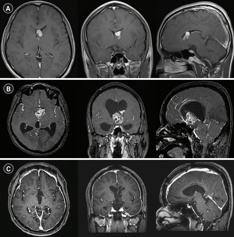
Fig. 1. (A) The axial/coronal/sagittal contrast-enhanced T1-weighted sequence from 16 years earlier showed a solid homogeneous lesion in the left lateral ventricle, extending toward the foramen of Monro without hydrocephalus. (B) Immediately preoperatively, the contrast-enhanced T1-weighted sequence showed marked heterogeneous enhancement and proximal aqueduct occlusion with marked hydrocephalus. Coronal and sagittal magnetic resonance imaging images showed marked dilatation of the lateral and third ventricles, with a normal-sized fourth ventricle. Note the downward bowing of the third ventricle floor with expanded infundibular and optic recesses. (C) Postoperative contrast-enhanced T1 axial scans showed no residual tumor with marked meningeal enhancement.
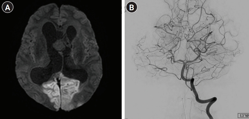
Fig. 2. (A) Diffusion-weighted image showed hyperintensity suggesting acute ischemic stroke in the region of the bilateral posterior cerebral artery (PCA). (B) Conventional angiography showed patency of the lumens of both PCAs, which have normal diameters.
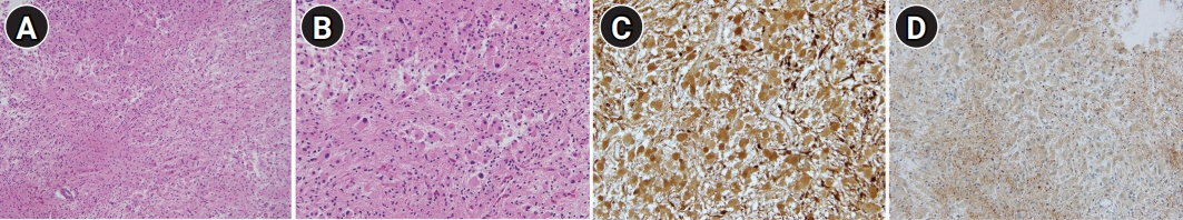
Fig. 3. (A, B) Microscopically, the tumor comprised mainly of large polygonal cells resembling astrocytes or ganglion cells. The tumor cells have abundant, finely granular eosinophilic cytoplasm with large round to oval nuclei and prominent nucleoli (H&E, A: ×100, B: ×200). (C, D) Tumor cells show positive immunoreactivity for S-100 and glial fibrillary acidic protein (immunohistochemical stain, ×200).
Reference
-
1. Henske EP, Jóźwiak S, Kingswood JC, Sampson JR, Thiele EA. Tuberous sclerosis complex. Nat Rev Dis Primers. 2016; 2:16035.
Article2. Crino PB, Nathanson KL, Henske EP. The tuberous sclerosis complex. N Engl J Med. 2006; 355:1345–56.
Article3. Roth J, Roach ES, Bartels U, Jóźwiak S, Koenig MK, Weiner HL, et al. Subependymal giant cell astrocytoma: diagnosis, screening, and treatment. Recommendations from the International Tuberous Sclerosis Complex Consensus Conference 2012. Pediatr Neurol. 2013; 49:439–44.
Article4. Jansen AC, Belousova E, Benedik MP, Carter T, Cottin V, Curatolo P, et al. Clinical characteristics of subependymal giant cell astrocytoma in tuberous sclerosis complex. Front Neurol. 2019; 10:705.
Article5. Von Haken MS, Aschoff AA, Diringer MN. Acute obstructive hydrocephalus. In : Hacke W, Hanley DF, Einhäupl KM, Bleck TP, Diringer MN, Ropper AH, editors. Neurocritical care. Berlin, Heidelberg: Springer Berlin Heidelberg;1994. p. 869–82.6. Jóźwiak S, Nabbout R, Curatolo P; Participants of the TSC Consensus Meeting for SEGA and Epilepsy Management. Management of subependymal giant cell astrocytoma (SEGA) associated with tuberous sclerosis complex (TSC): clinical recommendations. Eur J Paediatr Neurol. 2013; 17:348–52.
Article7. Sgouros S, Malluci C, Walsh AR, Hockley AD. Long-term complications of hydrocephalus. Pediatr Neurosurg. 1995; 23:127–32.
Article8. Gabor AJ, Brooks AG, Scobey RP, Parsons GH. Intracranial pressure during epileptic seizures. Electroencephalogr Clin Neurophysiol. 1984; 57:497–506.
Article9. Ansari M, Chang GY. Pathophysiology of stroke in the contralateral posterior cerebral artery distribution from a tentorial herniation. J Clin Neurol. 2017; 13:101–2.
Article10. Kuybu O, Tadi P, Dossani RH. Posterior cerebral artery stroke. Treasure Island, FL: StatPearls Publishing;2021.
- Full Text Links
-
- Actions
-
Cited
- CITED
-
- Close
- Share
-
- Similar articles
-
- A case of tuberous sclerosis developing with subependymal giant-cell astrocytoma
- A Case of Subependymal Giant Cell Astrocytoma in the Absence of Tuberous Sclerosis: Case Report
- A Case of Tuberous Sclerosis Complex Developing with Subependymal Giant-Cell Astrocytoma and Hydrocephalus
- A Case of Subependymal Giant Cell Astrocytoma associated with Tuberous Sclerosis(Case Report)
- Subependymal Giant Cell Astrocytoma in the tuberous Sclerosis
