
Anat Cell Biol.
2021 Dec;54(4):409-416. 10.5115/acb.21.113.
Comprehensive review of the incisive branch of the inferior alveolar nerve

- Affiliations
-
- 1Department of Oral and Maxillofacial Surgery, University of Pennsylvania, Philadelphia, PA, USA
- 2Harvard School of Dental Medicine, Harvard University, Boston, MA, USA
- 3Department of Restorative Dentistry and Biomaterials Sciences, Harvard School of Dental Medicine, Boston, MA, USA
- 4Department of Neurosurgery, Tulane Center for Clinical Neurosciences, Tulane University School of Medicine, New Orleans, LA, USA
- 5Department of Neurology, Tulane Center for Clinical Neurosciences, Tulane University School of Medicine, New Orleans, LA, USA
- 6Department of Anatomical Sciences, St. George’s University, St. George’s, Grenada
- 7Department of Structural & Cellular Biology, Tulane University School of Medicine, New Orleans, LA, USA
- 8Department of Neurosurgery and Ochsner Neuroscience Institute, Ochsner Health System, New Orleans, LA, USA
- 9Dental and Oral Medical Center, Kurume University School of Medicine, Kurume, Fukuoka, Japan
- 10Division of Gross and Clinical Anatomy, Department of Anatomy, Kurume University School of Medicine, Kurume, Fukuoka, Japan
- 11Department of Surgery, Tulane University School of Medicine, New Orleans, LA, USA
- KMID: 2523570
- DOI: http://doi.org/10.5115/acb.21.113
Abstract
- The incisive branch of the inferior alveolar nerve is a vital anatomical structure within the anterior mandible that has not been thoroughly defined and outlined in reports in the literature until recent years. Advances in radiological imaging, particularly the widespread use of cone-beam computed tomography has allowed for accurate visualization of the mandibular incisive canal (MIC) and its associated incisive branch of the inferior alveolar nerve. Surgical damage to the MIC, which could result in hemorrhage and sensory disturbance, may occur in commonly practiced oral and maxillofacial procedures, such as chin bone harvesting, implant placement, fracture repair and removal of pathologic entities of the anterior mandible. Knowledge of both the presence, dimensions and location of the incisive branch is a vital component to pre and peri-operative planning of oral and maxillofacial surgeries performed within the mandible, particularly within the interforaminal zone. In this article, the terminology, anatomy, imaging, surgical consideration, and pathology of the incisive branch will be discussed.
Figure
-

Fig. 1 Cadaveric example of the incisive branch of the inferior alveolar nerve and artery. Note the buccal cortical plate of the mandible is removed (right side).
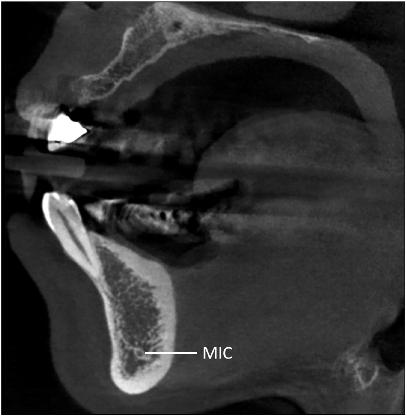
Fig. 2 Mandibular incisive canal (MIC) in the lower canine region in sagittal cone-beam computed tomography. Note the MIC is surrounded by the dense bone.

Fig. 3 Continuity of the mandibular incisive canal (MIC) and mandibular canal (MC) shown in axial cone-beam computed tomography. The bony canal to the mental foramen is also shown (arrow).
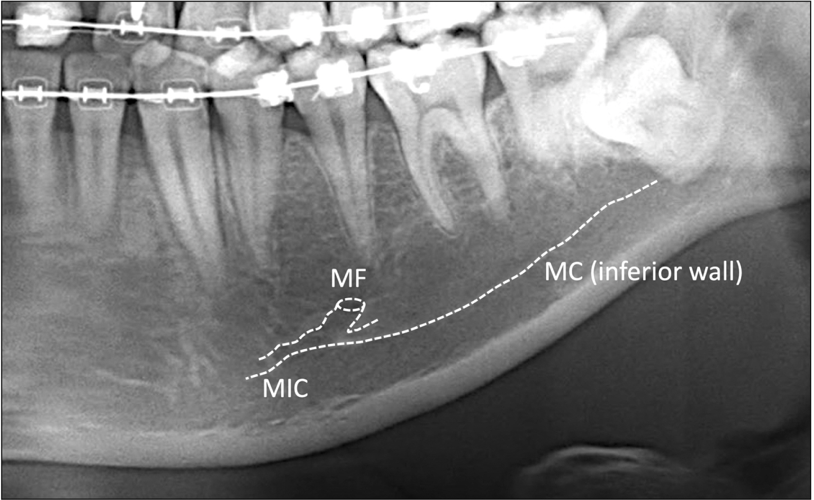
Fig. 4 Mandibular incisive canal (MIC) in panoramic radiograph. Note the initial part of the MIC is visible but not clear mesial to the first premolar. MF, mental foramen; MC, mandibular canal.
Reference
-
References
1. Wadu SG, Penhall B, Townsend GC. 1997; Morphological variability of the human inferior alveolar nerve. Clin Anat. 10:82–7. DOI: 10.1002/(SICI)1098-2353(1997)10:2<82::AID-CA2>3.0.CO;2-V. PMID: 9058013.
Article2. Iwanaga J, Matsushita Y, Decater T, Ibaragi S, Tubbs RS. 2021; Mandibular canal vs. inferior alveolar canal: evidence-based terminology analysis. Clin Anat. 34:209–17. DOI: 10.1002/ca.23648. PMID: 32644203.
Article3. Iwanaga J, Ibaragi S, Takeshita Y, Asaumi J, Horner K, Gest TR, Tubbs RS. 2021; Apr. 27. Mandibular canal versus inferior alveolar canal: a Delphi study. Clin Anat. [Epub]. https://doi.org/10.1002/ca.23740. DOI: 10.1002/ca.23740. PMID: 33905583.
Article4. Xu Y, Suo N, Tian X, Li F, Zhong G, Liu X, Bao Y, Song T, Tian H. 2015; Anatomic study on mental canal and incisive nerve canal in interforaminal region in Chinese population. Surg Radiol Anat. 37:585–9. DOI: 10.1007/s00276-014-1402-7. PMID: 25519298.
Article5. Rodella LF, Buffoli B, Labanca M, Rezzani R. 2012; A review of the mandibular and maxillary nerve supplies and their clinical relevance. Arch Oral Biol. 57:323–34. DOI: 10.1016/j.archoralbio.2011.09.007. PMID: 21996489.
Article6. Hur MS, Kim HC, Won SY, Hu KS, Song WC, Koh KS, Kim HJ. 2013; Topography and spatial fascicular arrangement of the human inferior alveolar nerve. Clin Implant Dent Relat Res. 15:88–95. DOI: 10.1111/j.1708-8208.2011.00335.x. PMID: 21414139.
Article7. von Arx T, Lozanoff S. von Arx T, Lozanoff S, editors. 2017. Mandibular incisive canal. Clinical Oral Anatomy: A Comprehensive Review for Dental Practitioners and Researchers. Springer International Publishing;Cham: p. 445–61. DOI: 10.1007/978-3-319-41993-0_21.
Article8. Nkenke E, Schultze-Mosgau S, Radespiel-Tröger M, Kloss F, Neukam FW. 2001; Morbidity of harvesting of chin grafts: a prospective study. Clin Oral Implants Res. 12:495–502. DOI: 10.1034/j.1600-0501.2001.120510.x. PMID: 11564110.
Article9. De Andrade E, Otomo-Corgel J, Pucher J, Ranganath KA, St George N Jr. 2001; The intraosseous course of the mandibular incisive nerve in the mandibular symphysis. Int J Periodontics Restorative Dent. 21:591–7. PMID: 11794570.10. Krasny A, Krasny N, Prescher A. 2012; Study of inferior dental canal and its contents using high-resolution magnetic resonance imaging. Surg Radiol Anat. 34:687–93. DOI: 10.1007/s00276-011-0910-y. PMID: 22143348.
Article11. Yang XW, Zhang FF, Li YH, Wei B, Gong Y. 2017; Characteristics of intrabony nerve canals in mandibular interforaminal region by using cone-beam computed tomography and a recommendation of safe zone for implant and bone harvesting. Clin Implant Dent Relat Res. 19:530–8. DOI: 10.1111/cid.12474. PMID: 28374431.
Article12. Jacobs R, Lambrichts I, Liang X, Martens W, Mraiwa N, Adriaensens P, Gelan J. 2007; Neurovascularization of the anterior jaw bones revisited using high-resolution magnetic resonance imaging. Oral Surg Oral Med Oral Pathol Oral Radiol Endod. 103:683–93. DOI: 10.1016/j.tripleo.2006.11.014. PMID: 17320428.
Article13. Hirsch JM, Brånemark PI. 1995; Fixture stability and nerve function after transposition and lateralization of the inferior alveolar nerve and fixture installation. Br J Oral Maxillofac Surg. 33:276–81. DOI: 10.1016/0266-4356(95)90037-3. PMID: 8555142.
Article14. Starkie C, Stewart D. 1931; The intra-mandibular course of the inferior dental nerve. J Anat. 65(Pt 3):319–23. PMID: 17104322. PMCID: PMC1248854.15. Stewart D, Wilson SL. 1928; Regional anesthesia and innervation of the teeth. Lancet. 212:809–11. DOI: 10.1016/S0140-6736(00)77724-5.
Article16. Mardinger O, Chaushu G, Arensburg B, Taicher S, Kaffe I. 2000; Anatomic and radiologic course of the mandibular incisive canal. Surg Radiol Anat. 22:157–61. DOI: 10.1007/s00276-000-0157-5. PMID: 11143307.
Article17. Parnia F, Moslehifard E, Hafezeqoran A, Mahboub F, Mojaver-Kahnamoui H. 2012; Characteristics of anatomical landmarks in the mandibular interforaminal region: a cone-beam computed tomography study. Med Oral Patol Oral Cir Bucal. 17:e420–5. DOI: 10.4317/medoral.17520. PMID: 22143718. PMCID: PMC3476108.
Article18. Gilis S, Dhaene B, Dequanter D, Loeb I. 2019; Mandibular incisive canal and lingual foramina characterization by cone-beam computed tomography. Morphologie. 103:48–53. DOI: 10.1016/j.morpho.2018.12.005. PMID: 30642812.
Article19. Kong N, Hui M, Miao F, Yuan H, Du Y, Chen N. 2016; Mandibular incisive canal in Han Chinese using cone beam computed tomography. Int J Oral Maxillofac Surg. 45:1142–6. DOI: 10.1016/j.ijom.2016.04.019. PMID: 27184354.
Article20. Apostolakis D, Brown JE. 2012; The anterior loop of the inferior alveolar nerve: prevalence, measurement of its length and a recommendation for interforaminal implant installation based on cone beam CT imaging. Clin Oral Implants Res. 23:1022–30. DOI: 10.1111/j.1600-0501.2011.02261.x. PMID: 22092440.
Article21. Uchida Y, Noguchi N, Goto M, Yamashita Y, Hanihara T, Takamori H, Sato I, Kawai T, Yosue T. 2009; Measurement of anterior loop length for the mandibular canal and diameter of the mandibular incisive canal to avoid nerve damage when installing endosseous implants in the interforaminal region: a second attempt introducing cone beam computed tomography. J Oral Maxillofac Surg. 67:744–50. DOI: 10.1016/j.joms.2008.05.352. PMID: 19304029.
Article22. Pires CA, Bissada NF, Becker JJ, Kanawati A, Landers MA. 2012; Mandibular incisive canal: cone beam computed tomography. Clin Implant Dent Relat Res. 14:67–73. DOI: 10.1111/j.1708-8208.2009.00228.x. PMID: 19673957.
Article23. Sahman H, Sekerci AE, Sisman Y, Payveren M. 2014; Assessment of the visibility and characteristics of the mandibular incisive canal: cone beam computed tomography versus panoramic radiography. Int J Oral Maxillofac Implants. 29:71–8. DOI: 10.11607/jomi.3304. PMID: 24451856.
Article24. Pereira-Maciel P, Tavares-de-Sousa E, Oliveira-Sales MA. 2015; The mandibular incisive canal and its anatomical relationships: a cone beam computed tomography study. Med Oral Patol Oral Cir Bucal. 20:e723–8. DOI: 10.4317/medoral.20644. PMID: 26449433. PMCID: PMC4670253.
Article25. Hunt DR, Jovanovic SA. 1999; Autogenous bone harvesting: a chin graft technique for particulate and monocortical bone blocks. Int J Periodontics Restorative Dent. 19:165–73. PMID: 10635182.26. Al-Ani O, Nambiar P, Ha KO, Ngeow WC. 2013; Safe zone for bone harvesting from the interforaminal region of the mandible. Clin Oral Implants Res. 24 Suppl A100:115–21. DOI: 10.1111/j.1600-0501.2011.02393.x. PMID: 22233422.
Article27. Sbordone L, Menchini-Fabris GB, Toti P, Sbordone C, Califano L, Guidetti F. 2009; Clinical survey of neurosensory side-effects of mandibular parasymphyseal bone harvesting. Int J Oral Maxillofac Surg. 38:139–45. DOI: 10.1016/j.ijom.2008.11.017. PMID: 19128942.
Article28. Pommer B, Tepper G, Gahleitner A, Zechner W, Watzek G. 2008; New safety margins for chin bone harvesting based on the course of the mandibular incisive canal in CT. Clin Oral Implants Res. 19:1312–6. DOI: 10.1111/j.1600-0501.2008.01590.x. PMID: 19040448.
Article29. Lee CY, Yanagihara LC, Suzuki JB. 2012; Brisk, pulsatile bleeding from the anterior mandibular incisive canal during implant surgery: a case report and use of an active hemostatic matrix to terminate acute bleeding. Implant Dent. 21:368–73. DOI: 10.1097/ID.0b013e318264d70d. PMID: 22968570.30. Mraiwa N, Jacobs R, Moerman P, Lambrichts I, van Steenberghe D, Quirynen M. 2003; Presence and course of the incisive canal in the human mandibular interforaminal region: two-dimensional imaging versus anatomical observations. Surg Radiol Anat. 25:416–23. DOI: 10.1007/s00276-003-0152-8. PMID: 13680184.
Article31. Kütük N, Demirbaş AE, Gönen ZB, Topan C, Kiliç E, Etöz OA, Alkan A. 2013; Anterior mandibular zone safe for implants. J Craniofac Surg. 24:e405–8. DOI: 10.1097/SCS.0b013e318292c7d5. PMID: 23851883.
Article32. Romanos GE, Greenstein G. 2009; The incisive canal. Considerations during implant placement: case report and literature review. Int J Oral Maxillofac Implants. 24:740–5. PMID: 19885417.33. Wismeijer D, van Waas MA, Vermeeren JI, Kalk W. 1997; Patients' perception of sensory disturbances of the mental nerve before and after implant surgery: a prospective study of 110 patients. Br J Oral Maxillofac Surg. 35:254–9. DOI: 10.1016/S0266-4356(97)90043-7. PMID: 9291263.
Article34. Tay AB, Lai JB, Lye KW, Wong WY, Nadkarni NV, Li W, Bautista D. 2015; Inferior alveolar nerve injury in trauma-induced mandible fractures. J Oral Maxillofac Surg. 73:1328–40. DOI: 10.1016/j.joms.2015.02.003. PMID: 25914133.
Article35. Pickrell BB, Serebrakian AT, Maricevich RS. 2017; Mandible fractures. Semin Plast Surg. 31:100–7. DOI: 10.1055/s-0037-1601374. PMID: 28496390. PMCID: PMC5423793.36. Ellis E 3rd. 2011; A study of 2 bone plating methods for fractures of the mandibular symphysis/body. J Oral Maxillofac Surg. 69:1978–87. DOI: 10.1016/j.joms.2011.01.032. PMID: 21549485.
Article37. Agnihotri A, Prabhu S, Thomas S. 2014; A comparative analysis of the efficacy of cortical screws as lag screws and miniplates for internal fixation of mandibular symphyseal region fractures: a randomized prospective study. Int J Oral Maxillofac Surg. 43:22–8. DOI: 10.1016/j.ijom.2013.07.001. PMID: 23928155.
Article38. Sun HH, Lin SC, Park CM, Elo JA. 2020; Asymptomatic intrabony radiolucency of the anterior mandible. Oral Surg Oral Med Oral Pathol Oral Radiol. 130:350–6. DOI: 10.1016/j.oooo.2019.05.002. PMID: 32173391.
Article39. Bilginaylar K, Orhan K, Uyanik LO. 2016; Mandibular incisive canal in relation to periapical surgery. Contemp Clin Dent. 7:79–81. DOI: 10.4103/0976-237X.177095. PMID: 27041907. PMCID: PMC4792062.
Article40. Perkins D, Stiharu TI, Swift JQ, Dao TV, Mainville GN. 2018; Intraosseous schwannoma of the jaws: an updated review of the literature and report of 2 new cases affecting the mandible. J Oral Maxillofac Surg. 76:1226–47. DOI: 10.1016/j.joms.2017.12.017. PMID: 29360457.
Article41. Rengaswamy V. 1978; Central neurilemmoma of the jaws. Review of literature and case report. Int J Oral Surg. 7:300–4. DOI: 10.1016/S0300-9785(78)80099-4. PMID: 100443.42. Iwanaga J, Singh V, Ohtsuka A, Hwang Y, Kim HJ, Moryś J, Ravi KS, Ribatti D, Trainor PA, Sañudo JR, Apaydin N, Şengül G, Albertine KH, Walocha JA, Loukas M, Duparc F, Paulsen F, Del Sol M, Adds P, Hegazy A, Tubbs RS. 2021; Acknowledging the use of human cadaveric tissues in research papers: recommendations from anatomical journal editors. Clin Anat. 34:2–4. DOI: 10.1002/ca.23671. PMID: 32808702.
Article
- Full Text Links
-
- Actions
-
Cited
- CITED
-
- Close
- Share
-
- Similar articles
-
- Updates on the Inferior Alveolar Nerve Block Anesthesia
- Mylohyoid foramen of mandible: a rare exit point of intra-mandibular origin of nerve to mylohyoid
- Diplopia after Inferior Alveolar Nerve Block Anesthesia: A Case Report
- Assessment of the anterior loop of the inferior alveolar nerve via cone-beam computed tomography
- Delayed paresthesia of inferior alveolar nerve after dental surgery: case report and related pathophysiology


