J Korean Assoc Oral Maxillofac Surg.
2017 Dec;43(6):395-400. 10.5125/jkaoms.2017.43.6.395.
Assessment of the anterior loop of the inferior alveolar nerve via cone-beam computed tomography
- Affiliations
-
- 1Department of Oral and Maxillofacial Surgery, Faculty of Dentistry, Mashhad University of Medical Sciences, Mashhad, Iran.
- 2Department of Periodontics, Faculty of Dentistry, Mashhad University of Medical Sciences, Mashhad, Iran.
- 3Department of Oral and Maxillofacial Radiology, Faculty of Dentistry, Mashhad University of Medical Sciences, Mashhad, Iran.
- KMID: 2398994
- DOI: http://doi.org/10.5125/jkaoms.2017.43.6.395
Abstract
OBJECTIVES
The aim of this study was to evaluate different anatomical variants of the anterior loop of the inferior alveolar nerve (IAN) via cone-beam computed tomography (CBCT).
MATERIALS AND METHODS
CBCT images of 71 patients (36 males and 35 females) were evaluated. We used the classification described by Solar for IAN evaluation. In this classification, three different types of IAN loops were introduced prior to emerging from the mental foramen. We classified patients according to this system and introduced a new, fourth type.
RESULTS
Type I was seen in 15 sites (10.6%), type II in 39 sites (27.5%), and type III in 50 sites (35.2%). We found a new type in 38 sites (26.8%) that constituted a fourth type.
CONCLUSION
We found that type III was the most common variant. In the fourth type, the IAN was not detectable because the main nerve was adjacent to the cortical plate and the incisive branch was thinner than the main branch and alongside it. In this type, more care is needed for surgeries including inferior alveolar and mental nerve transposition.
Keyword
MeSH Terms
Figure
-

Fig. 1 Both left and right mental foramina on the axial section and panoramic curve drawn on the axial section.
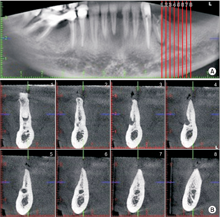
Fig. 2 A. Panoramic section showing type I on both sides; the guide plans for cross sections are also seen. B. Cross-sectional images showing type I.
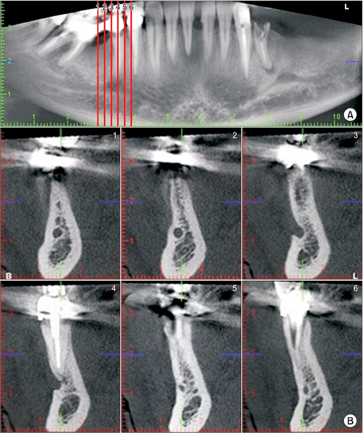
Fig. 3 A. Panoramic view of type IV. B. Cross-sectional images of type IV.
Reference
-
1. Mraiwa N, Jacobs R, van Steenberghe D, Quirynen M. Clinical assessment and surgical implications of anatomic challenges in the anterior mandible. Clin Implant Dent Relat Res. 2003; 5:219–225.
Article2. Jacobs R, Mraiwa N, vanSteenberghe D, Gijbels F, Quirynen M. Appearance, location, course, and morphology of the mandibular incisive canal: an assessment on spiral CT scan. Dentomaxillofac Radiol. 2002; 31:322–327.
Article3. Agbaje JO, Sun Y, De Munter S, Schepers S, Vrielinck L, Lambrichts I, et al. CBCT-based predictability of attachment of the neurovascular bundle to the proximal segment of the mandible during sagittal split osteotomy. Int J Oral Maxillofac Surg. 2013; 42:308–315.
Article4. Alqerban A, Jacobs R, Souza PC, Willems G. In-vitro comparison of 2 cone-beam computed tomography systems and panoramic imaging for detecting simulated canine impaction-induced external root resorption in maxillary lateral incisors. Am J Orthod Dentofacial Orthop. 2009; 136:764764.e1–764.e11. discussion 764-5.
Article5. White SC. Cone-beam imaging in dentistry. Health Phys. 2008; 95:628–637.
Article6. Chávez-Lomeli ME, Mansilla Lory J, Pompa JA, Kjaer I. The human mandibular canal arises from three separate canals innervating different tooth groups. J Dent Res. 1996; 75:1540–1544.
Article7. de Villiers H. The skull of the South African Negro: a biometrical and morphological study. Johannesburg: Wits University Press;1970.8. Ngeow WC, Dionysius DD, Ishak H, Nambiar P. A radiographic study on the visualization of the anterior loop in dentate subjects of different age groups. J Oral Sci. 2009; 51:231–237.
Article9. Arzouman MJ, Otis L, Kipnis V, Levine D. Observations of the anterior loop of the inferior alveolar canal. Int J Oral Maxillofac Implants. 1993; 8:295–300.10. Solar P, Ulm C, Frey G, Matejka M. A classification of the intraosseous paths of the mental nerve. Int J Oral Maxillofac Implants. 1994; 9:339–344.11. Kajan ZD, Salari A. Presence and course of the mandibular incisive canal and presence of the anterior loop in cone beam computed tomography images of an Iranian population. Oral Radiol. 2012; 28:55–61.
Article12. Vazquez L, Saulacic N, Belser U, Bernard JP. Efficacy of panoramic radiographs in the preoperative planning of posterior mandibular implants: a prospective clinical study of 1527 consecutively treated patients. Clin Oral Implants Res. 2008; 19:81–85.
Article13. Wismeijer D, van Waas MA, Vermeeren JI, Kalk W. Patients' perception of sensory disturbances of the mental nerve before and after implant surgery: a prospective study of 110 patients. Br J Oral Maxillofac Surg. 1997; 35:254–259.
Article14. Kalender A, Orhan K, Aksoy U. Evaluation of the mental foramen and accessory mental foramen in Turkish patients using cone-beam computed tomography images reconstructed from a volumetric rendering program. Clin Anat. 2012; 25:584–592.
Article15. Kuzmanovic DV, Payne AG, Kieser JA, Dias GJ. Anterior loop of the mental nerve: a morphological and radiographic study. Clin Oral Implants Res. 2003; 14:464–471.
Article16. Chen JC, Lin LM, Geist JR, Chen JY, Chen CH, Chen YK. A retrospective comparison of the location and diameter of the inferior alveolar canal at the mental foramen and length of the anterior loop between American and Taiwanese cohorts using CBCT. Surg Radiol Anat. 2013; 35:11–18.
Article17. Parnia F, Moslehifard E, Hafezeqoran A, Mahboub F, Mojaver-Kahnamoui H. Characteristics of anatomical landmarks in the mandibular interforaminal region: a cone-beam computed tomography study. Med Oral Patol Oral Cir Bucal. 2012; 17:e420–e425.
Article18. Danforth RA, Dus I, Mah J. 3-D volume imaging for dentistry: a new dimension. J Calif Dent Assoc. 2003; 31:817–823.19. Haghanifar S, Rokouei M. Radiographic evaluation of the mental foramen in a selected Iranian population. Indian J Dent Res. 2009; 20:150–152.
Article20. Bushong SC. Radiologic science for technologists: physics, biology, and protection. 8th ed. New York: Elsevier;2004. p. 29–30.21. Katakami K, Mishima A, Shiozaki K, Shimoda S, Hamada Y, Kobayashi K. Characteristics of accessory mental foramina observed on limited cone-beam computed tomography images. J Endod. 2008; 34:1441–1445.
Article22. Koh KJ, Kim KA. Observation of the anterior loop and mental foramen of the mandibular canal using cone beam computed tomography. Korean J Oral Maxillofac Radiol. 2009; 39:81–87.23. Demir A, Izgi E, Pekiner FN. Anterior loop of the mental foramen in a Turkish subpopulation with dentate patients: a cone beam computed tomography study. J Marmara Univ Inst Health Sci. 2015; 5:231–238.
Article24. Rosenquist B. Is there an anterior loop of the inferior alveolar nerve? Int J Periodontics Restorative Dent. 1996; 16:40–45.25. Kaya Y, Sencimen M, Sahin S, Okcu KM, Dogan N, Bahcecitapar M. Retrospective radiographic evaluation of the anterior loop of the mental nerve: comparison between panoramic radiography and spiral computerized tomography. Int J Oral Maxillofac Implants. 2008; 23:919–925.26. Benninger B, Miller D, Maharathi A, Carter W. Dental implant placement investigation: is the anterior loop of the mental nerve clinically relevant? J Oral Maxillofac Surg. 2011; 69:182–185.
Article27. Mraiwa N, Jacobs R, Moerman P, Lambrichts I, van Steenberghe D, Quirynen M. Presence and course of the incisive canal in the human mandibular interforaminal region: two-dimensional imaging versus anatomical observations. Surg Radiol Anat. 2003; 25:416–423.
Article28. Bavitz JB, Harn SD, Hansen CA, Lang M. An anatomical study of mental neurovascular bundle-implant relationships. Int J Oral Maxillofac Implants. 1993; 8:563–567.29. Mardinger O, Chaushu G, Arensburg B, Taicher S, Kaffe I. Anterior loop of the mental canal: an anatomical-radiologic study. Implant Dent. 2000; 9:120–125.30. Yu SK, Kim S, Kang SG, Kim JH, Lim KO, Hwang SI, et al. Morphological assessment of the anterior loop of the mandibular canal in Koreans. Anat Cell Biol. 2015; 48:75–80.
Article31. Uchida Y, Noguchi N, Goto M, Yamashita Y, Hanihara T, Takamori H, et al. Measurement of anterior loop length for the mandibular canal and diameter of the mandibular incisive canal to avoid nerve damage when installing endosseous implants in the interforaminal region: a second attempt introducing cone beam computed tomography. J Oral Maxillofac Surg. 2009; 67:744–750.
Article32. Oguz O, Bozkir MG. Evaluation of location of mandibular and mental foramina in dry, young, adult human male, dentulous mandibles. West Indian Med J. 2002; 51:14–16.33. Rosa MB, Sotto-Maior BS, Machado Vde C, Francischone CE. Retrospective study of the anterior loop of the inferior alveolar nerve and the incisive canal using cone beam computed tomography. Int J Oral Maxillofac Implants. 2013; 28:388–392.
Article34. Apostolakis D, Brown JE. The dimensions of the mandibular incisive canal and its spatial relationship to various anatomical landmarks of the mandible: a study using cone beam computed tomography. Int J Oral Maxillofac Implants. 2013; 28:117–124.
Article35. Lee JK, Kim YG. An anatomical study on the mandibular medial surface by CBCT analysis for safer implant placement. J Korean Assoc Oral Maxillofac Surg. 2011; 37:43–48.
Article
- Full Text Links
-
- Actions
-
Cited
- CITED
-
- Close
- Share
-
- Similar articles
-
- Removal of a broken needle using three-dimensional computed tomography: a case report
- Assessment of the relationship between the mandibular third molar and the mandibular canal using panoramic radiograph and cone beam computed tomography
- Cone-beam computed tomography measurement of the position of the inferior alveolar nerve canal in mandibular prognathism
- Observation of the anterior loop and mental foramen of the mandibular canal using cone beam computed tomography
- An alternative approach to extruding a vertically impacted lower third molar using an orthodontic miniscrew: A case report with cone-beam CT follow-up
