Intracranial Tumors Associated With IgG4-Related Disease
- Affiliations
-
- 1Departments of Neurosurgery, Yonsei University College of Medicine, Seoul, Korea
- 2Departments of Pathology, Yonsei University College of Medicine, Seoul, Korea
- 3Pituitary Tumor Center, Severance Hospital, Seoul, Korea
- 4Brain Tumor Center, Severance Hospital, Seoul, Korea
- KMID: 2522226
- DOI: http://doi.org/10.14791/btrt.2021.9.e17
Abstract
- IgG4-related disease (IgG4-RD) is an immune-mediated inflammatory condition which is characterized by dense lymphoplasmacytic infiltrations with a predominance of IgG4 plasma cells in the affected tissue. Although pachymeninx and pituitary gland are the most common sites where IgG4-RD infiltrates, the associations with IgG4-RD and a true intracranial tumor have not been yet reported in literature. Herein, we report two cases with intracranial tumors associated with IgG4-RD; a 36-year-old male patient with a huge meningioma and another 54-year old woman with a pituitary macroadenoma. Pathological examination revealed their tumors were substantially infiltrated by IgG4 plasma cells indicating its possible relation with IgG4-RD.
Keyword
Figure
-
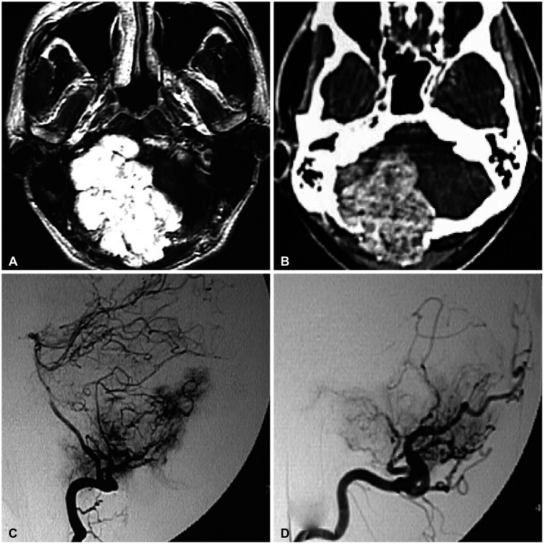
Fig. 1 Preoperative radiological findings of a 36-year-old male patient (Case 1). A: MRI shows a large extra-axial tumor on the right posterior fossa. B: CT reveals extensive destruction of the occipital bone. C and D: Cerebral angiography demonstrates a strong vascular supply from the right occipital artery, right posterior inferior cerebellar artery, and right posterior meningeal artery.
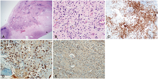
Fig. 2 Pathological findings of a 36-year-old male patient (Case 1). A: The first case shows a well demarcated extra-axial dura-based mass (H&E, ×12). B: High power view shows neoplastic meningothelial proliferation with many areas of plasmacytic infiltration (H&E, ×200). C: In addition, the meningothelial cells show EMA immunoreactivity (EMA, ×100). D and E: On immunohistochemistry, a high IgG4 (IgG4, ×100)/IgG (IgG, ×100) ratio is noted.
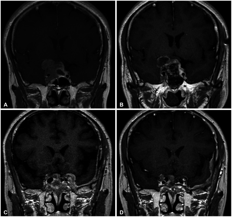
Fig. 3 Radiological findings of a 54-year-old female patient (Case 2). A: MRI shows invasive adenoma invading right cavernous sinus. B: Postoperative MRI shows subtotal removal status of pituitary adenoma. C: MRI shows newly developed bulging contour in the left cavernous sinus with adjacent dural thickening. D: After repetitive administration of steroid, follow up MRI shows decreased dural thickening lesion in the left cavernous sinus.
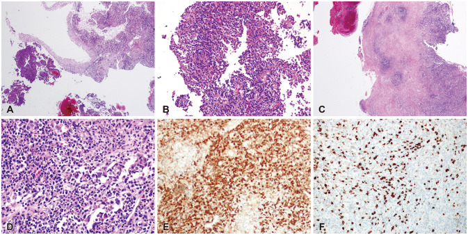
Fig. 4 Pathological findings of a 54-year-old female patient (Case 2). A: Low power (H&E, ×12) shows a portion of pituitary adenoma (left lower) and dura with inflammation (right upper). B: High power view (H&E, ×100) shows a classic histological findings of pituitary adenoma C: Dura with dense inflammation (H&E, ×40) is noted. D: High power view (H&E, ×200) shows dense inflammatory cell infiltrates with many plasma cells. E and F: The IgG4 positive plasma cells/IgG positive cells ratio was 24.8% based on IgG (E, ×200) and IgG4 (F, ×200) staining.
Reference
-
1. Stone JH, Khosroshahi A, Deshpande V, et al. Recommendations for the nomenclature of IgG4-related disease and its individual organ system manifestations. Arthritis Rheum. 2012; 64:3061–3067. PMID: 22736240.2. Baptista B, Casian A, Gunawardena H, D’Cruz D, Rice CM. Neurological manifestations of IgG4-related disease. Curr Treat Options Neurol. 2017; 19:14. PMID: 28374231.3. Umehara H, Okazaki K, Masaki Y, et al. Comprehensive diagnostic criteria for IgG4-related disease (IgG4-RD), 2011. Mod Rheumatol. 2012; 22:21–30. PMID: 22218969.4. Deshpande V, Zen Y, Chan JK, et al. Consensus statement on the pathology of IgG4-related disease. Mod Pathol. 2012; 25:1181–1192. PMID: 22596100.5. Wallace ZS, Deshpande V, Mattoo H, et al. IgG4-related disease: clinical and laboratory features in one hundred twentyfive patients. Arthritis Rheumatol. 2015; 67:2466–2475. PMID: 25988916.6. Wallace ZS, Stone JH. An update on IgG4-related disease. Curr Opin Rheumatol. 2015; 27:83–90. PMID: 25415530.7. Stone JH, Zen Y, Deshpande V. IgG4-related disease. N Engl J Med. 2012; 366:539–551. PMID: 22316447.8. Vanegas-Garcia AL, Calle-Lopez Y, Zapata CH, Alvarez-Espinal DM, Saavedra-Gonzalez YA, Arango-Viana JC. Central nervous system in IgG4-related disease: case report and literature review. Rev Neurol. 2016; 63:119–124. PMID: 27412018.9. Yamamoto M, Takahashi H, Ohara M, et al. A case of Mikulicz’s disease (IgG4-related plasmacytic disease) complicated by autoimmune hypophysitis. Scand J Rheumatol. 2006; 35:410–411. PMID: 17062446.10. Lindstrom KM, Cousar JB, Lopes MBS. IgG4-related meningeal disease: clinico-pathological features and proposal for diagnostic criteria. Acta Neuropathol. 2010; 120:765–776. PMID: 20844883.11. Mattoo H, Mahajan VS, Maehara T, et al. Clonal expansion of CD4(+) cytotoxic T lymphocytes in patients with IgG4-related disease. J Allergy Clin Immunol. 2016; 138:825–838. PMID: 26971690.12. Chen Y, Lin W, Yang H, et al. Aberrant expansion and function of follicular helper T cell subsets in IgG4-related disease. Arthritis Rheumatol. 2018; 70:1853–1865. PMID: 29781221.13. Ahn SS, Song JJ, Park YB, Lee SW. Malignancies in Korean patients with immunoglobulin G4 related disease. Int J Rheum Dis. 2017; 20:1028–1035. PMID: 28544157.14. Wallace ZS, Wallace CJ, Lu N, Choi HK, Stone JH. Association of IgG4-related disease with history of malignancy. Arthritis Rheumatol. 2016; 68:2283–2289. PMID: 27273903.15. Poo SX, Tham CSW, Smith C, et al. IgG4-related disease in a multi-ethnic community: clinical characteristics and association with malignancy. QJM. 2019; 112:763–769. PMID: 31225617.16. Hirano K, Tada M, Sasahira N, et al. Incidence of malignancies in patients with IgG4-related disease. Intern Med. 2014; 53:171–176. PMID: 24492683.17. Yamamoto M, Takahashi H, Tabeya T, et al. Risk of malignancies in IgG4-related disease. Mod Rheumatol. 2012; 22:414–418. PMID: 21894525.18. Tang H, Yang H, Zhang P, et al. Malignancy and IgG4-related disease: the incidence, related factors and prognosis from a prospective cohort study in China. Sci Rep. 2020; 10:4910. PMID: 32188869.19. Chiba T, Marusawa H, Ushijima T. Inflammation-associated cancer development in digestive organs: mechanisms and roles for genetic and epigenetic modulation. Gastroenterology. 2012; 143:550–563. PMID: 22796521.20. Daveau M, Pavie-Fischer J, Rivat L, et al. IgG4 subclass in malignant melanoma. J Natl Cancer Inst. 1977; 58:189–192. PMID: 833869.21. Karagiannis P, Gilbert AE, Josephs DH, et al. IgG4 subclass antibodies impair antitumor immunity in melanoma. J Clin Invest. 2013; 123:1457–1474. PMID: 23454746.22. Karagiannis P, Villanova F, Josephs DH, et al. Elevated IgG4 in patient circulation is associated with the risk of disease progression in melanoma. Oncoimmunology. 2015; 4:e1032492. PMID: 26451312.23. Harada K, Nakanuma Y. Cholangiocarcinoma with respect to IgG4 reaction. Int J Hepatol. 2014; 2014:803876. PMID: 25132998.24. Raina A, Krasinskas AM, Greer JB, et al. Serum immunoglobulin G fraction 4 levels in pancreatic cancer: elevations not associated with autoimmune pancreatitis. Arch Pathol Lab Med. 2008; 132:48–53. PMID: 18181673.25. Harshyne LA, Nasca BJ, Kenyon LC, Andrews DW, Hooper DC. Serum exosomes and cytokines promote a T-helper cell type 2 environment in the peripheral blood of glioblastoma patients. Neuro Oncol. 2016; 18:206–215. PMID: 26180083.26. Aalberse RC, Stapel SO, Schuurman J, Rispens T. Immunoglobulin G4: an odd antibody. Clin Exp Allergy. 2009; 39:469–477. PMID: 19222496.27. Kimura Y, Harada K, Nakanuma Y. Pathologic significance of immunoglobulin G4-positive plasma cells in extrahepatic cholangiocarcinoma. Hum Pathol. 2012; 43:2149–2156. PMID: 22647350.28. Crescioli S, Correa I, Karagiannis P, et al. IgG4 characteristics and functions in cancer immunity. Curr Allergy Asthma Rep. 2016; 16:7. PMID: 26742760.29. Mahajan VS, Mattoo H, Deshpande V, Pillai SS, Stone JH. IgG4-related disease. Annu Rev Pathol. 2014; 9:315–347. PMID: 24111912.
- Full Text Links
-
- Actions
-
Cited
- CITED
-
- Close
- Share
-
- Similar articles
-
- IgG4-Related Hypertrophic Pachymeningitis Mimicking Cerebral Venous Thrombosis
- Advances in IgG4-related Hepatobiliary Disease
- Role of Endoscopic Procedures in the Diagnosis of IgG4-Related Pancreatobiliary Disease
- IgG4-Related Intracranial Hypertrophic Pachymeningitis : A Case Report and Review of the Literature
- A Case of IgG4-Related Disease with Pachymeningitis and Periaortitis
