Mapping of folic acid in the children brainstem
- Affiliations
-
- 1Laboratory of Neurosciences, School of Medicine, Universidad de Santander, Bucaramanga, Colombia
- 2Institute of Neurosciences of Castilla y León (INCYL), Laboratory of Neuroanatomy of the Peptidergic Systems, University of Salamanca, Salamanca, Spain
- 3Grupo GIR BMD (Bases Moleculares del Desarrollo), University of Salamanca, Salamanca, Spain
- KMID: 2521047
- DOI: http://doi.org/10.5115/acb.21.016
Abstract
- Using highly specific antisera, the neuroanatomical distribution of folic acid (FA) and retinoic acid (RA) has been studied for the first time in the children brainstem. Neither immunoreactive structures containing RA nor immunoreactive fibers containing FA were found. FA-immunoreactive perikarya (fusiform, small/medium in size, one short dendrite) were only found in the pons in three regions: central gray, reticular formation, and locus coeruleus. The number of cell bodies decreased with age. In the first case studied (2 years), a moderate density of cell bodies was observed in the central gray and reticular formation, whereas a low density was found in the locus coeruleus. In the second case (6 years), a low density of these perikarya was observed in the central gray, reticular formation, and locus coeruleus. In the third case (7 years), a low density of FA-immunoreactive cell bodies was found in the central gray and reticular formation, whereas in the locus coeruleus no immunoreactive cell bodies were observed. The distribution of FA in the central nervous system of humans and monkeys is different and, in addition, in these species the vitamin was located in different parts of the nerve cells. The restricted distribution of FA suggests that the vitamin is involved in specific physiological mechanisms.
Keyword
Figure
-
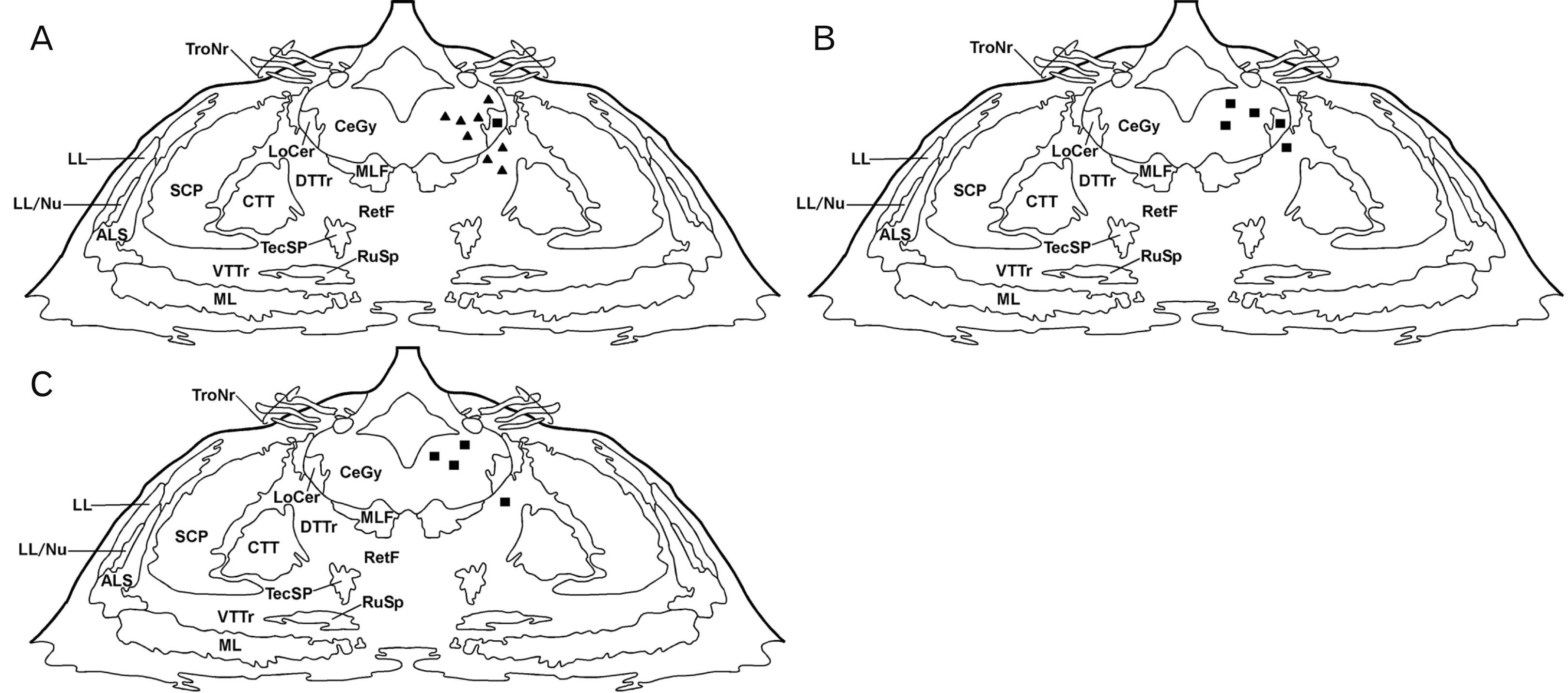
Fig. 1 Distribution of folic acid (FA)-immunoreactive cell bodies in frontal planes of the human pons. Two (A), six (B), and seven years (C). Cell bodies are represented by closed triangles (moderate density) and closed squares (low density). ALS, anterolateral system; CeGy, central gray; CTT, central tegmental tract; DTTr, dorsal trigeminothalamic tract; LL, lateral lemniscus; LL/Nu, lateral lemniscus nucleus; LoCer, locus coeruleus; ML, medial lemniscus; MLF, medial longitudinal fasciculus; SCP, superior cerebellar peduncle; RetF, reticular formation; RuSp, rubrospinal tract; TecSp, tectospinal tract; TroNr, trochlear nerve, VTTr, ventral trigeminothalamic tract.
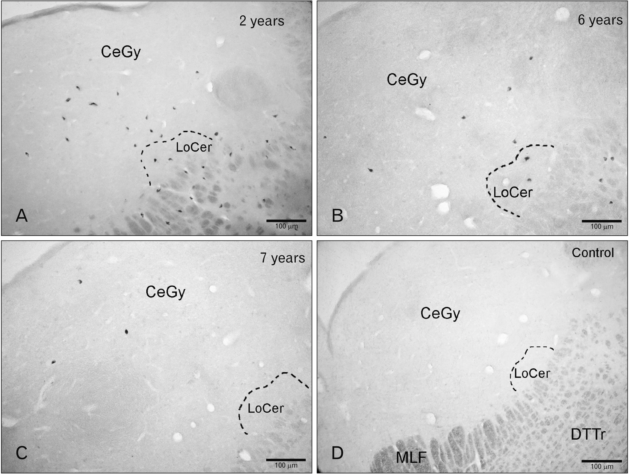
Fig. 2 Folic acid (FA) in the children pons. (A) Cell bodies containing FA in the central gray (CeGy), reticular formation (RetF), and locus coeruleus (LoCer) (2 years) (A) and 6 years (B). (C) Immunoreactive cell bodies in the CeGy (7 years). (D) Histological control (preabsorption of the anti-FA with an excess of synthetic FA). DTTr, dorsal trigeminothalamic tract; MLF, medial longitudinal fasciculus.
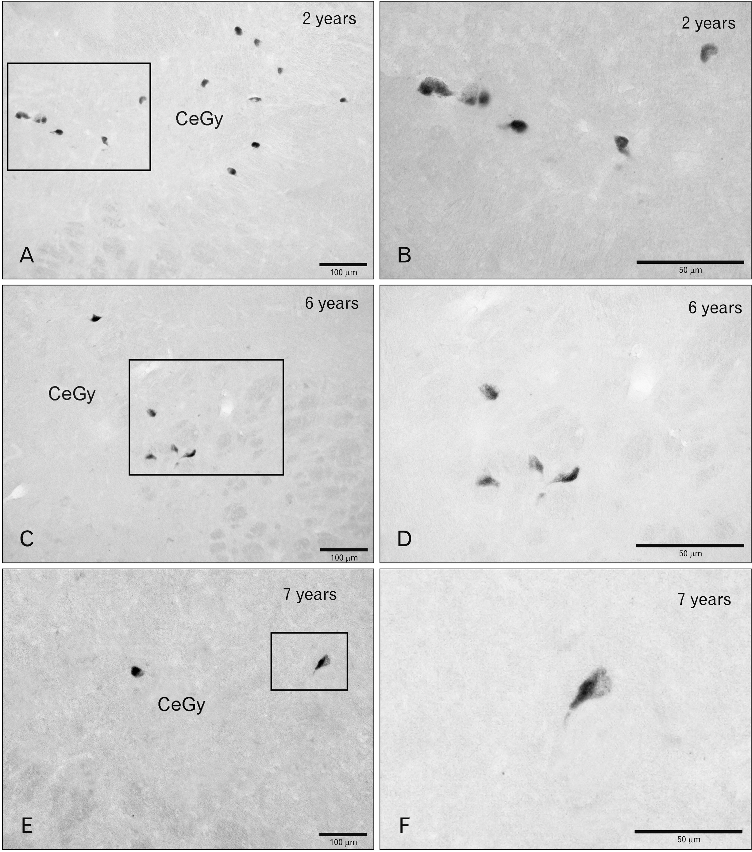
Fig. 3 Folic acid (FA)-immunoreacive cell bodies in the children pons central gray (CeGy). Two (A), six (C), and seven (E) years. (B, D, F) Higher magnifications of the regions delimited by rectangles in A, C, and E.
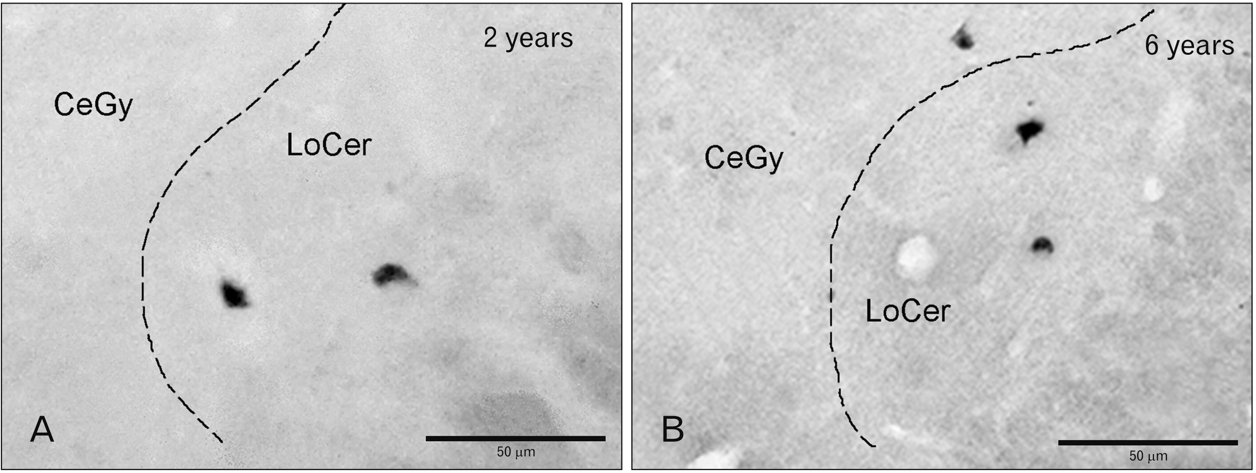
Fig. 4 Folic acid (FA)-immunoreactive perikarya in the children pons. (A) Locus coeruleus (LoCer) (2 years). (B) LoCer and central gray (CeGy) (6 years).
Reference
-
References
1. Abdollahifar MA, Azad N, Sajadi E, Shams Mofarahe Z, Zare F, Moradi A, Rezaee F, Gholamin M, Abdi S. 2019; Vitamin C restores ovarian follicular reservation in a mouse model of aging. Anat Cell Biol. 52:196–203. DOI: 10.5115/acb.2019.52.2.196. PMID: 31338237. PMCID: PMC6624328.
Article2. Hong JM, Kim JH, Kang JS, Lee WJ, Hwang YI. 2016; Vitamin C is taken up by human T cells via sodium-dependent vitamin C transporter 2 (SVCT2) and exerts inhibitory effects on the activation of these cells in vitro. Anat Cell Biol. 49:88–98. DOI: 10.5115/acb.2016.49.2.88. PMID: 27382510. PMCID: PMC4927435.3. Coveñas R, Mangas A, Bodet D, Duleu S, Marcos P, Karakas B, Geffard M. 2011; Frontiers in vitamin research: new antibodies, new data. ScientificWorldJournal. 11:1226–1242. DOI: 10.1100/tsw.2011.115. PMID: 21666992. PMCID: PMC5720100.
Article4. Coveñas R, Mangas A, Bodet D, Duleu S, Marcos P, Geffard M. Jackson CM, editor. 2011. Vitamin C in the monkey brain. Vitamin C: Nutrition, Side Effects, and Supplements. Nova Science Publishers;New York: p. 275–88.5. Coveñas R, González-Fuentes J, Rivas-Infante E, Lagartos-Donate MJ, Mangas A, Geffard M, Arroyo-Jiménez MM, Cebada-Sánchez S, Insausti R, Marcos P. 2015; Developmental study of vitamin C distribution in children's brainstems by immunohistochemistry. Ann Anat. 201:65–78. DOI: 10.1016/j.aanat.2015.06.001. PMID: 26226232.
Article6. Mangas A, Coveñas R, Geffard K, Geffard M, Marcos P, Insausti R, Dabadie MP. 2004; Folic acid in the monkey brain: an immunocytochemical study. Neurosci Lett. 362:258–61. DOI: 10.1016/j.neulet.2004.03.034. PMID: 15158027.
Article7. Mangas A, Coveñas R, Geffard K, Geffard M, Marcos P, Insausti R, Glaize G, Dabadie MP. 2006; Riboflavin-like inmunoreactive fibers in the monkey brain. Anat Embryol (Berl). 211:267–72. DOI: 10.1007/s00429-006-0080-6. PMID: 16456676.
Article8. Mangas A, Coveñas R, Geffard K, Geffard M, Marcos P, Insausti R, Dabadie MP. 2006; Thiamine-like fibers in the monkey brain: an immunocytochemical study. Life Sci. 79:1121–8. DOI: 10.1016/j.lfs.2006.03.017. PMID: 16624330.
Article9. Mangas A, Coveñas R, Bodet D, Duleu S, Marcos P, Geffard M. 2009; Vitamins in the monkey brain: an immunocytochemical study. J Chem Neuroanat. 38:1–8. DOI: 10.1016/j.jchemneu.2009.05.007. PMID: 19477264.
Article10. Mangas A, Bodet D, Duleu S, Yajeya J, Geffard M, Coveñas R. 2012; Direct visualization of retinoic acid in the rat hypothalamus: an immunohistochemical study. Neurosci Lett. 509:64–8. DOI: 10.1016/j.neulet.2011.12.053. PMID: 22230896.
Article11. Mangas A, Yajeya J, Gonzalez N, Husson M, Geffard M, Coveñas R. 2016; Detection of pantothenic acid-immunoreactive neurons in the rat lateral septal nucleus by a newly developed antibody. Folia Histochem Cytobiol. 54:186–192. DOI: 10.5603/FHC.a2016.0024. PMID: 27966211.
Article12. Chang S, Lu X, Wang S, Wang Z, Huo J, Huang J, Shangguan S, Li S, Zou J, Bao Y, Guo J, Wang F, Niu B, Zhang T, Qiu Z, Wu J, Wang L. 2019; The effect of folic acid deficiency on FGF pathway via Brachyury regulation in neural tube defects. FASEB J. 33:4688–702. DOI: 10.1096/fj.201801536R. PMID: 30592646.13. Li W, Ma Y, Li Z, Lv X, Wang X, Zhou D, Luo S, Wilson JX, Huang G. 2019; Folic acid decreases astrocyte apoptosis by preventing oxidative stress-induced telomere attrition. Int J Mol Sci. 21:62. DOI: 10.3390/ijms21010062. PMID: 31861819. PMCID: PMC6981374.
Article14. Liu H, Cao J, Zhang H, Qin S, Yu M, Zhang X, Wang X, Gao Y, Wilson JX, Huang G. 2013; Folic acid stimulates proliferation of transplanted neural stem cells after focal cerebral ischemia in rats. J Nutr Biochem. 24:1817–22. DOI: 10.1016/j.jnutbio.2013.04.002. PMID: 23850087.
Article15. Pei P, Cheng X, Yu J, Shen J, Li X, Wu J, Wang S, Zhang T. 2019; Folate deficiency induced H2A ubiquitination to lead to downregulated expression of genes involved in neural tube defects. Epigenetics Chromatin. 12:69. DOI: 10.1186/s13072-019-0312-7. PMID: 31722724. PMCID: PMC6852770.
Article16. Zhao Y, Huang G, Chen S, Gou Y, Dong Z, Zhang X. 2016; Folic acid deficiency increases brain cell injury via autophagy enhancement after focal cerebral ischemia. J Nutr Biochem. 38:41–9. DOI: 10.1016/j.jnutbio.2016.08.009. PMID: 27721115.
Article17. Liu H, Tian T, Qin S, Li W, Zhang X, Wang X, Gao Y, Huang G. 2015; Folic acid deficiency enhances abeta accumulation in APP/PS1 mice brain and decreases amyloid-associated miRNAs expression. J Nutr Biochem. 26:1502–8. DOI: 10.1016/j.jnutbio.2015.07.020. PMID: 26345540.
Article18. Zhuo JM, Praticò D. 2010; Acceleration of brain amyloidosis in an Alzheimer's disease mouse model by a folate, vitamin B6 and B12-deficient diet. Exp Gerontol. 45:195–201. DOI: 10.1016/j.exger.2009.12.005. PMID: 20005283. PMCID: PMC2826592.
Article19. Haghdoost-Yazdi H, Fraidouni N, Faraji A, Jahanihashemi H, Sarookhani M. 2012; High intake of folic acid or complex of B vitamins provides anti-Parkinsonism effect: no role for serum level of homocysteine. Behav Brain Res. 233:375–81. DOI: 10.1016/j.bbr.2012.05.011. PMID: 22610053.
Article20. ivastav S Sr, Singh SK, Yadav AK, ikrishna S Sr. 2015; Folic acid supplementation rescues anomalies associated with knockdown of parkin in dopaminergic and serotonergic neurons in Drosophila model of Parkinson's disease. Biochem Biophys Res Commun. 460:780–5. DOI: 10.1016/j.bbrc.2015.03.106. PMID: 25824034.21. Bremner JD, McCaffery P. 2008; The neurobiology of retinoic acid in affective disorders. Prog Neuropsychopharmacol Biol Psychiatry. 32:315–31. DOI: 10.1016/j.pnpbp.2007.07.001. PMID: 17707566. PMCID: PMC2704911.
Article22. Das BC, Thapa P, Karki R, Das S, Mahapatra S, Liu TC, Torregroza I, Wallace DP, Kambhampati S, Van Veldhuizen P, Verma A, Ray SK, Evans T. 2014; Retinoic acid signaling pathways in development and diseases. Bioorg Med Chem. 22:673–83. DOI: 10.1016/j.bmc.2013.11.025. PMID: 24393720. PMCID: PMC4447240.
Article23. Dev S, Adler AJ, Edwards RB. 1993; Adult rabbit brain synthesizes retinoic acid. Brain Res. 632:325–8. DOI: 10.1016/0006-8993(93)91170-W. PMID: 8149239.
Article24. Luo T, Wagner E, Crandall JE, Dräger UC. 2004; A retinoic-acid critical period in the early postnatal mouse brain. Biol Psychiatry. 56:971–80. DOI: 10.1016/j.biopsych.2004.09.020. PMID: 15601608.
Article25. Kane MA, Chen N, Sparks S, Napoli JL. 2005; Quantification of endogenous retinoic acid in limited biological samples by LC/MS/MS. Biochem J. 388(Pt 1):363–9. DOI: 10.1042/BJ20041867. PMID: 15628969. PMCID: PMC1186726.
Article26. Kane MA, Folias AE, Wang C, Napoli JL. 2008; Quantitative profiling of endogenous retinoic acid in vivo and in vitro by tandem mass spectrometry. Anal Chem. 80:1702–8. DOI: 10.1021/ac702030f. PMID: 18251521. PMCID: PMC4086453.27. Duque E, Mangas A, Salinas P, Díaz-Cabiale Z, Narváez JA, Coveñas R. 2013; Mapping of alpha-neo-endorphin- and neurokinin B-immunoreactivity in the human brainstem. Brain Struct Funct. 218:131–49. DOI: 10.1007/s00429-012-0388-3. PMID: 22318412.
Article28. Guntern R, Vallet PG, Bouras C, Constantinidis J. 1989; An improved immunohistostaining procedure for peptides in human brain. Experientia. 45:159–61. DOI: 10.1007/BF01954858. PMID: 2646140.
Article29. Shi SR, Taylor CR. Shi SR, Taylor CR, editors. 2010. Standardization of immunohistochemistry based on antigen retrieval technique. Antigen Retrieval Immunohistochemistry Based Research and Diagnostics. John Wiley & Sons;Hoboken: p. 87–99. DOI: 10.1002/9780470875612.ch5.
Article30. Coveñas R, González-Fuentes J, Rivas-Infante E, Lagartos-Donate MJ, Cebada-Sánchez S, Arroyo-Jiménez MM, Insausti R, Marcos P. 2014; Developmental study of the distribution of hypoxia-induced factor-1 alpha and microtubule-associated protein 2 in children's brainstem: comparison between controls and cases with signs of perinatal hypoxia. Neuroscience. 271:77–98. DOI: 10.1016/j.neuroscience.2014.04.018. PMID: 24780770.
Article31. Haines DE. 2012. Neuroanatomy: an atlas of structures, sections and systems. 2nd ed. Urban & Schwartzenberg;Baltimore:32. Broeke J, Mateos J, Pascau J. 2015. Image processing with ImageJ. 2nd ed. Packt Publishing;Birmingham:33. Sánchez ML, Díaz-Cabiale Z, Narváez JA, Manso B, Salinas P, Rivada E, Smith V, Coveñas R. 2016; Mapping of methionine-enkephalin-arg6-gly7-leu8 in the human diencephalon. Neuroscience. 334:245–58. DOI: 10.1016/j.neuroscience.2016.08.010. PMID: 27531857.
Article34. Coveñas R, Martin F, Belda M, Smith V, Salinas P, Rivada E, Diaz-Cabiale Z, Narvaez JA, Marcos P, Tramu G, Gonzalez-Baron S. 2003; Mapping of neurokinin-like immunoreactivity in the human brainstem. BMC Neurosci. 4:3. DOI: 10.1186/1471-2202-4-3. PMID: 12617753. PMCID: PMC149367.35. Morse NL. 2012; Benefits of docosahexaenoic acid, folic acid, vitamin D and iodine on foetal and infant brain development and function following maternal supplementation during pregnancy and lactation. Nutrients. 4:799–840. DOI: 10.3390/nu4070799. PMID: 22852064. PMCID: PMC3407995.
Article36. Dean JH, Pauly R, Stevenson RE. 2020; Neural tube defects and associated anomalies before and after folic acid fortification. J Pediatr. 226:186–94.E4. DOI: 10.1016/j.jpeds.2020.07.002. PMID: 32634404.
Article37. Copp AJ, Adzick NS, Chitty LS, Fletcher JM, Holmbeck GN, Shaw GM. 2015; Spina bifida. Nat Rev Dis Primers. 1:15007. DOI: 10.1038/nrdp.2015.7. PMID: 27189655. PMCID: PMC4898641.
Article38. Sable P, Dangat K, Kale A, Joshi S. 2011; Altered brain neurotrophins at birth: consequence of imbalance in maternal folic acid and vitamin B₁₂ metabolism. Neuroscience. 190:127–34. DOI: 10.1016/j.neuroscience.2011.05.010. PMID: 21640168.39. Kim GB, Chen Y, Kang W, Guo J, Payne R, Li H, Wei Q, Baker J, Dong C, Zhang S, Wong PK, Rizk EB, Yan J, Yang J. 2018; The critical chemical and mechanical regulation of folic acid on neural engineering. Biomaterials. 178:504–16. DOI: 10.1016/j.biomaterials.2018.03.059. PMID: 29657092. PMCID: PMC6328061.
Article40. Radziejewska A, Chmurzynska A. 2019; Folate and choline absorption and uptake: their role in fetal development. Biochimie. 158:10–9. DOI: 10.1016/j.biochi.2018.12.002. PMID: 30529042.
Article41. Song X, Fan X, Li X, Kennedy D, Pang L, Quan M, Chen X, Gao J, Zhang W, Zhang J, Lv L. 2014; Serum levels of BDNF, folate and homocysteine: in relation to hippocampal volume and psychopathology in drug naïve, first episode schizophrenia. Schizophr Res. 159:51–5. DOI: 10.1016/j.schres.2014.07.033. PMID: 25128453.
Article42. Cheng M, Yang L, Dong Z, Wang M, Sun Y, Liu H, Wang X, Sai N, Huang G, Zhang X. 2019; Folic acid deficiency enhanced microglial immune response via the Notch1/nuclear factor kappa B p65 pathway in hippocampus following rat brain I/R injury and BV2 cells. J Cell Mol Med. 23:4795–807. DOI: 10.1111/jcmm.14368. PMID: 31087489. PMCID: PMC6584545.
Article
- Full Text Links
-
- Actions
-
Cited
- CITED
-
- Close
- Share
-
- Similar articles
-
- A case of polyneuropathy associated with folic acid deficiency
- A Case of Megaloblastic Anemia Induced by Folic Acid Deficiency in a Child with Cerebral Palsy
- Intraoperative Monitoring and Mapping of the Functional Integrity of the Brainstem
- Folic acid metabolism and the side effect of the methotrexate in rheumatoid arthritis
- Influence of folic acid knowledge on effective folic acid intake in Chinese pregnant women: a cross-sectional study
