Anat Cell Biol.
2021 Sep;54(3):315-320. 10.5115/acb.20.194.
Radiological study of fabella in Omani subjects at a tertiary care center
- Affiliations
-
- 1College of Medicine and Health Sciences, Sultan Qaboos University, Muscat, Oman.
- 2Department of Human and Clinical Anatomy, College of Medicine and Health Sciences, Sultan Qaboos University, Muscat, Oman.
- 3Radiology Residency Program, Oman Medical Specialty Board, Muscat, Oman.
- 4Department of Radiology and Molecular Imaging, College of Medicine and Health Sciences, Sultan Qaboos University, Muscat, Oman.
- 5Department of Family Medicine & Public Health, College of Medicine and Health Sciences, Sultan Qaboos University, Muscat, Oman.
- KMID: 2521044
- DOI: http://doi.org/10.5115/acb.20.194
Abstract
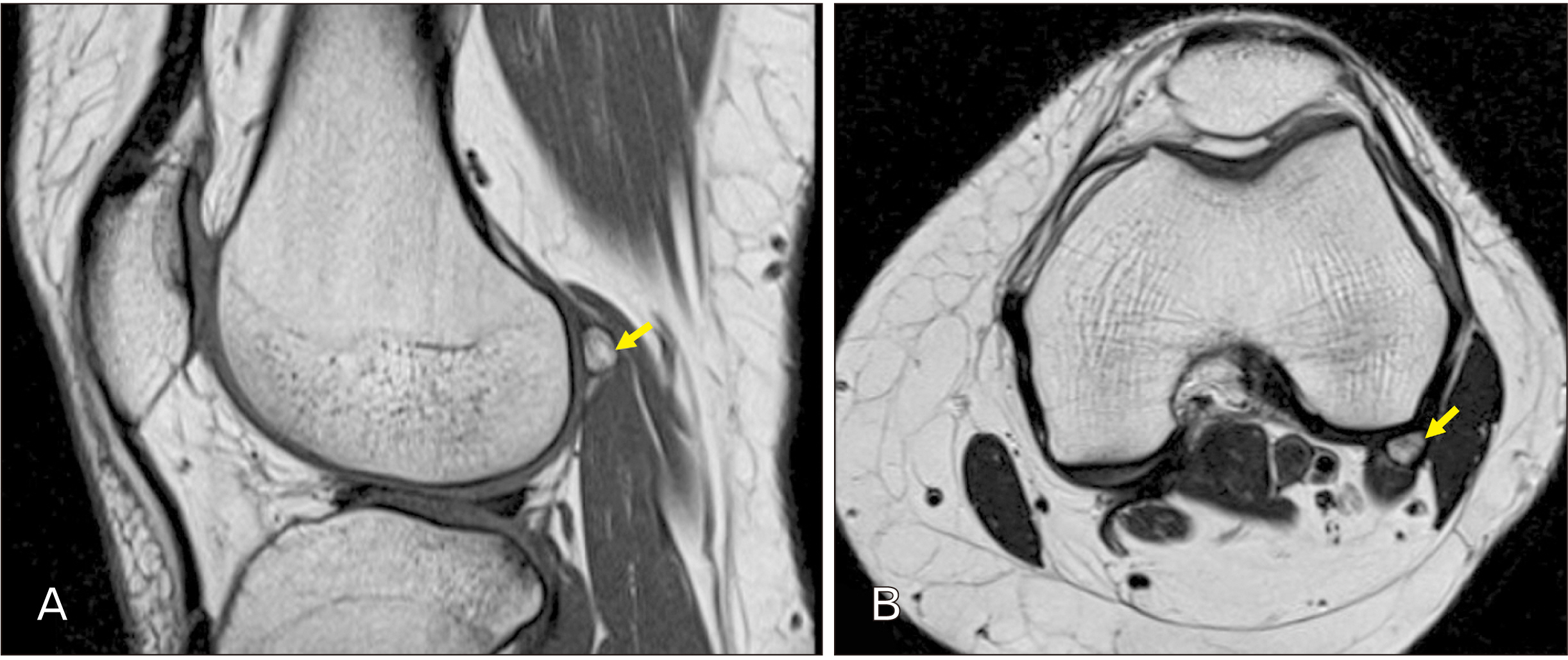
- Ethnic diversity is associated with variability in the prevalence rates of fabella. We aimed to evaluate the prevalence and the radiological features of fabella in Omani patients. This is a retrospective analysis of hospital electronic database of patients referred for radiological investigations (radiographs and magnetic resonance imaging) of the knee, at a tertiary care referral center. Descriptive statistics were performed to determine the prevalence of fabella. Chi-square test was used to determine the association between sex or age with respect to the presence of fabella. A total of 813 knee radiographs were reviewed for the presence of fabella. Fabella was found in 24.1% of total cases. A statistically significant sex difference was observed with respect to the presence of fabella in left knees in males (P<0.01). The presence of fabella was significantly associated with age groups for the right (P<0.05) and left knees (P<0.01). In magnetic resonance imaging film reviews, all the identified fabellae (20.2%) were bony structures and were located within the lateral head of the gastrocnemius muscle. There were no cartilaginous fabellae detected. The current study revealed a prevalence of 24.1% of fabella in Omani subjects which is almost similar to the results as seen in Caucasian ethnic populations.
Keyword
Figure
Reference
-
References
1. Duncan W, Dahm DL. 2003; Clinical anatomy of the fabella. Clin Anat. 16:448–9. DOI: 10.1002/ca.10137. PMID: 12903068.
Article2. Berthaume MA, Bull AMJ. 2020; Human biological variation in sesamoid bone prevalence: the curious case of the fabella. J Anat. 236:228–42. DOI: 10.1111/joa.13091. PMID: 31623020. PMCID: PMC6956444.
Article3. Theodorou SJ, Theodorou DJ, Resnick D. 2005; Painful stress fractures of the fabella in patients with total knee arthroplasty. AJR Am J Roentgenol. 185:1141–4. DOI: 10.2214/AJR.04.1230. PMID: 16247123.
Article4. Hauser NH, Hoechel S, Toranelli M, Klaws J, Müller-Gerbl M. 2015; Functional and structural details about the fabella: what the important stabilizer looks like in the central European population. Biomed Res Int. 2015:343728. DOI: 10.1155/2015/343728. PMID: 26413516. PMCID: PMC4564579.
Article5. Kawashima T, Takeishi H, Yoshitomi S, Ito M, Sasaki H. 2007; Anatomical study of the fabella, fabellar complex and its clinical implications. Surg Radiol Anat. 29:611–6. DOI: 10.1007/s00276-007-0259-4. PMID: 17882346.
Article6. Sarin VK, Erickson GM, Giori NJ, Bergman AG, Carter DR. 1999; Coincident development of sesamoid bones and clues to their evolution. Anat Rec. 257:174–80. DOI: 10.1002/(SICI)1097-0185(19991015)257:5<174::AID-AR6>3.0.CO;2-O. PMID: 10597342.
Article7. Eyal S, Rubin S, Krief S, Levin L, Zelzer E. 2019; Common cellular origin and diverging developmental programs for different sesamoid bones. Development. 146:dev167452. DOI: 10.1242/dev.167452. PMID: 30745426. PMCID: PMC6919532.
Article8. Phukubye P, Oyedele O. 2011; The incidence and structure of the fabella in a South African cadaver sample. Clin Anat. 24:84–90. DOI: 10.1002/ca.21049. PMID: 20830786.
Article9. Zipple JT, Hammer RL, Loubert PV. 2003; Treatment of fabella syndrome with manual therapy: a case report. J Orthop Sports Phys Ther. 33:33–9. DOI: 10.2519/jospt.2003.33.1.33. PMID: 12570284.
Article10. Pop TS, Pop AM, Olah P, Trâmbiţaş C. 2018; Prevalence of the fabella and its association with pain in the posterolateral corner of the knee: a cross-sectional study in a Romanian population. Medicine (Baltimore). 97:e13333. DOI: 10.1097/MD.0000000000013333. PMID: 30461651. PMCID: PMC6392660.11. Silva JG, Chagas CAA, Torres DFM, Servidio L, Vilela AC, Chagas WA. 2010; Morphological analyisis of the fabella in Brazilians. Int J Morphol. 28:105–10. DOI: 10.4067/S0717-95022010000100015.
Article12. Zeng SX, Dong XL, Dang RS, Wu GS, Wang JF, Wang D, Huang HL, Guo XD. 2012; Anatomic study of fabella and its surrounding structures in a Chinese population. Surg Radiol Anat. 34:65–71. DOI: 10.1007/s00276-011-0828-4. PMID: 21626275.
Article13. Yu JS, Salonen DC, Hodler J, Haghighi P, Trudell D, Resnick D. 1996; Posterolateral aspect of the knee: improved MR imaging with a coronal oblique technique. Radiology. 198:199–204. DOI: 10.1148/radiology.198.1.8539378. PMID: 8539378.
Article14. Minowa T, Murakami G, Kura H, Suzuki D, Han SH, Yamashita T. 2004; Does the fabella contribute to the reinforcement of the posterolateral corner of the knee by inducing the development of associated ligaments? J Orthop Sci. 9:59–65. DOI: 10.1007/s00776-003-0739-2. PMID: 14767706.
Article15. Chew CP, Lee KH, Koh JS, Howe TS. 2014; Incidence and radiological characteristics of fabellae in an Asian population. Singapore Med J. 55:198–201. DOI: 10.11622/smedj.2014052. PMID: 24763835. PMCID: PMC4291947.
Article16. Berthaume MA, Di Federico E, Bull AMJ. 2019; Fabella prevalence rate increases over 150 years, and rates of other sesamoid bones remain constant: a systematic review. J Anat. 235:67–79. DOI: 10.1111/joa.12994. PMID: 30994938. PMCID: PMC6579948.17. Ghimire I, Maharjan S, Pokharel G, Subedi K. 2017; Evaluation of occurrence of sesamoid bones in the lower extremity radiographs. J Chitwan Med Coll. 7:11–4. DOI: 10.3126/jcmc.v7i2.22995.
Article18. Kaneko K, Serizawa M, Genda N, Kikkawa F. 1966; Einige Betrachtungen über die Fabella des Menschen. Acta Anat Nippon. 42:85–8. German.19. Robertson A, Jones SC, Paes R, Chakrabarty G. 2004; The fabella: a forgotten source of knee pain? Knee. 11:243–5. DOI: 10.1016/S0968-0160(03)00103-0. PMID: 15194103.
Article20. Hur JW, Lee S, Jun JB. 2020; The prevalence of fabella and its association with the osteoarthritic severity of the knee in Korea. Clin Rheumatol. 39:3625–9. DOI: 10.1007/s10067-020-05078-4. PMID: 32556935.
Article21. Patel A, Singh R, Johnson B, Smith A. 2013; Compression neuropathy of the common peroneal nerve by the fabella. BMJ Case Rep. 2013:bcr2013202154. DOI: 10.1136/bcr-2013-202154. PMID: 24293541. PMCID: PMC3847518.
Article22. Cesmebasi A, Spinner RJ, Smith J, Bannar SM, Finnoff JT. 2016; Role of sonography in the diagnosis and treatment of common peroneal neuropathy secondary to fabellae. J Ultrasound Med. 35:441–7. DOI: 10.7863/ultra.15.04003. PMID: 26782165.
Article23. Ando Y, Miyamoto Y, Tokimura F, Nakazawa T, Hamaji H, Kanetaka M, Koshiishi A, Hirabayashi K, Anamizu Y, Miyazaki T. 2017; A case report on a very rare variant of popliteal artery entrapment syndrome due to an enlarged fabella associated with severe knee osteoarthritis. J Orthop Sci. 22:164–8. DOI: 10.1016/j.jos.2015.06.025. PMID: 26740435.
Article24. Franceschi F, Longo UG, Ruzzini L, Leonardi F, Rojas M, Gualdi G, Denaro V. 2007; Dislocation of an enlarged fabella as uncommon cause of knee pain: a case report. Knee. 14:330–2. DOI: 10.1016/j.knee.2007.03.007. PMID: 17490883.25. Kwee TC, Heggelman B, Gaasbeek R, Nix M. 2016; Fabella fractures after total knee arthroplasty with correction of valgus malalignment. Case Rep Orthop. 2016:4749871. DOI: 10.1155/2016/4749871. PMID: 27340579. PMCID: PMC4908254.
Article26. Zhou F, Zhang F, Deng G, Bi C, Wang J, Wang Q, Wang Q. 2017; Fabella fracture with radiological imaging: a case report. Trauma Case Rep. 12:19–23. DOI: 10.1016/j.tcr.2017.10.010. PMID: 29644278. PMCID: PMC5887092.
Article27. Kim T, Chung H, Lee H, Choi Y, Son JH. 2018; A case report and literature review on fabella syndrome after high tibial osteotomy. Medicine (Baltimore). 97:e9585. DOI: 10.1097/MD.0000000000009585. PMID: 29369174. PMCID: PMC5794358.
Article28. Egerci OF, Kose O, Turan A, Kilicaslan OF, Sekerci R, Keles-Celik N. 2017; Prevalence and distribution of the fabella: a radiographic study in Turkish subjects. Folia Morphol (Warsz). 76:478–83. DOI: 10.5603/FM.a2016.0080. PMID: 28026849.
Article29. Kaplan EB. 1961; The fabellofibular and short lateral ligaments of the knee joint. J Bone Joint Surg Am. 43-A:169–79. DOI: 10.2106/00004623-196143020-00002. PMID: 13830884.
Article30. Piyawinijwong S, Sirisathira N, icharoenvej S Sr. 2012; The fabella, fabellofibular and short lateral ligaments: an anatomical study in Thais cadavers. Siriraj Med J. 64(Suppl 1):S15–8.31. Tabira Y, Saga T, Takahashi N, Watanabe K, Nakamura M, Yamaki K. 2013; Influence of a fabella in the gastrocnemius muscle on the common fibular nerve in Japanese subjects. Clin Anat. 26:893–902. DOI: 10.1002/ca.22153. PMID: 22933414.
Article32. Takebe K, Kita K, Hirohata K. 1983; Radiological and anatomical observation on fabella. Orthop Surg. 34:1163–70. Japanese.33. Pritchett JW. 1984; The incidence of fabellae in osteoarthrosis of the knee. J Bone Joint Surg Am. 66:1379–80. DOI: 10.2106/00004623-198466090-00009. PMID: 6501334.
Article34. Raheem O, Philpott J, Ryan W, O'Brien M. 2007; Anatomical variations in the anatomy of the posterolateral corner of the knee. Knee Surg Sports Traumatol Arthrosc. 15:895–900. DOI: 10.1007/s00167-007-0301-4. PMID: 17641923.
Article35. Jin ZW, Shibata S, Abe H, Jin Y, Li XW, Murakami G. 2017; A new insight into the fabella at knee: the foetal development and evolution. Folia Morphol (Warsz). 76:87–93. DOI: 10.5603/FM.a2016.0048. PMID: 27665955.
Article36. Ehara S. 2014; Potentially symptomatic fabella: MR imaging review. Jpn J Radiol. 32:1–5. DOI: 10.1007/s11604-013-0253-1. PMID: 24158650.
Article37. Kato Y, Oshida M, Ryu K, Horaguchi T, Seki M, Tokuhashi Y. The incidence and structure of the fabella in Japanese population. Anatomical study, radiographic study, and clinical cases. Paper presented at: ORS 2012 Annual Meeting. 2012; San Francisco, USA.38. Merolla G, Dave AC, Paladini P, Campi F, Porcellini G. 2015; Ossifying tendinitis of the rotator cuff after arthroscopic excision of calcium deposits: report of two cases and literature review. J Orthop Traumatol. 16:67–73. DOI: 10.1007/s10195-014-0309-8. PMID: 25017026. PMCID: PMC4348528.
Article39. Pountain G. 1992; Musculoskeletal pain in Omanis, and the relationship to joint mobility and body mass index. Br J Rheumatol. 31:81–5. DOI: 10.1093/rheumatology/31.2.81. PMID: 1737235.
Article


