Post-Stroke Cognitive Impairment: Pathophysiological Insights into Brain Disconnectome from Advanced Neuroimaging Analysis Techniques
- Affiliations
-
- 1Department of Neurology, Asan Medical Center, University of Ulsan College of Medicine, Seoul, Korea
- 2Department of Biomedical Engineering, Sungkyunkwan University, Suwon, Korea
- 3Center for Neuroscience Imaging Research, Institute for Basic Science, Suwon, Korea
- 4Department of Intelligent Precision Healthcare Convergence, Sungkyunkwan University, Suwon, Korea
- KMID: 2520909
- DOI: http://doi.org/10.5853/jos.2021.02376
Abstract
- The neurological symptoms of stroke have traditionally provided the foundation for functional mapping of the brain. However, there are many unresolved aspects in our understanding of cerebral activity, especially regarding high-level cognitive functions. This review provides a comprehensive look at the pathophysiology of post-stroke cognitive impairment in light of recent findings from advanced imaging techniques. Combining network neuroscience and clinical neurology, our research focuses on how changes in brain networks correlate with post-stroke cognitive prognosis. More specifically, we first discuss the general consequences of stroke lesions due to damage of canonical resting-state large-scale networks or changes in the composition of the entire brain. We also review emerging methods, such as lesion-network mapping and gradient analysis, used to study the aforementioned events caused by stroke lesions. Lastly, we examine other patient vulnerabilities, such as superimposed amyloid pathology and blood-brain barrier leakage, which potentially lead to different outcomes for the brain network compositions even in the presence of similar stroke lesions. This knowledge will allow a better understanding of the pathophysiology of post-stroke cognitive impairment and provide a theoretical basis for the development of new treatments, such as neuromodulation.
Keyword
Figure
-
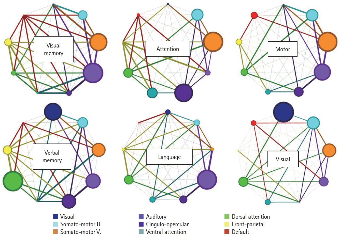
Figure 1. Contribution of functional connections within a resting-state network and between resting-state network pairs for each functional connectivity-behavior prediction. Each circle refers to a representative resting-state network, and its size is proportional to the contribution of within-network connections. Line thickness is proportional to the weighting of between-network connectivity. Adapted from Siegel et al.,[32] with permission from National Academy of Sciences.
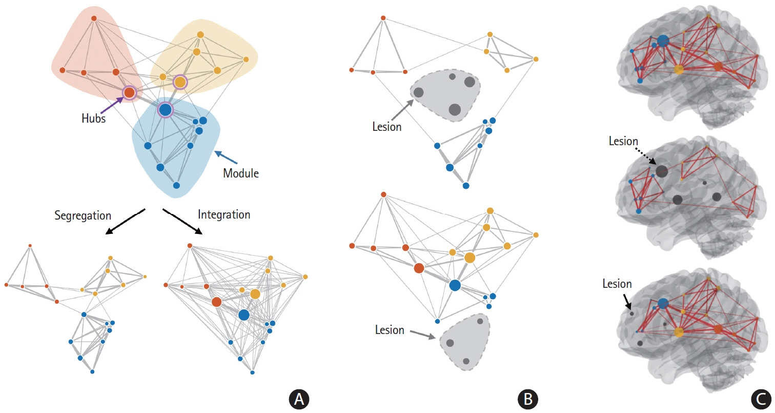
Figure 2. Integration and segregation in the brain as a complex system. (A) Schematic representation of functional segregation and integration. (B) Stroke lesions can cause different changes in segregation and integration depending on where they occur: upper panel, maintained segregation and reduced integration; bottom panel, decreased segregation while maintaining integration. (C) Stroke lesions can have widespread effects in the brain if they occur in the hubs (dashed arrow).

Figure 3. Dispersion caused by lesions and the subsequent compensation processes. The intra-network connectivity of the lesion-occurring module is weakened and dispersed; however, the compensation for this can enhance the between-network connectivity with functionally adjacent networks.
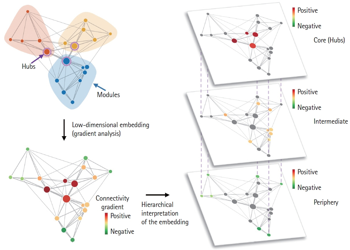
Figure 4. Hierarchy in brain connectivity. A hierarchy exists in functional brain connections. Figure 2A can be represented according to the connectivity hierarchy via a gradient analysis. In the lower left and right panels, the principal connectivity gradient explaining most of the variance in the connectivity data was represented as separate layers. It usually follows the well-known axis of unimodal (e.g., visual, somatomotor networks; periphery; green-colored circles) to heteromodal (e.g., default mode network; core; red-colored circles) hierarchy of the brain.
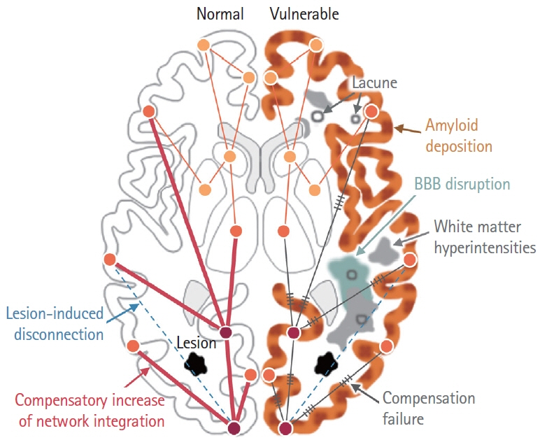
Figure 5. Effects of vulnerability factors on stroke-induced brain network changes. Prognosis after stroke can be affected by multiple risk factors, including amyloid decomposition, blood-brain barrier (BBB) disruption, and whiter matter hyperintensities. Left: Brain without vulnerability. Stroke lesion (black polygon) induces disconnection of brain regions (dashed skyblue line), followed by compensatory increase of brain network integration. Line thickness is proportional to connectivity strength. Circles indicate different brain regions. Right: Brain with vulnerability. Amyloid decomposition (brown speckles), white matter hyperintensities (gray polygon), BBB disruption (cyan polygon) disrupt compensatory brain network integration, indicated as gray line with hatch marks. Lacune are also marked as gray colored open circles.
Reference
-
References
1. Broca P. Localisations des fonctions cérébrales. Siège de la faculté du langage articulé. Bull Soc Anthropol Paris. 1863; 4:200–204.2. Wernicke C. The symptom-complex of aphasia. In : Campbell TH, editor. Disease of the Nervous System. London: Cassell;1908. p. 265–324.3. Ladino LD, Rizvi S, Téllez-Zenteno JF. The Montreal procedure: the legacy of the great Wilder Penfield. Epilepsy Behav. 2018; 83:151–161.
Article4. Verdelho A, Wardlaw J, Pavlovic A, Pantoni L, Godefroy O, Duering M, et al. Cognitive impairment in patients with cerebrovascular disease: a white paper from the links between stroke ESO Dementia Committee. Eur Stroke J. 2021; 6:5–17.
Article5. Munsch F, Sagnier S, Asselineau J, Bigourdan A, Guttmann CR, Debruxelles S, et al. Stroke location is an independent predictor of cognitive outcome. Stroke. 2016; 47:66–73.
Article6. Duering M, Zieren N, Hervé D, Jouvent E, Reyes S, Peters N, et al. Strategic role of frontal white matter tracts in vascular cognitive impairment: a voxel-based lesion-symptom mapping study in CADASIL. Brain. 2011; 134(Pt 8):2366–2375.
Article7. Weaver NA, Zhao L, Biesbroek JM, Kuijf HJ, Aben HP, Bae HJ, et al. The Meta VCI Map consortium for meta-analyses on strategic lesion locations for vascular cognitive impairment using lesion-symptom mapping: design and multicenter pilot study. Alzheimers Dement (Amst). 2019; 11:310–326.
Article8. Biesbroek JM, Lim JS, Weaver NA, Arikan G, Kang Y, Kim BJ, et al. Anatomy of phonemic and semantic fluency: a lesion and disconnectome study in 1231 stroke patients. Cortex. 2021; 143:148–163.
Article9. Biesbroek JM, van Zandvoort MJ, Kuijf HJ, Weaver NA, Kappelle LJ, Vos PC, et al. The anatomy of visuospatial construction revealed by lesion-symptom mapping. Neuropsychologia. 2014; 62:68–76.
Article10. Weaver NA, Kuijf HJ, Aben HP, Abrigo J, Bae HJ, Barbay M, et al. Strategic infarct locations for post-stroke cognitive impairment: a pooled analysis of individual patient data from 12 acute ischaemic stroke cohorts. Lancet Neurol. 2021; 20:448–459.
Article11. Hillis AE, Beh YY, Sebastian R, Breining B, Tippett DC, Wright A, et al. Predicting recovery in acute poststroke aphasia. Ann Neurol. 2018; 83:612–622.
Article12. Weaver NA, Kancheva AK, Lim JS, Biesbroek JM, Wajer IMH, Kang Y, et al. Post-stroke cognitive impairment on the MiniMental State Examination primarily relates to left middle cerebral artery infarcts. Int J Stroke. 2021 Jan 20 [Epub]. https://doi.org/10.1177/1747493020984552.
Article13. Salvalaggio A, De Filippo De Grazia M, Zorzi M, Thiebaut de Schotten M, Corbetta M. Post-stroke deficit prediction from lesion and indirect structural and functional disconnection. Brain. 2020; 143:2173–2188.
Article14. Fox MD. Mapping symptoms to brain networks with the human connectome. N Engl J Med. 2018; 379:2237–2245.
Article15. Carrera E, Tononi G. Diaschisis: past, present, future. Brain. 2014; 137(Pt 9):2408–2422.
Article16. Duering M, Righart R, Csanadi E, Jouvent E, Hervé D, Chabriat H, et al. Incident subcortical infarcts induce focal thinning in connected cortical regions. Neurology. 2012; 79:2025–2028.
Article17. Alstott J, Breakspear M, Hagmann P, Cammoun L, Sporns O. Modeling the impact of lesions in the human brain. PLoS Comput Biol. 2009; 5:e1000408.
Article18. van den Heuvel MP, Sporns O. Rich-club organization of the human connectome. J Neurosci. 2011; 31:15775–15786.
Article19. Lim JS, Kim N, Jang MU, Han MK, Kim S, Baek MJ, et al. Cortical hubs and subcortical cholinergic pathways as neural substrates of poststroke dementia. Stroke. 2014; 45:1069–1076.
Article20. Griffis JC, Metcalf NV, Corbetta M, Shulman GL. Structural disconnections explain brain network dysfunction after stroke. Cell Rep. 2019; 28:2527.e9–2540.e9.
Article21. Shen J, Tozer DJ, Markus HS, Tay J. Network efficiency mediates the relationship between vascular burden and cognitive impairment: a diffusion tensor imaging study in UK Biobank. Stroke. 2020; 51:1682–1689.22. Thiebaut de Schotten M, Foulon C, Nachev P. Brain disconnections link structural connectivity with function and behaviour. Nat Commun. 2020; 11:5094.
Article23. Greicius MD, Krasnow B, Reiss AL, Menon V. Functional connectivity in the resting brain: a network analysis of the default mode hypothesis. Proc Natl Acad Sci U S A. 2003; 100:253–258.
Article24. Bressler SL, Menon V. Large-scale brain networks in cognition: emerging methods and principles. Trends Cogn Sci. 2010; 14:277–290.
Article25. Greicius M. Resting-state functional connectivity in neuropsychiatric disorders. Curr Opin Neurol. 2008; 21:424–430.
Article26. Lee JJ, Kim HJ, Čeko M, Park BY, Lee SA, Park H, et al. A neuroimaging biomarker for sustained experimental and clinical pain. Nat Med. 2021; 27:174–182.
Article27. Sporns O. Contributions and challenges for network models in cognitive neuroscience. Nat Neurosci. 2014; 17:652–660.
Article28. Farahani FV, Karwowski W, Lighthall NR. Application of graph theory for identifying connectivity patterns in human brain networks: a systematic review. Front Neurosci. 2019; 13:585.
Article29. Matthews PM, Hampshire A. Clinical concepts emerging from fMRI functional connectomics. Neuron. 2016; 91:511–528.
Article30. Dacosta-Aguayo R, Iturria-Medina Y, FernándezAndújar M, López-Cancio E, Cáceres C, et al. Impairment of functional integration of the default mode network correlates with cognitive outcome at three months after stroke. Hum Brain Mapp. 2015; 36:577–590.31. Mesulam MM. From sensation to cognition. Brain. 1998; 121(Pt 6):1013–1052.
Article32. Siegel JS, Ramsey LE, Snyder AZ, Metcalf NV, Chacko RV, Weinberger K, et al. Disruptions of network connectivity predict impairment in multiple behavioral domains after stroke. Proc Natl Acad Sci U S A. 2016; 113:E4367–E4376.
Article33. Rubinov M, Sporns O. Complex network measures of brain connectivity: uses and interpretations. Neuroimage. 2010; 52:1059–1069.
Article34. Bullmore E, Sporns O. Complex brain networks: graph theoretical analysis of structural and functional systems. Nat Rev Neurosci. 2009; 10:186–198.
Article35. Gordon EM, Laumann TO, Gilmore AW, Newbold DJ, Greene DJ, Berg JJ, et al. Precision functional mapping of individual human brains. Neuron. 2017; 95:791.e7–807.e7.
Article36. Siegel JS, Seitzman BA, Ramsey LE, Ortega M, Gordon EM, Dosenbach NUF, et al. Re-emergence of modular brain networks in stroke recovery. Cortex. 2018; 101:44–59.
Article37. Wig GS. Segregated systems of human brain networks. Trends Cogn Sci. 2017; 21:981–996.
Article38. Grefkes C, Fink GR. Connectivity-based approaches in stroke and recovery of function. Lancet Neurol. 2014; 13:206–216.
Article39. Gratton C, Nomura EM, Pérez F, D’Esposito M. Focal brain lesions to critical locations cause widespread disruption of the modular organization of the brain. J Cogn Neurosci. 2012; 24:1275–1285.
Article40. Tao Y, Rapp B. The effects of lesion and treatment-related recovery on functional network modularity in post-stroke dysgraphia. Neuroimage Clin. 2019; 23:101865.
Article41. Chan MY, Park DC, Savalia NK, Petersen SE, Wig GS. Decreased segregation of brain systems across the healthy adult lifespan. Proc Natl Acad Sci U S A. 2014; 111:E4997–E5006.
Article42. Nomura EM, Gratton C, Visser RM, Kayser A, Perez F, D’Esposito M. Double dissociation of two cognitive control networks in patients with focal brain lesions. Proc Natl Acad Sci U S A. 2010; 107:12017–12022.
Article43. Aben HP, Biessels GJ, Weaver NA, Spikman JM, Visser-Meily JMA, de Kort PLM, et al. Extent to which network hubs are affected by ischemic stroke predicts cognitive recovery. Stroke. 2019; 50:2768–2774.
Article44. Guo J, Biswal BB, Han S, Li J, Yang S, Yang M, et al. Altered dynamics of brain segregation and integration in poststroke aphasia. Hum Brain Mapp. 2019; 40:3398–3409.
Article45. Wang Y, Wang C, Miao P, Liu J, Wei Y, Wu L, et al. An imbalance between functional segregation and integration in patients with pontine stroke: a dynamic functional network connectivity study. Neuroimage Clin. 2020; 28:102507.
Article46. Xu X, Tang R, Zhang L, Cao Z. Altered topology of the structural brain network in patients with post-stroke depression. Front Neurosci. 2019; 13:776.
Article47. Blaschke SJ, Hensel L, Minassian A, Vlachakis S, Tscherpel C, Vay SU, et al. Translating functional connectivity after stroke: functional magnetic resonance imaging detects comparable network changes in mice and humans. Stroke. 2021; 52:2948–2960.
Article48. van Meer MP, Otte WM, van der Marel K, Nijboer CH, Kavelaars A, van der Sprenkel JW, et al. Extent of bilateral neuronal network reorganization and functional recovery in relation to stroke severity. J Neurosci. 2012; 32:4495–4507.
Article49. Margulies DS, Ghosh SS, Goulas A, Falkiewicz M, Huntenburg JM, Langs G, et al. Situating the default-mode network along a principal gradient of macroscale cortical organization. Proc Natl Acad Sci U S A. 2016; 113:12574–12579.
Article50. Bayrak Ş, Khalil AA, Villringer K, Fiebach JB, Villringer A, Margulies DS, et al. The impact of ischemic stroke on connectivity gradients. Neuroimage Clin. 2019; 24:101947.
Article51. Bethlehem RAI, Paquola C, Seidlitz J, Ronan L, Bernhardt B, Consortium CC, et al. Dispersion of functional gradients across the adult lifespan. Neuroimage. 2020; 222:117299.
Article52. Laumann TO, Ortega M, Hoyt CR, Seider NA, Snyder AZ, Dosenbach NU, et al. Brain network reorganisation in an adolescent after bilateral perinatal strokes. Lancet Neurol. 2021; 20:255–256.
Article53. Ramsey LE, Siegel JS, Lang CE, Strube M, Shulman GL, Corbetta M. Behavioural clusters and predictors of performance during recovery from stroke. Nat Hum Behav. 2017; 1:0038.
Article54. Specht K, Zahn R, Willmes K, Weis S, Holtel C, Krause BJ, et al. Joint independent component analysis of structural and functional images reveals complex patterns of functional reorganisation in stroke aphasia. Neuroimage. 2009; 47:2057–2063.
Article55. Hu Y, Li X, Wang L, Han B, Nie S. T-distribution stochastic neighbor embedding for fine brain functional parcellation on rs-fMRI. Brain Res Bull. 2020; 162:199–207.
Article56. Bahrami M, Lyday RG, Casanova R, Burdette JH, Simpson SL, Laurienti PJ. Using low-dimensional manifolds to map relationships between dynamic brain networks. Front Hum Neurosci. 2019; 13:430.
Article57. Yourganov G, Fridriksson J, Rorden C, Gleichgerrcht E, Bonilha L. Multivariate connectome-based symptom mapping in poststroke patients: networks supporting language and speech. J Neurosci. 2016; 36:6668–6679.
Article58. De Baene W, Rijnen SJM, Gehring K, Meskal I, Rutten GM, Sitskoorn MM. Lesion symptom mapping at the regional level in patients with a meningioma. Neuropsychology. 2019; 33:103–110.
Article59. Thothathiri M, Kimberg DY, Schwartz MF. The neural basis of reversible sentence comprehension: evidence from voxelbased lesion symptom mapping in aphasia. J Cogn Neurosci. 2012; 24:212–222.
Article60. Rondina JM, Filippone M, Girolami M, Ward NS. Decoding post-stroke motor function from structural brain imaging. Neuroimage Clin. 2016; 12:372–380.
Article61. Newbold DJ, Laumann TO, Hoyt CR, Hampton JM, Montez DF, Raut RV, et al. Plasticity and spontaneous activity pulses in disused human brain circuits. Neuron. 2020; 107:580.e6–589.e6.
Article62. Greene DJ, Marek S, Gordon EM, Siegel JS, Gratton C, Laumann TO, et al. Integrative and network-specific connectivity of the basal ganglia and thalamus defined in individuals. Neuron. 2020; 105:742.e6–758.e6.
Article63. Darby RR, Laganiere S, Pascual-Leone A, Prasad S, Fox MD. Finding the imposter: brain connectivity of lesions causing delusional misidentifications. Brain. 2017; 140:497–507.
Article64. Laganiere S, Boes AD, Fox MD. Network localization of hemichorea-hemiballismus. Neurology. 2016; 86:2187–2195.
Article65. Boes AD, Prasad S, Liu H, Liu Q, Pascual-Leone A, Caviness VS Jr, et al. Network localization of neurological symptoms from focal brain lesions. Brain. 2015; 138(Pt 10):3061–3075.
Article66. Cohen AL, Ferguson MA, Fox MD. Lesion network mapping predicts post-stroke behavioural deficits and improves localization. Brain. 2021; 144:e35.
Article67. Cohen AL, Fox MD. Reply: the influence of sample size and arbitrary statistical thresholds in lesion-network mapping. Brain. 2020; 143:e41.
Article68. Salvalaggio A, Pini L, De Filippo De Grazia M, Thiebaut De Schotten M, Zorzi M, Corbetta M. Reply: lesion network mapping: where do we go from here? Brain. 2021; 144:e6.
Article69. Holmes AJ, Hollinshead MO, O’Keefe TM, Petrov VI, Fariello GR, Wald LL, et al. Brain Genomics Superstruct Project initial data release with structural, functional, and behavioral measures. Sci Data. 2015; 2:150031.
Article70. Liu W, Wong A, Au L, Yang J, Wang Z, Leung EY, et al. Influence of amyloid-beta on cognitive decline after stroke/transient ischemic attack: three-year longitudinal study. Stroke. 2015; 46:3074–3080.71. Ye BS, Seo SW, Kim JH, Kim GH, Cho H, Noh Y, et al. Effects of amyloid and vascular markers on cognitive decline in subcortical vascular dementia. Neurology. 2015; 85:1687–1693.
Article72. Mok VCT, Lam BYK, Wang Z, Liu W, Au L, Leung EYL, et al. Delayed-onset dementia after stroke or transient ischemic attack. Alzheimers Dement. 2016; 12:1167–1176.
Article73. Ly JV, Rowe CC, Villemagne VL, Zavala JA, Ma H, Sahathevan R, et al. Subacute ischemic stroke is associated with focal 11C PiB positron emission tomography retention but not with global neocortical Abeta deposition. Stroke. 2012; 43:1341–1346.74. Garcia-Alloza M, Gregory J, Kuchibhotla KV, Fine S, Wei Y, Ayata C, et al. Cerebrovascular lesions induce transient betaamyloid deposition. Brain. 2011; 134(Pt 12):3697–3707.75. Toombs J, Zetterberg H. In the blood: biomarkers for amyloid pathology and neurodegeneration in Alzheimer’s disease. Brain Commun. 2020; 2:fcaa054.
Article76. Thrippleton MJ, Backes WH, Sourbron S, Ingrisch M, van Osch MJP, Dichgans M, et al. Quantifying blood-brain barrier leakage in small vessel disease: review and consensus recommendations. Alzheimers Dement. 2019; 15:840–858.
Article77. Maillard P, Fletcher E, Singh B, Martinez O, Johnson DK, Olichney JM, et al. Cerebral white matter free water: a sensitive biomarker of cognition and function. Neurology. 2019; 92:e2221–e2231.78. Maillard P, Mitchell GF, Himali JJ, Beiser A, Fletcher E, Tsao CW, et al. Aortic stiffness, increased white matter free water, and altered microstructural integrity: a continuum of injury. Stroke. 2017; 48:1567–1573.79. Planetta PJ, Ofori E, Pasternak O, Burciu RG, Shukla P, DeSimone JC, et al. Free-water imaging in Parkinson’s disease and atypical parkinsonism. Brain. 2016; 139(Pt 2):495–508.
Article80. Bocti C, Swartz RH, Gao FQ, Sahlas DJ, Behl P, Black SE. A new visual rating scale to assess strategic white matter hyperintensities within cholinergic pathways in dementia. Stroke. 2005; 36:2126–2131.
Article81. Uh J, Yezhuvath U, Cheng Y, Lu H. In vivo vascular hallmarks of diffuse leukoaraiosis. J Magn Reson Imaging. 2010; 32:184–190.
Article82. Topakian R, Barrick TR, Howe FA, Markus HS. Blood-brain barrier permeability is increased in normal-appearing white matter in patients with lacunar stroke and leucoaraiosis. J Neurol Neurosurg Psychiatry. 2010; 81:192–197.
Article83. Vernooij MW, Ikram MA, Vrooman HA, Wielopolski PA, Krestin GP, Hofman A, et al. White matter microstructural integrity and cognitive function in a general elderly population. Arch Gen Psychiatry. 2009; 66:545–553.
Article84. van Veluw SJ, Zwanenburg JJ, Engelen-Lee J, Spliet WG, Hendrikse J, Luijten PR, et al. In vivo detection of cerebral cortical microinfarcts with high-resolution 7T MRI. J Cereb Blood Flow Metab. 2013; 33:322–329.
Article85. van Veluw SJ, Shih AY, Smith EE, Chen C, Schneider JA, Wardlaw JM, et al. Detection, risk factors, and functional consequences of cerebral microinfarcts. Lancet Neurol. 2017; 16:730–740.
Article86. Wang Z, van Veluw SJ, Wong A, Liu W, Shi L, Yang J, et al. Risk factors and cognitive relevance of cortical cerebral microinfarcts in patients with ischemic stroke or transient ischemic attack. Stroke. 2016; 47:2450–2455.
Article87. Wardlaw JM, Smith EE, Biessels GJ, Cordonnier C, Fazekas F, Frayne R, et al. Neuroimaging standards for research into small vessel disease and its contribution to ageing and neurodegeneration. Lancet Neurol. 2013; 12:822–838.
Article88. Smith EE, Beaudin AE. New insights into cerebral small vessel disease and vascular cognitive impairment from MRI. Curr Opin Neurol. 2018; 31:36–43.
Article89. Lo JW, Crawford JD, Desmond DW, Godefroy O, Jokinen H, Mahinrad S, et al. Profile of and risk factors for poststroke cognitive impairment in diverse ethnoregional groups. Neurology. 2019; 93:e2257–e2271.
Article90. Lo JW, Crawford JD, Samaras K, Desmond DW, Köhler S, Staals J, et al. Association of prediabetes and type 2 diabetes with cognitive function after stroke: a STROKOG Collaboration Study. Stroke. 2020; 51:1640–1646.
- Full Text Links
-
- Actions
-
Cited
- CITED
-
- Close
- Share
-
- Similar articles
-
- Neuroimaging Markers for Alzheimer's Disease and Mild Cognitive Impairment in Alzheimer's Disease Neuroimaging Initiative (ADNI)
- Advances in Functional Connectomics in Neuroscience : A Focus on Post-Traumatic Stress Disorder
- The Study of the Signal Detection Test of the Traffic Accident Patients with and without Brain Lesions
- Update on Stroke Rehabilitation for Non-Motor Impairment
- Stroke Connectome and Its Implications for Cognitive and Behavioral Sequela of Stroke
