Detection of nerve fibers in the eutopic endometrium of women with endometriosis, uterine fibroids and adenomyosis
- Affiliations
-
- 1Department of Obstetrics & Gynaecology, All India Institute of Medical Sciences, Jodhpur, India
- 2Department of Pathology, All India Institute of Medical Sciences, Jodhpur, India
- 3Department of Community Medicine and Family Medicine, All India Institute of Medical Sciences, Jodhpur, India
- KMID: 2520265
- DOI: http://doi.org/10.5468/ogs.21114
Abstract
Objective
The primary objective of this study was to establish the presence of nerve fibers in the eutopic endometrium of women with endometriosis and to determine whether these nerve fibers are exclusive to endometriosis or are also found in other pelvic pathologies associated with dysmenorrhea.
Methods
Endometrial tissue was obtained by aspiration (Pipelle), endometrial curettage, or following hysterectomy in women with endometriosis confirmed through histopathological examination, leiomyomas, and adenomyosis. The eutopic endometrium was subjected to immunohistochemical staining to detect PGP 9.5, which is a highly specific pan-neuronal marker. The nerve fiber density was correlated with the patient’s pain score, as indicated by the Visual Analog Scale. A control group was formed by staining the endometrium of women presenting with dysmenorrhea but without the above-mentioned disorders.
Results
Nerve fibers were observed in sections of the endo-myometrium (in the deep endometrium) in 68% of patients with endometriosis who underwent hysterectomy or a deep endometrial biopsy. Nerve fibers were not observed in the aspirated endometrium of women with endometriosis. Only 13.7% of women with adenomyosis and 3.3% of women with fibroids had nerve fibers in their endometrium. Nerve fiber density was correlated with pain score in women with endometriosis.
Conclusion
Nerve fibers were found in the functional layer of eutopic endometrium in women with endometriosis; hence, we concluded that the presence of nerve fibers in the eutopic endometrium could diagnose endometriosis with a fairly good specificity of 92.7%. However, the absence of nerve fibers does not always exclude the disease.
Keyword
Figure
-
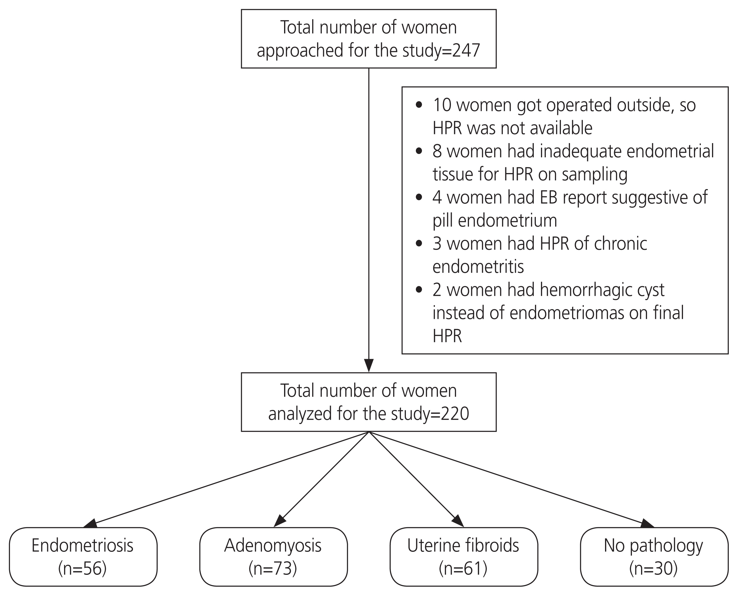
Fig. 1 Patient flow following recruitment in the study. HPR, histopathological report; EB, endometrial biopsy.

Fig. 2 The endometrium shows nerve fibers highlighted by the protein gene product (PGP) 9.5 antibody (black arrow) and endometrial gland (red asterisks) in a case of endometriosis (immunohistochemical staining, ×40).
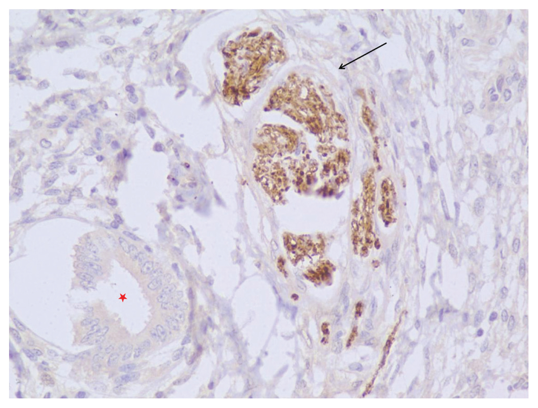
Fig. 3 High power view (immunohistochemical staining, ×40) of a proliferative endometrium (red asterisk) showing nerve fibers highlighted by the protein gene product (PGP) 9.5 antibody (black arrow) in a case of endometriosis.
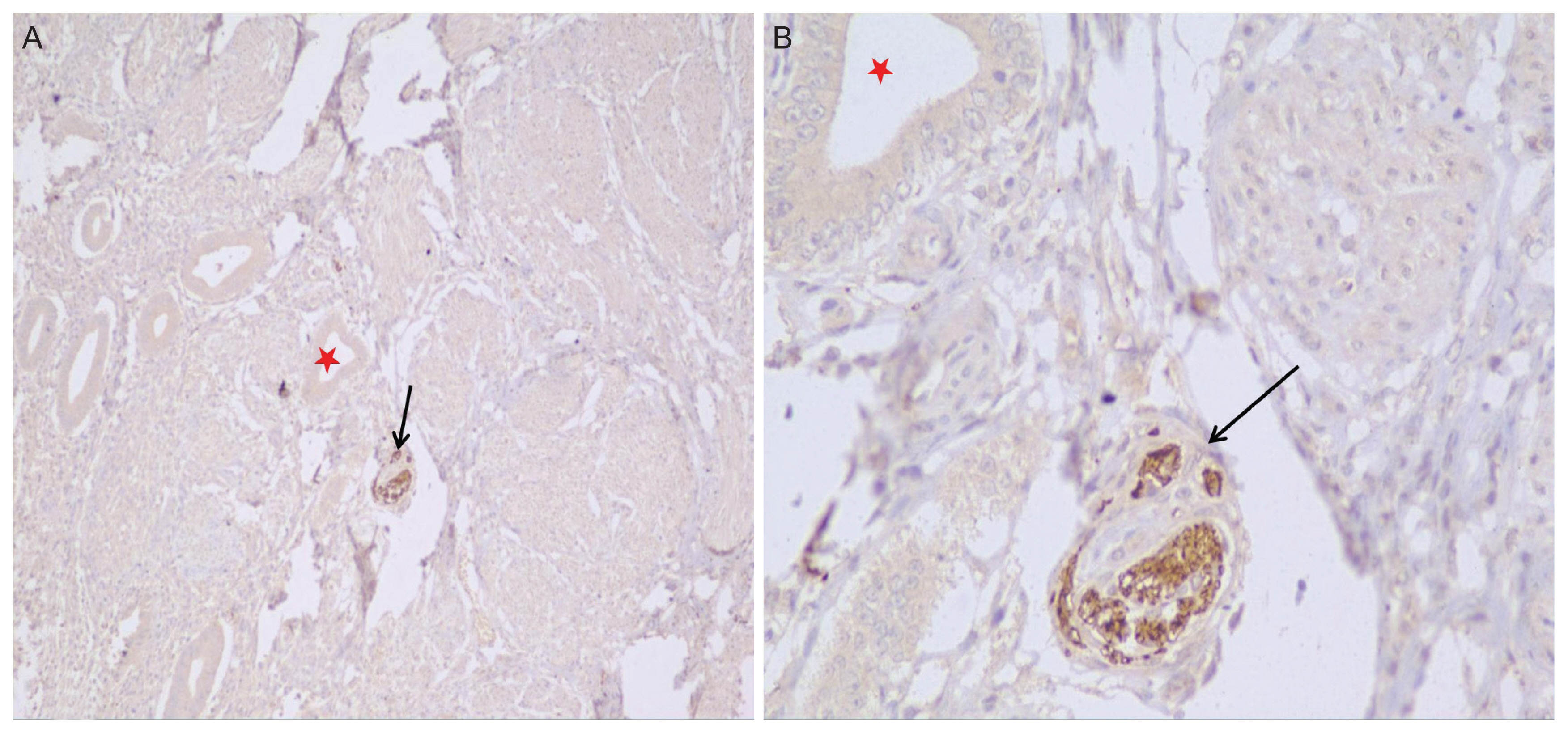
Fig. 4 (A) The endometrium shows nerve fibers highlighted by the protein gene product (PGP) 9.5 antibody (black arrows) in a case of adenomyosis. (B) High power view of the image in (A) where the upper right corner shows the myometrium (red asterisks) (immunohistochemical staining, ×40).
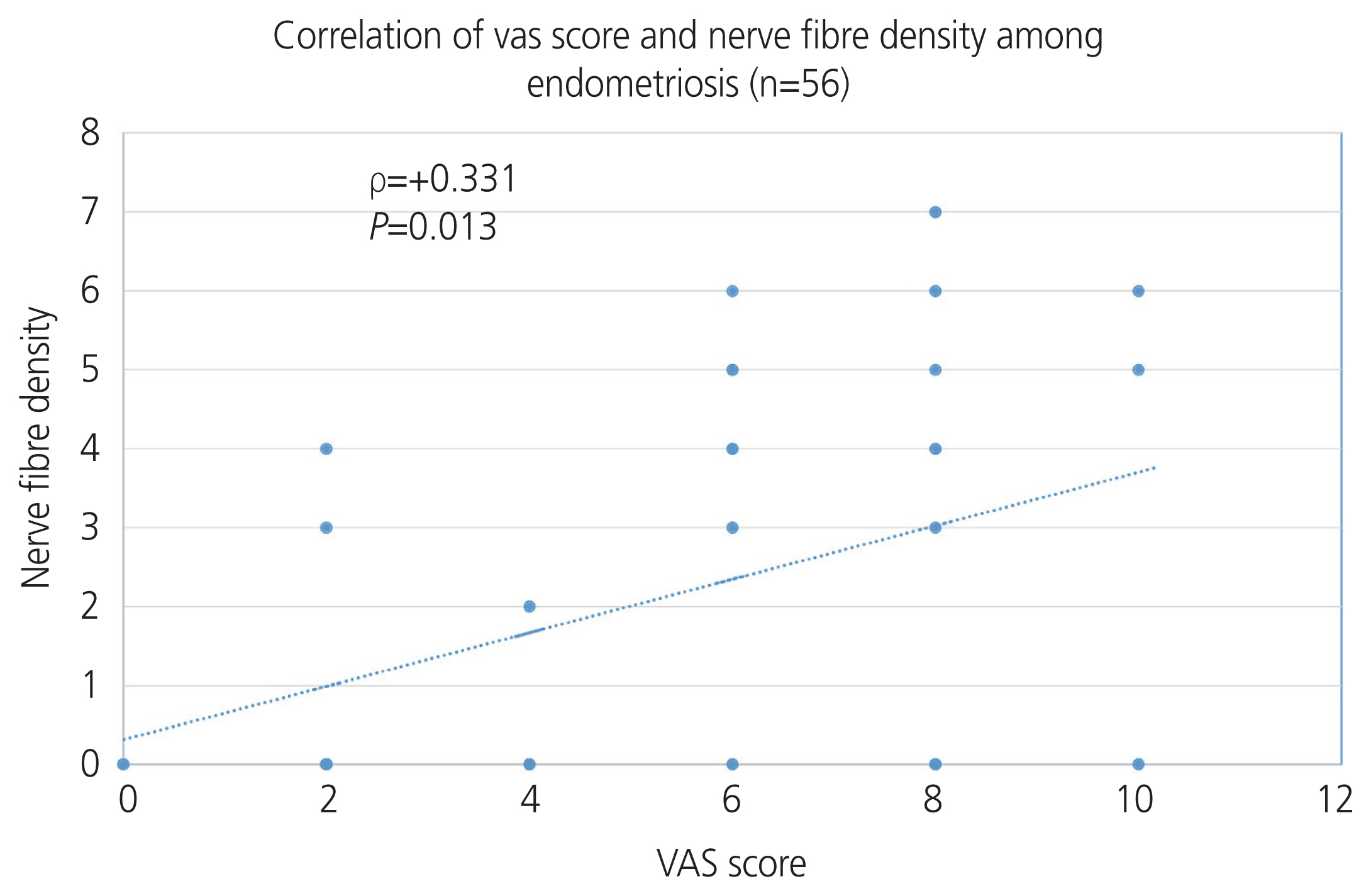
Fig. 5 Correlation of the Visual Analog Scale (VAS) score and nerve fiber density among patients with endometriosis.
Cited by 1 articles
-
Development of an endometriosis self-assessment tool for patient
Hyun-Hee Cho, Young-Sub Yoon
Obstet Gynecol Sci. 2022;65(3):256-265. doi: 10.5468/ogs.21252.
Reference
-
References
1. Ferland LV, Shah DK, Kvaskoff M, Zondervan K, Missmer SA. Epidemiological and clinical risk factors for endometriosis. D’Hooghe T, editor. Biomarkers for endometriosis. Cham (CH): Springer;2015. p. 95–121.2. Parasar P, Ozcan P, Terry KL. Endometriosis: epidemiology, diagnosis and clinical management. Curr Obstet Gynecol Rep. 2017; 6:34–41.
Article3. Kiesel L, Sourouni M. Diagnosis of endometriosis in the 21st century. Climacteric. 2019; 22:296–302.
Article4. Nisenblat V, Bossuyt PM, Farquhar C, Johnson N, Hull ML. Imaging modalities for the non-invasive diagnosis of endometriosis. Cochrane Database Syst Rev. 2016; 2:CD009591.
Article5. Kobayashi H, Yamada Y, Morioka S, Niiro E, Shigemitsu A, Ito F. Mechanism of pain generation for endometriosis-associated pelvic pain. Arch Gynecol Obstet. 2014; 289:13–21.
Article6. Yadav G, Radhakrishnan G, Singh N, Radhika AG. Detection of endometrial nerve fibres-a novel technique to diagnose endometriosis. J Endometr Pelvic Pain Disord. 2013; 5:144–50.7. Elgafor El Sharkwy IA. Combination of non-invasive and semi-invasive tests for diagnosis of minimal to mild endometriosis. Arch Gynecol Obstet. 2013; 288:793–7.
Article8. Ellett L, Readman E, Newman M, McIlwaine K, Villegas R, Jagasia N, et al. Are endometrial nerve fibres unique to endometriosis? A prospective case-control study of endometrial biopsy as a diagnostic test for endometriosis in women with pelvic pain. Hum Reprod. 2015; 30:2808–15.
Article9. Boujenah J, Hugues JN, Sifer C, Bricou A, Cédrin-Durnerin I, Sonigo C, et al. Endometriosis Fertility Index, or classification of the American Society of Reproductive Medicine for postoperative endometriosis patients with infertility: Which is more relevant? Gynecol Obstet Fertil. 2015; 43:806–9.10. Leslie C, Ma T, McElhinney B, Leake R, Stewart CJ. Is the detection of endometrial nerve fibers useful in the diagnosis of endometriosis? Int J Gynecol Pathol. 2013; 32:149–55.
Article11. Bektaş YT, Sakin Ö, Pirimoğlu ZM, Yılmaz AO, Başak K, Büyükbayrak EE, et al. Diagnostic value of endometrial nerves in the endometrial tissue of patients with endometriosis. South Clin Ist Euras. 2017; 28:261–5.12. Gupta D, Hull ML, Fraser I, Miller L, Bossuyt PM, Johnson N, et al. Endometrial biomarkers for the non-invasive diagnosis of endometriosis. Cochrane Database Syst Rev. 2016; 4:CD012165.
Article
- Full Text Links
-
- Actions
-
Cited
- CITED
-
- Close
- Share
-
- Similar articles
-
- Increased expression of nuclear factor kappa-B p65 subunit in adenomyosis
- Innervation in women with uterine myoma and adenomyosis
- Survivin and Bcl-2 expression in eutopic endometrium with and without endometriosis
- VEGF Expression Patterns in Eutopic Endometrium of Patients with Endometriosis
- mRNA Expression Differences of uPA, uPAR in Eutopic Endometrium of Advanced Stage Endometriosis Patients
