Circulating microRNAs as biomarkers in bile-derived exosomes of cholangiocarcinoma
- Affiliations
-
- 1Division of Hepatobiliary and Pancreatic Surgery, Department of Surgery, Keimyung University Dongsan Medical Center, Daegu, Korea
- 2Department of Microbiology, Keimyung University School of Medicine, Daegu, Korea
- KMID: 2519844
- DOI: http://doi.org/10.4174/astr.2021.101.3.140
Abstract
- Purpose
In this pilot study, using next-generation sequencing and integrated messenger RNA (mRNA) sequencing, we investigated circulating microRNA (miRNA) expression profiling from bile-derived exosomes to identify dysregulated miRNA signatures and oncogenic pathways and determine their effects on targeted mRNAs in cholangiocarcinoma (CCA). Moreover, we explored the possibility that genetic analysis using bile-derived exosomes may replace gene analysis using tissue.
Methods
Bile was collected from a patient with perihilar CCA before curative resection. As a control, bile was collected from a patient with a common bile duct stone. Exosomes were isolated from the bile, and we performed next-generation miRNA sequencing using isolated exosomes. To evaluate miRNA-mRNA interactions, mRNA sequencing was performed using bile fluid in both patients.
Results
We identified 22 differentially expressed miRNAs. More than 65% of the predicted mRNA targets of those miRNAs were actually differentially expressed between control and CCA bile samples. In functional pathway analysis, targets of 22 miRNAs were primarily enriched in mitogen-activated protein kinase, platelet derived growth factor, vascular endothelial growth factor, epidermal growth factor receptor, and p53 signaling. In particular, in the functional assessment of miRNAmRNA interactions, RAS pathways, including downstream pathways (PI3K-AKT-mTOR and RAS-RAF-MEK-ERK), were determined to be enriched.
Conclusion
Circulating miRNAs in bile-derived exosomes provide new information for the development of miRNA analysis in CCA. These miRNAs may represent the oncogenic characteristics of CCA tissue, enabling them to be used instead of tissue samples for the diagnosis of CCA. Further research investigating circulating miRNAs in bile exosomes may lead to more rational, targeted approaches to treatment.
Keyword
Figure
-

Fig. 1 Heat map of the 1-way hierarchical clustering using Z-score for normalized value (log2 based) based on 52 mature microRNAs satisfying |fold change| ≥ 2 and raw. P < 0.05.
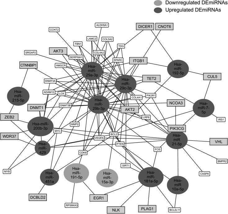
Fig. 2 MicroRNA (miRNA)-messenger RNA (mRNA) regulatory network. Among 22 differentially expressed miRNAs (DEmiRNAs), 14 netDEmiRNA had a network of miRNAs–mRNAs that has a threshold for the minimum number of miRNA-target interactions of 4 and false discovery rate < 0.01. Bold blue boxes indicate differentially expressed mRNAs between normal and cancer bile samples. CCNT2, cyclin T2; ALDH5A1, aldehyde dehydrogenase 5 family member A1; DICER1, dicer 1, ribonuclease III; CNOT6, CCR4-NOT transcription complex subunit 6; ITGB1, integrin subunit beta 1; AKT3, AKT serine/threonine kinase 3; CTNNBIP1, catenin beta interacting protein 1; DNMT1, DNA methyltransferase 1; ZEB2, zinc finger E-box binding homeobox 2; WDR37, WD repeat domain 37; MYB, MYB proto-oncogene, transcription factor; DCBLD2, discoidin, CUB and LCCL domain containing 2; RPS6KA3, ribosomal protein S6 kinase A3; EGR1, early growth response protein 1; NLK, nemo like kinase; PLAG1, PLAG1 zinc finger; BCL2L11, BCL2 like 11; CASP8, caspase 8; BMPR2, bone morphogenetic protein receptor type 2; VHL, Von Hippel-Lindau syndrome; IRS1, insulin receptor substrate 1; CUL5, cullin 5; FOS, Fos protooncogene; PIK3CG, phosphatidylinositol-4,5-bisphosphate 3-kinase catalytic subunit gamma; NCOA3, nuclear receptor coactivator 3; SIRT1, sirtuin 1; PTEN, Phosphatase and tensin homolog; BCL2, B-cell lymphoma 2; VEGFA, vascular endothelial growth factor A; CDK6, cyclin dependent kinase 6; MYC, MYC proto-oncogene; MMP2, matrix metallopeptidase 2; DNMT3B, DNA methyltransferase 3 beta; LOX, lysyl oxidase; ADAM12, ADAM metallopeptidase domain 12; HMGCR, 3-hydroxy-3-methylglutaryl-CoA reductase; SERPINH1, serpin family H member 1; COL4A2, collagen type IV alpha 2 chain; FBN1, fibrillin 1; LAMC2, laminin subunit gamma 2; COL5A2, collagen type V alpha 2 chain; TDG, thymine-DNA glycosylase; SPARC, secreted protein acidic and rich in cysteine; TET2, Tet methylcytosine dioxygenase 2; IRS1, insulin receptor substrate 1; BMPR2, bone morphogenetic protein receptor type 2; PIK3R1, phosphoinositide-3-kinase regulatory subunit 1; COL4A1, collagen type IV alpha 1 chain; AKT2, AKT serine/threonine kinase 2; LRP6, LDL receptor related protein 6.
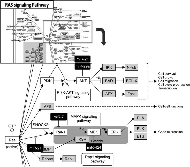
Fig. 3 RAS pathway in functional assessment of microRNA (miRNA)-messenger RNA (mRNA) interaction. Highlighted red-colored and yellow-colored boxes indicate differentially expressed miRNA and mRNA signaling in the present study, respectively. This image was obtained by Kyoto Encyclopedia of Genes and Genomes with copyright permission (Kanehisa [14]). RAS, rat sarcoma virus; IKK, inhibitor of nuclear factor-κB (IκB) kinase; NF-κB, nuclear factor kappa-light-chain-enhancer of activated B cells; PI3K, phosphoinositide 3-kinase; PIP3, phosphoinositide 3-kinase; AKT, protein kinase B; BAD, BCL2 associated agonist of cell death; BCL-X, B-cell lymphoma-extra large; AFX, FOXO4; FasL, Fas ligand; AF6, ALL1-fused gene from chromosome 6 protein; Raf-1, rapidly accelerated fibrosarcoma-1; MAPK, mitogen-activated protein kinase; MEK, mitogen-activated protein kinase kinase; ERK, extracellular-signal-regulated kinase; KSR, kinase suppressor of Ras; PLA, poly-lactic acid; ELK, ETS transcription factor; ETS, electron transport chain; Repac, another Epac subfamily member called.
Reference
-
1. DeOliveira ML, Cunningham SC, Cameron JL, Kamangar F, Winter JM, Lillemoe KD, et al. Cholangiocarcinoma: thirty-one-year experience with 564 patients at a single institution. Ann Surg. 2007; 245:755–762. PMID: 17457168.2. Keller S, Ridinger J, Rupp AK, Janssen JW, Altevogt P. Body f luid derived exosomes as a novel template for clinical diagnostics. J Transl Med. 2011; 9:86. PMID: 21651777.
Article3. Sagredo AI, Sepulveda SA, Roa JC, Oróstica LJ. Exosomes in bile as potential pancreatobiliary tumor biomarkers. Trans Cancer Res. 2017; 6 Suppl 8:S1371–S1383.
Article4. Gobbo J, Marcion G, Cordonnier M, Dias AM, Pernet N, Hammann A, et al. Restoring anticancer immune response by targeting tumor-derived exosomes with a HSP70 peptide aptamer. J Natl Cancer Inst. 2015; 108.
Article5. Subra C, Grand D, Laulagnier K, Stella A, Lambeau G, Paillasse M, et al. Exosomes account for vesicle-mediated transcellular transport of activatable phospholipases and prost ag landins. J Lipid Res. 2010; 51:2105–2120. PMID: 20424270.6. Prieto D, Sotelo N, Seija N, Sernbo S, Abreu C, Durán R, et al. S100-A9 protein in exosomes from chronic lymphocytic leukemia cells promotes NF-κB activity during disease progression. Blood. 2017; 130:777–788. PMID: 28596424.
Article7. Zhang H, Deng T, Ge S, Liu Y, Bai M, Zhu K, et al. Exosome circRNA secreted from adipocytes promotes the growth of hepatocellular carcinoma by targeting deubiquitination-related USP7. Oncogene. 2019; 38:2844–2859. PMID: 30546088.
Article8. Février B, Raposo G. Exosomes: endosomal-derived vesicles shipping extracellular messages. Curr Opin Cell Biol. 2004; 16:415–421. PMID: 15261674.
Article9. Kitdumrongthum S, Metheetrairut C, Charoensawan V, Ounjai P, Janpipatkul K, Panvongsa W, et al. Dysregulated microRNA expression prof i les in cholangiocarcinoma cel l -der ived exosomes. Life Sci. 2018; 210:65–75. PMID: 30165035.10. Kosaka N, Yoshioka Y, Fujita Y, Ochiya T. Versatile roles of extracellular vesicles in cancer. J Clin Invest. 2016; 126:1163–1172. PMID: 26974161.
Article11. Li L, Masica D, Ishida M, Tomuleasa C, Umegaki S, Kalloo AN, et al. Human bile contains microRNA-laden extracellular vesicles that can be used for cholangiocarcinoma diagnosis. Hepatology. 2014; 60:896–907. PMID: 24497320.
Article12. Kern F, Fehlmann T, Solomon J, Schwed L, Grammes N, Backes C, et al. miEAA 2.0: integrating multi-species microRNA enrichment analysis and workf low management systems. Nucleic Acids Res. 2020; 48:W521–W528. PMID: 32374865.13. Huangda W, Sherman BT, Lempicki RA. Bioinformatics enrichment tools: paths toward the comprehensive functional analysis of large gene lists. Nucleic Acids Res. 2009; 37:1–13. PMID: 19033363.14. Iwakawa HO, Tomari Y. The functions of mic roRNAs: mRNA decay and translational repression. Trends Cell Biol. 2015; 25:651–665. PMID: 26437588.15. Kanehisa M. Toward understanding the origin and evolution of cellular organisms. Protein Sci. 2019; 28:1947–1951. PMID: 31441146.
Article16. Peng Y, Croce CM. The role of microRNAs in human cancer. Signal Transduct Target Ther. 2016; 1:15004. PMID: 29263891.
Article17. Kawahigashi Y, Mishima T, Mizuguchi Y, Arima Y, Yokomuro S, Kanda T, et al. MicroRNA profiling of human intrahepatic cholangiocarcinoma cell lines reveals biliary epithelial cell-specific microRNAs. J Nippon Med Sch. 2009; 76:188–197. PMID: 19755794.
Article18. Plieskatt JL, Rinaldi G, Feng Y, Peng J, Yonglitthipagon P, Easley S, et al. Distinct miRNA signatures associate with subtypes of cholangiocarcinoma from infection with the tumourigenic liver fluke Opisthorchis viverrini. J Hepatol. 2014; 61:850–858. PMID: 25017828.
Article19. Meng F, Henson R, Lang M, Wehbe H, Maheshwari S, Mendell JT, et al. Involvement of human micro-RNA in growth and response to chemotherapy in human cholangiocarcinoma cell lines. Gastroenterology. 2006; 130:2113–2129. PMID: 16762633.
Article20. Kishimoto T, Eguchi H, Nagano H, Kobayashi S, Akita H, Hama N, et al. Plasma miR-21 is a novel diagnostic biomarker for biliary tract cancer. Cancer Sci. 2013; 104:1626–1631. PMID: 24118467.
Article21. Wang S, Yin J, Li T, Yuan L, Wang D, He J, et al. Upregulated circulating miR-150 is associated with the risk of intrahepatic chol ang ioc arc inoma. Oncol Rep. 2015; 33:819–825. PMID: 25482320.22. Silakit R, Loilome W, Yongvanit P, Chusorn P, Techasen A, Boonmars T, et al. Circulating miR-192 in liver fluke-associated cholangiocarcinoma patients: a prospective prognostic indicator. J Hepatobiliary Pancreat Sci. 2014; 21:864–872. PMID: 25131257.
Article23. Creemers EE, Tijsen AJ, Pinto YM. Circulating microRNAs: novel biomarkers and extracel lular communicators in cardiovascular disease. Circ Res. 2012; 110:483–495. PMID: 22302755.24. Zheng B, Jeong S, Zhu Y, Chen L, Xia Q. miRNA and lncRNA as biomarkers in cholangiocarcinoma(CCA). Oncotarget. 2017; 8:100819–100830. PMID: 29246025.
Article25. Selaru FM, Olaru AV, Kan T, David S, Cheng Y, Mori Y, et al. MicroRNA-21 is overexpressed in human cholangiocarcinoma and regulates programmed cell death 4 and tissue inhibitor of metalloproteinase 3. Hepatology. 2009; 49:1595–1601. PMID: 19296468.
Article26. Ahn KS, O'Brien D, Kang YN, Mounajjed T, Kim YH, Kim TS, et al. Prognostic subclass of intrahepatic cholangiocarcinoma by integrative molecular-clinical analysis and potential targeted approach. Hepatol Int. 2019; 13:490–500. PMID: 31214875.
Article27. Andersen JB, Spee B, Blechacz BR, Avital I, Komuta M, Barbour A, et al. Genomic and genetic characterization of cholangiocarcinoma identifies therapeutic targets for tyrosine kinase inhibitors. Gastroenterology. 2012; 142:1021–1031. PMID: 22178589.
Article28. Gao L, Yang X, Zhang H, Yu M, Long J, Yang T. Inhibition of miR-10a-5p suppresses cholangiocarcinoma cell growth through downregulation of Akt pathway. Onco Targets Ther. 2018; 11:6981–6994. PMID: 30410355.
Article
- Full Text Links
-
- Actions
-
Cited
- CITED
-
- Close
- Share
-
- Similar articles
-
- Circulating Plasma and Exosomal microRNAs as Indicators of Drug-Induced Organ Injury in Rodent Models
- Exosomes and Microvesicles as Biomarkers in Metabolic Diseases
- Quantitative Analysis of Exosomes From Murine Lung Cancer Cells by Flow Cytometry
- Exosomes as the source of biomarkers of metabolic diseases
- Surveillance and Early Diagnosis of Pancreatic Cancer
