Carbon-ion radiotherapy in osteosarcoma of the mandible: a case report
- Affiliations
-
- 1Department of Oral & Maxillofacial Surgery, College of Dentistry, Yonsei University, Seoul, Korea
- KMID: 2519828
- DOI: http://doi.org/10.5125/jkaoms.2021.47.4.315
Abstract
- Carbon-ion radiotherapy (CIRT) is on the rise as a treatment choice for malignant tumor. Compared to conventional radiotherapy, particle beams have different physical and biological properties. Particle beam provides a low entry dose, deposits most of the energy at the endpoint of the flight path, and forms an asymptotic dose peak (the “Bragg peak”). Compared to protons, carbon with its larger mass decreases beam scattering, resulting in a sharper dose distribution border. We report a 50-year-old male who underwent CIRT without surgical resection on osteosarcoma of the mandible. After CIRT, the patient’s pain was gone, and the malignant mass remained stable with accompanying necrosis. Nine months later, however, magnetic resonance imaging demonstrated progression of the left mandibular osteosarcoma with pulmonary metastases. After multidisciplinary discussion, concurrent chemoradiotherapy was conducted. While necrotic bone segments came out of the mandible during subsequent periodic outpatient visits, the tumor itself was stable. Thirty months after his first visit and diagnosis, the patient is waiting for chemotherapy. Although CIRT is superior in treating radioresistant hypoxic disease, CIRT is in its infancy, so care must be taken for its indications and complications.
Figure
-
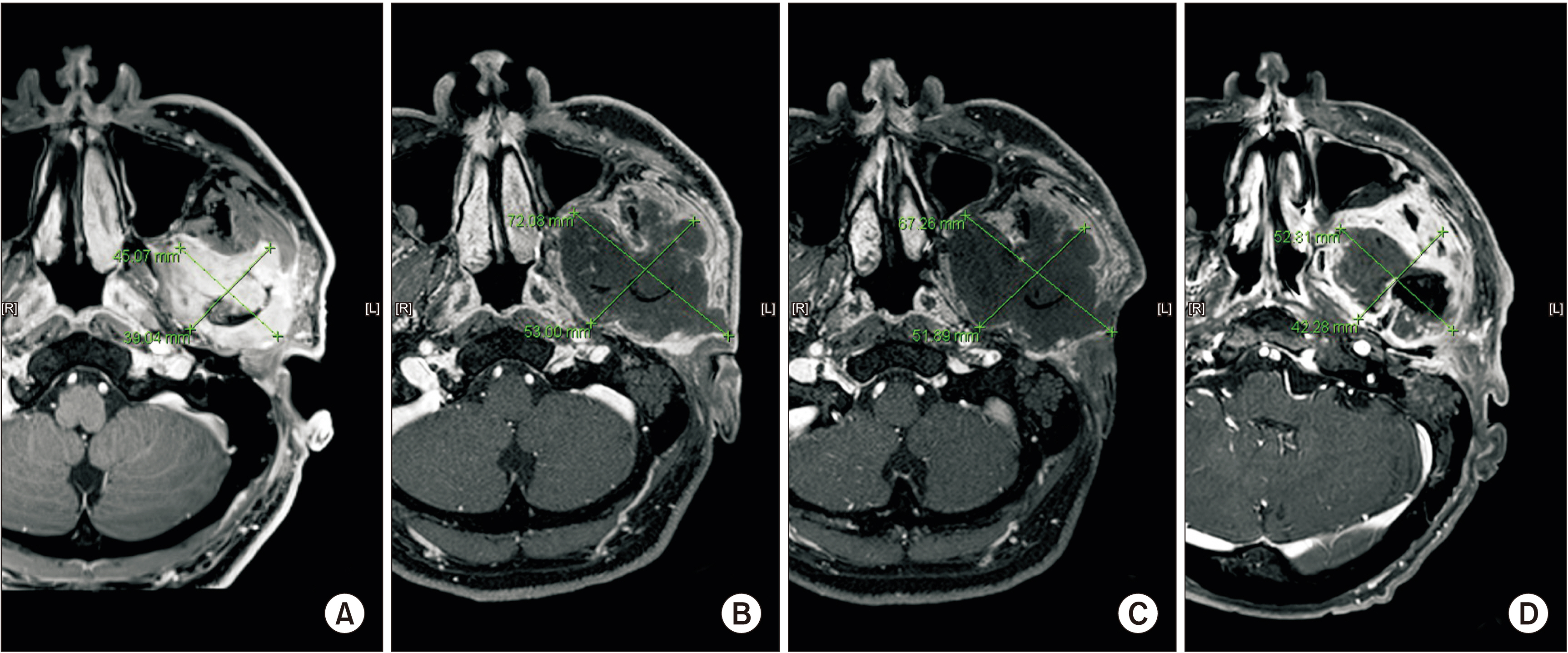
Fig. 1 Measured tumor size in magnetic resonance imaging. A. October 2016, before carbon-ion radiotherapy (CIRT). 4.5 cm×3.9 cm. B. April 2017, 3 months after CIRT. 7.2 cm×5.3 cm. C. October 2017, 9 months after CIRT. No change in overall size of tumor. D. October 2018, 21 months after CIRT. Suspected recurrence of tumor.

Fig. 2 A. Apirl 2017. The patient had newly-developed symptoms such as dysesthesia on left buccal cheek, limited mouth opening, xerostomia, oral ulcers and ipsilateral otitis externa with hearing loss. B. January 2018. Peroral biopsy of ulcerative soft tissue mass was done.

Fig. 3 A. November 2016. Mass with increased 18F-fluorodeoxyglucose (FDG) uptake in the left masticator space, suggesting malignancy. No sign of lymph node or distant metastasis was observed. B. October 2017, 9 months after carbon-ion radiotherapy (CIRT). C. February 2018, 13 months after CIRT. D. October 2018, 21 months after CIRT. Recurrent tumor with metastatic lymphadenopathy was suspected.
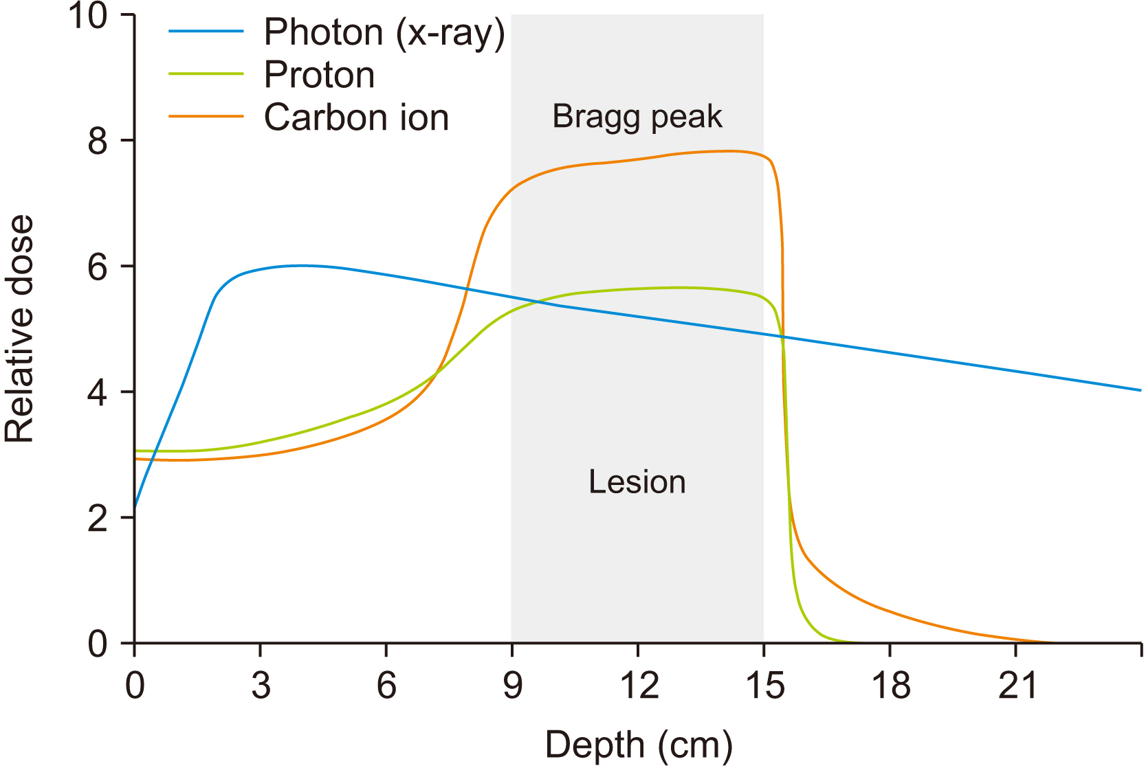
Fig. 4 A comparison of the physics of X-ray and particle beam doses. The proton and carbon ion beams have a significantly lower entrance dose, and no exit dose.

Fig. 5 A. October 2016. Osteolytic lesion on left mandibular condyle with pathologic fracture. B. March 2018. C. November 2018. Necrotic change was noted. D. January 2019.
Reference
-
References
1. Lim S, Lee S, Rha SY, Rho JK. 2016; Cranofacial osteosarcoma: single institutional experience in Korea. Asia Pac J Clin Oncol. 12:e149–53. https://doi.org/10.1111/ajco.12072 . DOI: 10.1111/ajco.12072. PMID: 23718845.
Article2. Sturgis EM, Potter BO. 2003; Sarcomas of the head and neck region. Curr Opin Oncol. 15:239–52. https://doi.org/10.1097/00001622-200305000-00011 . DOI: 10.1097/00001622-200305000-00011. PMID: 12778019.
Article3. Schwarz R, Bruland O, Cassoni A, Schomberg P, Bielack S. Jaffe N, Bruland OS, Bielack S, editors. 2010. The role of radiotherapy in oseosarcoma. Pediatric and adolescent osteosarcoma. Cancer treatment and research. Springer;Boston: p. 147–64.
Article4. Jeong HI, Lee MJ, Nam W, Cha IH, Kim HJ. 2017; Osteosarcoma of the jaws in Koreans: analysis of 26 cases. J Korean Assoc Oral Maxillofac Surg. 43:312–7. https://doi.org/10.5125/jkaoms.2017.43.5.312 . DOI: 10.5125/jkaoms.2017.43.5.312. PMID: 29142865. PMCID: PMC5685860.
Article5. Machak GN, Tkachev SI, Solovyev YN, Sinyukov PA, Ivanov SM, Kochergina NV, et al. 2003; Neoadjuvant chemotherapy and local radiotherapy for high-grade osteosarcoma of the extremities. Mayo Clin Proc. 78:147–55. https://doi.org/10.4065/78.2.147 . DOI: 10.4065/78.2.147. PMID: 12583525.
Article6. Delaney G, Jacob S, Featherstone C, Barton M. 2005; The role of radiotherapy in cancer treatment: estimating optimal utilization from a review of evidence-based clinical guidelines. Cancer. 104:1129–37. https://doi.org/10.1002/cncr.21324 . DOI: 10.1002/cncr.21324. PMID: 16080176.
Article7. Neville B, Damm DD, Allen C, Chi A. 2015. Oral and maxillofacial pathology. 4th ed. Saunders;Philadelphia:8. Krishnamurthy A, Palaniappan R. 2018; Osteosarcomas of the head and neck region: a case series with a review of literature. J Maxillofac Oral Surg. 17:38–43. https://doi.org/10.1007/s12663-017-1017-8 . DOI: 10.1007/s12663-017-1017-8. PMID: 29382992. PMCID: PMC5772028.
Article9. Makary RF, Gopinath A, Markiewicz MR, Fernandes R. 2017; Margin analysis: sarcoma of the head and neck. Oral Maxillofac Surg Clin North Am. 29:355–66. https://doi.org/10.1016/j.coms.2017.04.002 . DOI: 10.1016/j.coms.2017.04.002. PMID: 28709534.
Article10. Kimura Y, Tomihara K, Tachinami H, Imaue S, Nakamori K, Fujiwara K, et al. 2017; Conventional osteosarcoma of the mandible successfully treated with radical surgery and adjuvant chemotherapy after responding poorly to neoadjuvant chemotherapy: a case report. J Med Case Rep. 11:210. https://doi.org/10.1186/s13256-017-1386-0 . DOI: 10.1186/s13256-017-1386-0. PMID: 28764797. PMCID: PMC5540298.
Article11. Ebner DK, Kamada T. 2016; The emerging role of carbon-ion radiotherapy. Front Oncol. 6:140. https://doi.org/10.3389/fonc.2016.00140 . DOI: 10.3389/fonc.2016.00140. PMID: 27376030. PMCID: PMC4894867.
Article12. Kong L, Gao J, Hu J, Lu R, Yang J, Qiu X, et al. 2019; Carbon ion radiotherapy boost in the treatment of glioblastoma: a randomized phase I/III clinical trial. Cancer Commun (Lond). 39:5. https://doi.org/10.1186/s40880-019-0351-2 . DOI: 10.1186/s40880-019-0351-2. PMID: 30786916. PMCID: PMC6383247.
Article13. Durante M, Paganetti H. 2016; Nuclear physics in particle therapy: a review. Rep Prog Phys. 79:096702. https://doi.org/10.1088/0034-4885/79/9/096702 . DOI: 10.1088/0034-4885/79/9/096702. PMID: 27540827.
Article14. Mohamad O, Yamada S, Durante M. 2018; Clinical indications for carbon ion radiotherapy. Clin Oncol (R Coll Radiol). 30:317–29. https://doi.org/10.1016/j.clon.2018.01.006 . DOI: 10.1016/j.clon.2018.01.006. PMID: 29402598.
Article15. Blattmann C, Oertel S, Schulz-Ertner D, Rieken S, Haufe S, Ewerbeck V, et al. 2010; Non-randomized therapy trial to determine the safety and efficacy of heavy ion radiotherapy in patients with non-resectable osteosarcoma. BMC Cancer. 10:96. https://doi.org/10.1186/1471-2407-10-96 . DOI: 10.1186/1471-2407-10-96. PMID: 20226028. PMCID: PMC2846886.
Article16. Mohamad O, Makishima H, Kamada T. 2018; Evolution of carbon ion radiotherapy at the National Institute of Radiological Sciences in Japan. Cancers (Basel). 10:66. https://doi.org/10.3390/cancers10030066 . DOI: 10.3390/cancers10030066. PMID: 29509684. PMCID: PMC5876641.
Article17. Ballo MT, Zagars GK, Cormier JN, Hunt KK, Feig BW, Patel SR, et al. 2004; Interval between surgery and radiotherapy: effect on local control of soft tissue sarcoma. Int J Radiat Oncol Biol Phys. 58:1461–7. https://doi.org/10.1016/j.ijrobp.2003.09.079 . DOI: 10.1016/j.ijrobp.2003.09.079. PMID: 15050324.
Article18. Jingu K, Tsujii H, Mizoe JE, Hasegawa A, Bessho H, Takagi R, et al. 2012; ; Organizing Committee for the Working Group for Head-and-Neck Cancer. Carbon ion radiation therapy improves the prognosis of unresectable adult bone and soft-tissue sarcoma of the head and neck. Int J Radiat Oncol Biol Phys. 82:2125–31. https://doi.org/10.1016/j.ijrobp.2010.08.043 .
Article19. Kohyama K, Yamada K, Sugiura H, Hyodo I, Ozawa T, Hasegawa Y, et al. 2015; Salvage surgery and microsurgical reconstruction for recurrence of skull base osteosarcoma after carbon ion radiotherapy. Nagoya J Med Sci. 77:667–73. PMID: 26663946. PMCID: PMC4664599.20. Sasahara G, Koto M, Ikawa H, Hasegawa A, Takagi R, Okamoto Y, et al. 2014; Effects of the dose-volume relationship on and risk factors for maxillary osteoradionecrosis after carbon ion radiotherapy. Radiat Oncol. 9:92. https://doi.org/10.1186/1748-717X-9-92 . DOI: 10.1186/1748-717X-9-92. PMID: 24708583. PMCID: PMC3992144.
Article
