J Korean Neurosurg Soc.
2021 Sep;64(5):784-790. 10.3340/jkns.2020.0320.
Physiologic Cervical Alignment Change between Cervical Spine X-ray and Computed Tomography
- Affiliations
-
- 1Department of Neurosurgery, St. Vincent Hospital, College of Medicine, The Catholic University of Korea, Suwon, Korea
- 2Department of Neurosurgery, Eunpyeong St. Mary’s Hospital, College of Medicine, The Catholic University of Korea, Seoul, Korea
- KMID: 2519705
- DOI: http://doi.org/10.3340/jkns.2020.0320
Abstract
Objective
: The purpose of this study was to investigate the correlations among various radiological parameters used to determine cervical alignment from cervical spine radiographs (X-CS) and cervical spine computed tomography (CT-CS), both within and between modalities.
Methods
: This study included 168 patients (≤60 years old) without a definite whole spine deformity who underwent CT-CS and X-CS. We measured occipital slope (O-s), C1 slope, C2 slope, C7 slope, sella turcica - C7 sagittal vertical axis (StC7-SVA), spinocranial angle, T1 slope, and C27-SVA. We calculated the O-C2 angle, O-C7 angle, and C2-7 angle from the measured parameters and conducted correlation analyses among multiple parameters.
Results
: The intrinsic correlation features among multiple cervical parameters were very similar for both X-CS and CT-CS. The two SVA parameters (C27-SVA and StC7-SVA) were mainly influenced by the upper cervical slope parameters (r=|0.13–0.74|) rather than the lower slope cervical parameters (r=|0.08–0.13|). The correlation between X-CS and CT-CS for each radiological parameter was statistically significant (r=0.26–0.44) except for O-s (r=0.10) and StC7-SVA (r=0.11).
Conclusion
: The correlation patterns within X-CS and CT-CS were very similar in this study. The correlation between X-ray and CT was statistically significant for most radiological parameters, and the correlation score increased when the horizontal gaze was consistently maintained. The lower cervical parameters were not statistically associated with translation-related parameters (C2-7 SVA and StC7-SVA). Therefore, the upper cervical segment may be a better predictor for determining head and neck translation.
Keyword
Figure
-
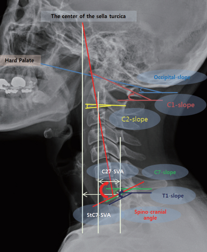
Fig. 1. The measured cervical parameters in this study; occipital slope, C1-slope, C2-slope, C7-slope, T1-slope, C2-C7-sagital vertical axis (C27-SVA), sella turcia-C7 sagittal vertical axis (StC7-SVA) and spino-cranial angle.
Reference
-
References
1. Ames CP, Blondel B, Scheer JK, Schwab FJ, Le Huec JC, Massicotte EM, et al. Cervical radiographical alignment: comprehensive assessment techniques and potential importance in cervical myelopathy. Spine (Phila Pa 1976). 38(22 Suppl 1):S149–S160. 2013.2. Beltsios M, Savvidou O, Mitsiokapa EA, Mavrogenis AF, Kaspiris A, Efstathopoulos N, et al. Sagittal alignment of the cervical spine after neck injury. Eur J Orthop Surg Traumatol 23 Suppl. 1:S47–S51. 2013.
Article3. Cheng J, Liu P, Sun D, Ma Z, Liu J, Wang Z, et al. Correlation of cervical and thoracic inlet sagittal parameters by MRI and radiography in patients with cervical spondylosis. Medicine (Baltimore). 98:e14393. 2019.
Article4. Janusz P, Tyrakowski M, Glowka P, Offoha R, Siemionow K. Influence of cervical spine position on the radiographic parameters of the thoracic inlet alignment. Eur Spine J. 24:2880–2884. 2015.
Article5. Jouibari MF, Le Huec JC, Ranjbar Hameghavandi MH, Moghadam N, Farahbakhsh F, Khadivi M, et al. Comparison of cervical sagittal parameters among patients with neck pain and healthy controls: a comparative cross-sectional study. Eur Spine J. 28:2319–2324. 2019.
Article6. Jun HS, Chang IB, Song JH, Kim TH, Park MS, Kim SW, et al. Is it possible to evaluate the parameters of cervical sagittal alignment on cervical computed tomographic scans? Spine (Phila Pa 1976). 39:E630–E636. 2014.
Article7. Kim JT, Lee HJ, Choi DY, Shin MH, Hong JT. Sequential alignment change of the cervical spine after anterior cervical discectomy and fusion in the lower cervical spine. Eur Spine J. 25:2223–2232. 2016.
Article8. Le Huec JC, Saddiki R, Franke J, Rigal J, Aunoble S. Equilibrium of the human body and the gravity line: the basics. Eur Spine J 20 Suppl. 5:558–563. 2011.
Article9. Lee HD, Jeon CH, Chung NS, Kwon HJ. Comparative analysis of three imaging modalities for evaluation of cervical sagittal alignment parameters: a validity and reliability study. Spine (Phila Pa 1976). 42:1901–1907. 2017.
Article10. Lee HJ, Kim JH, Kim IS, Hong JT. Physiologic cervical alignment change between whole spine radiographs and normal standing cervical radiographs. World Neurosurg. 122:e1222–e1227. 2019.
Article11. Lee SH, Kim KT, Seo EM, Suk KS, Kwack YH, Son ES. The influence of thoracic inlet alignment on the craniocervical sagittal balance in asymptomatic adults. J Spinal Disord Tech. 25:E41–E47. 2012.
Article12. Liu W, Fan J, Bai J, Tang P, Chen J, Luo Y, et al. Magnetic resonance imaging: a possible alternative to a standing lateral radiograph for evaluating cervical sagittal alignment in patients with cervical disc herniation? Medicine (Baltimore). 96:e8194. 2017.13. Núñez-Pereira S, Hitzl W, Bullmann V, Meier O, Koller H. Sagittal balance of the cervical spine: an analysis of occipitocervical and spinopelvic interdependence, with C-7 slope as a marker of cervical and spinopelvic alignment. J Neurosurg Spine. 23:16–23. 2015.
Article14. Park JH, Cho CB, Song JH, Kim SW, Ha Y, Oh JK. T1 slope and cervical sagittal alignment on cervical CT radiographs of asymptomatic persons. J Korean Neurosurg Soc. 53:356–359. 2013.
Article15. Qiao J, Zhu F, Liu Z, Xu L, Zhu Z, Qian B, et al. Measurement of thoracic inlet alignment on MRI: reliability and the influence of body position. Clin Spine Surg. 30:E377–E380. 2017.16. Scheer JK, Tang JA, Smith JS, Acosta FL Jr, Protopsaltis TS, Blondel B, et al. Cervical spine alignment, sagittal deformity, and clinical implications: a review. J Neurosurg Spine. 19:141–159. 2013.17. Tang JA, Scheer JK, Smith JS, Deviren V, Bess S, Hart RA, et al. The impact of standing regional cervical sagittal alignment on outcomes in posterior cervical fusion surgery. Neurosurgery. 71:662–669. discussion 669. 2012.
Article18. Weng C, Wang J, Tuchman A, Wang J, Fu C, Hsieh PC, et al. Influence of T1 slope on the cervical sagittal balance in degenerative cervical spine: an analysis using kinematic MRI. Spine (Phila Pa 1976). 41:185–190. 2016.
Article
- Full Text Links
-
- Actions
-
Cited
- CITED
-
- Close
- Share
-
- Similar articles
-
- Clinical Analysis of Lower Cervical Spine-Injuried Patients
- Cervical Sagittal Alignment: Literature Review and Future Directions
- The Sagittal Balance of Cervical Spine : Comprehensive Review of Recent Update
- Sagittal Fracture of Cervical Spine: Case Report
- Surgical Impact on Global Sagittal Alignment and Health-Related Quality of Life Following Cervical Kyphosis Correction Surgery: Systematic Review
