Sarcomatoid urothelial carcinoma arising in the female urethral diverticulum
- Affiliations
-
- 1Department of Pathology, Ewha Womans University Seoul Hospital, Seoul, Korea
- KMID: 2518430
- DOI: http://doi.org/10.4132/jptm.2021.04.23
Abstract
- A sarcomatoid variant of urothelial carcinoma in the female urethral diverticulum has not been reported previously. A 66-year-old woman suffering from dysuria presented with a huge urethral mass invading the urinary bladder and vagina. Histopathological examination of the resected specimen revealed predominantly undifferentiated pleomorphic sarcoma with sclerosis. Only a small portion of conventional urothelial carcinoma was identified around the urethral diverticulum, which contained glandular epithelium and villous adenoma. The patient showed rapid systemic recurrence and resistance to immune checkpoint inhibitor therapy despite high expression of programmed cell death ligand-1. We report the first case of urethral diverticular carcinoma with sarcomatoid features.
Figure
-

Fig. 1. Enhanced abdominopelvic computed tomography. (A) Axial image taken 7 months prior to presentation showed a urethral diverticulum (asterisk) at the level of the symphysis pubis. (B) Preoperative image revealed a large urethral mass (UB, urinary bladder; arrow, urinary catheter within the urethra).
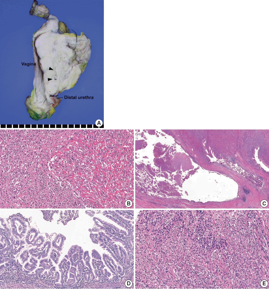
Fig. 2. Histopathological findings. (A) Gross examination revealed a 10-cm-sized, hard white urethral mass invading the uterus, vagina, urinary bladder, and perivesical fat. The cut surface showed necrosis and cystic space (arrowheads). (B) Microscopically, the majority of the tumor was composed of pleomorphic spindle cells with occasional collagen deposition. Intratumoral lymphoplasmacytic infiltration was noted. (C, D) The cystic space was lined focally by glandular epithelium (C) and associated villous adenoma (D). (E) A conventional urothelial carcinoma component was minimally present, and areas of epithelial-to-mesenchymal transition were noted.
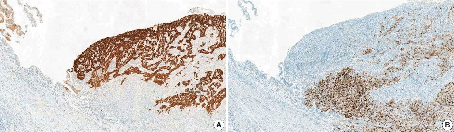
Fig. 3. Results of immunohistochemical staining. (A) Immunostaining for cytokeratin 7 highlighted the glandular epithelium (left side) and urothelial carcinoma component, whereas there was no staining of the sarcoma component. (B) Programmed death ligand-1 SP142 (Ventana Medical Systems, Tucson, AZ, USA) immunostaining showed diffuse positivity (90%) in tumor-infiltrating immune cells (ICs) of the sarcoma component, while ICs of the carcinoma component were negative.
Reference
-
References
1. Ahmed K, Dasgupta R, Vats A, et al. Urethral diverticular carcinoma: an overview of current trends in diagnosis and management. Int Urol Nephrol. 2010; 42:331–41.
Article2. O’Connor E, Iatropoulou D, Hashimoto S, Takahashi S, Ho DH, Greenwell T. Urethral diverticulum carcinoma in females: a case series and review of the English and Japanese literature. Transl Androl Urol. 2018; 7:703–29.3. Derksen JW, Visser O, de la Riviere GB, Meuleman EJ, Heldeweg EA, Lagerveld BW. Primary urethral carcinoma in females: an epidemiologic study on demographical factors, histological types, tumour stage and survival. World J Urol. 2013; 31:147–53.
Article4. Rajan N, Tucci P, Mallouh C, Choudhury M. Carcinoma in female urethral diverticulum: case reports and review of management. J Urol. 1993; 150:1911–4.
Article5. Gu L, Ai Q, Cheng Q, et al. Sarcomatoid variant urothelial carcinoma of the bladder: a systematic review and meta-analysis of the clinicopathological features and survival outcomes. Cancer Cell Int. 2020; 20:550.
Article6. Lembo F, Subba E, Lagana AS, Vitale SG, Valenti G, Magno C. Intradiverticular sarcomatoid carcinoma of the bladder: an overview starting from a peculiar case. Urol J. 2016; 13:2800–2.7. Wang Y, Liu H, Wang P. Primary sarcomatoid urothelial carcinoma of the ureter: a case report and review of the literature. World J Surg Oncol. 2018; 16:77.
Article8. Rashid S, Akhtar M. Sarcomatoid variant of urothelial carcinoma of the renal pelvis with inferior vena cava tumour thrombus: a case report and literature review. Case Rep Pathol. 2018; 2018:1837510.
Article9. D’Arrigo L, Costa A, Fraggetta F, Pennisi M, Pepe P, Aragona F. Carcinosarcoma of the female urethra. Urol Int. 2016; 96:370–2.
Article10. Moch H, Humphrey PA, Ulbright TM, Reuter VE. WHO classification of tumours of the urinary system and male genital organs. 4th ed. Lyon: International Agency for Research on Cancer;2016. p. 92.11. Bostwick DG, Cheng L. Urologic surgical pathology. 3rd ed. Philadelphia: Saunders-Elsevier;2014. p. 298–9.12. Sanfrancesco J, McKenney JK, Leivo MZ, Gupta S, Elson P, Hansel DE. Sarcomatoid urothelial carcinoma of the bladder: analysis of 28 cases with emphasis on clinicopathologic features and markers of epithelial-to-mesenchymal transition. Arch Pathol Lab Med. 2016; 140:543–51.
Article13. Balar AV, Galsky MD, Rosenberg JE, et al. Atezolizumab as first-line treatment in cisplatin-ineligible patients with locally advanced and metastatic urothelial carcinoma: a single-arm, multicentre, phase 2 trial. Lancet. 2017; 389:67–76.
Article14. Powles T, Duran I, van der Heijden MS, et al. Atezolizumab versus chemotherapy in patients with platinum-treated locally advanced or metastatic urothelial carcinoma (IMvigor211): a multicentre, openlabel, phase 3 randomised controlled trial. Lancet. 2018; 391:748–57.
Article15. Li H, Zhang Q, Shuman L, et al. Evaluation of PD-L1 and other immune markers in bladder urothelial carcinoma stratified by histologic variants and molecular subtypes. Sci Rep. 2020; 10:1439.
Article
- Full Text Links
-
- Actions
-
Cited
- CITED
-
- Close
- Share
-
- Similar articles
-
- A Localized Sarcomatoid Carcinoma of a Urinary Bladder Diverticulum
- A Case of Female Urethral Diverticulum Combined with Multiple Stones
- Sarcomatoid Urothelial Carcinoma of the Renal Pelvis with Extremely Aggressive Clinical Behavior
- Three Cases of Urethral Diverticulum in the Female
- A case of urethral diverticulum combined with stone
