Anat Cell Biol.
2021 Jun;54(2):289-291. 10.5115/acb.21.086.
Bicipital origin and the course of the plantaris muscle
- Affiliations
-
- 1Department of Anatomy, College of Medicine, University of Ulsan, Seoul, Korea
- 2Clinical Anatomy Education Center, Asan Medical Center, Seoul, Korea
- KMID: 2516914
- DOI: http://doi.org/10.5115/acb.21.086
Abstract
- The plantaris muscle (PM) has a small fusiform muscle belly and a long slender tendon sandwiched between the soleus (SM) and gastrocnemius muscle (GM). During routine dissection for research, an additional PM in the popliteal region of a 75-year-old Korean female was discovered. Two distinct PMs were present, the superior PM (sPM) and inferior PM (iPM). While the sPM originates from the lower lateral supracondylar ridge and the knee capsule, the iPM originates from the femoral condyle and sPM tendon splitting into two parts at the distal belly. The lateral side of the iPM tendon travels between GM and SM and ends at the calcaneal tendon. sPM and the medial side of the iPM tendon run along with the sPM tendon and inserts at the fascia at the inner surface of proximal 1/3 of the medial head of GM. This case report introduces a new variation of the PM that should be taken into consideration.
Figure
-
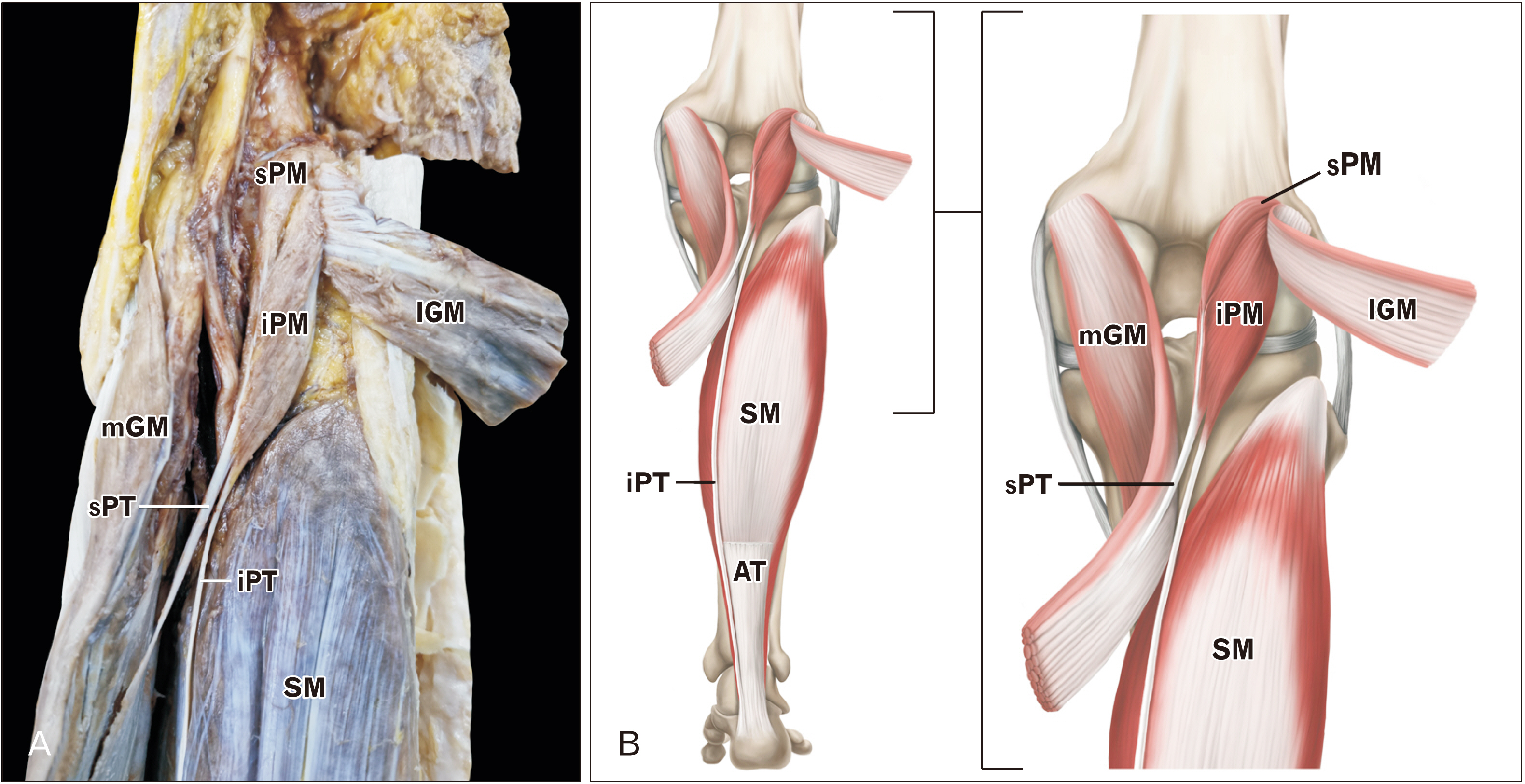
Fig. 1 Posterior view of the lower right leg. (A) The course of additional plantaris muscle/tendon unit. (B) Schematic drawing of plantaris muscle/tendon unit. AT, Achilles tendon (cut); iPM, inferior plantaris muscle; iPT, inferior plantaris tendon; lGM, lateral head of gastrocnemius muscle (cut); mGM, medial head of gastrocnemius muscle (cut); SM, soleus muscle (cut); sPM, superior plantaris muscle; sPT, superior plantaris tendon.
Reference
-
References
1. Olewnik Ł, Zielinska N, Karauda P, Tubbs RS, Polguj M. 2020; A three-headed plantaris muscle: evidence that the plantaris is not a vestigial muscle? Surg Radiol Anat. 42:1189–93. DOI: 10.1007/s00276-020-02478-8. PMID: 32382814. PMCID: PMC7366563.
Article2. Harwin JR, Richardson ML. 2016; "Tennis leg": gastrocnemius injury is a far more common cause than plantaris rupture. Radiol Case Rep. 12:120–3. DOI: 10.1016/j.radcr.2016.10.012. PMID: 28228893. PMCID: PMC5310238.
Article3. Olewnik Ł, Wysiadecki G, Podgórski M, Polguj M, Topol M. 2018; The plantaris muscle tendon and its relationship with the Achilles tendinopathy. Biomed Res Int. 2018:9623579. DOI: 10.1155/2018/9623579. PMID: 29955614. PMCID: PMC6000875.
Article4. Helms CA, Fritz RC, Garvin GJ. 1995; Plantaris muscle injury: evaluation with MR imaging. Radiology. 195:201–3. DOI: 10.1148/radiology.195.1.7892469. PMID: 7892469.
Article5. Olewnik Ł, Wysiadecki G, Polguj M, Topol M. 2017; Anatomic study suggests that the morphology of the plantaris tendon may be related to Achilles tendonitis. Surg Radiol Anat. 39:69–75. DOI: 10.1007/s00276-016-1682-1. PMID: 27155667. PMCID: PMC5309290.
Article6. Hadimani GA, Bagoji I. 2018; Accessory belly of the plantaris muscle and its clinical relevance. South East Asia J Med Sci. 2:5–6.7. Kurtys K, Gonera B, Olewnik Ł, Karauda P, Tubbs RS, Polguj M. 2020; Nov. 7. Is the plantaris muscle the most undefined human skeletal muscle? Anat Sci Int. [Epub]. https://doi.org/10.1007/s12565-020-00586-4. DOI: 10.1007/s12565-020-00586-4. PMID: 33159667. PMCID: PMC8139894.
Article8. Olewnik Ł, Podgórski M, Polguj M, Topol M. 2018; The plantaris muscle - rare relations to the neurovascular bundle in the popliteal fossa. Folia Morphol (Warsz). 77:785–8. DOI: 10.5603/FM.a2018.0039. PMID: 29651792.
Article9. Olewnik Ł, Wysiadecki G, Polguj M, Topol M. 2017; The report on the co-occurrence of two different rare anatomic variations of the plantaris muscle tendon on both sides of an individual. Folia Morphol (Warsz). 76:331–3. DOI: 10.5603/FM.a2016.0069. PMID: 27813626.
Article10. Aragao JA, Reis FP, Guerra DR, Cabral RH. 2010; The occurrence of the plantaris muscle and its muscle-tendon relationship in adult human cadavers. Int J Morphol. 28:255–8. DOI: 10.4067/S0717-95022010000100037.
Article11. Olewnik Ł, Kurtys K, Gonera B, Podgórski M, Sibiński M, Polguj M. 2020; Proposal for a new classification of plantaris muscle origin and its potential effect on the knee joint. Ann Anat. 231:151506. DOI: 10.1016/j.aanat.2020.151506. PMID: 32173563.
Article12. Rohilla S, Jain N, Yadav R. 2013; Plantaris rupture: why is it important? BMJ Case Rep. 2013:bcr2012007840. DOI: 10.1136/bcr-2012-007840. PMID: 23345486. PMCID: PMC3604295.
Article13. Park KR, Cho J, Choi YJ, Kim D, Kwon HW, Lee M, Yoon KH, Park J. 2020; Complex variations of plantaris muscle origin, course and insertion: a cadaveric case report. Anat Biol Anthropol. 33:143–7. DOI: 10.11637/aba.2020.33.3.143.
Article14. Herzog RJ. 2011; Accessory plantaris muscle: anatomy and prevalence. HSS J. 7:52–6. DOI: 10.1007/s11420-010-9175-y. PMID: 22294958. PMCID: PMC3026110.
Article15. Rana KK, Das S, Verma R. 2006; Double plantaris muscle: a cadaveric study with clinical importance. Int J Morphol. 24:495–8. DOI: 10.4067/S0717-95022006000400032.
Article
- Full Text Links
-
- Actions
-
Cited
- CITED
-
- Close
- Share
-
- Similar articles
-
- Variations for Incomplete Formation of the Plantaris Muscle: A Case Report
- Complex Variations of Plantaris Muscle Origin, Course and Insertion: A Cadaveric Case Report
- The Co-existence of the Gastrocnemius Tertius and Accessory Soleus Muscles
- Effect of exercise training following hypokinesia on the length and circumference of atrophied soleus and plantaris muscle in rats
- Effect of Periodic Walking on the Type II Muscle of Growing Suspended Rats
