J Korean Med Sci.
2011 Oct;26(10):1378-1381. 10.3346/jkms.2011.26.10.1378.
The Co-existence of the Gastrocnemius Tertius and Accessory Soleus Muscles
- Affiliations
-
- 1Department of Anatomy, Faculty of Medicine, Akdeniz University, Antalya, Turkey. levent@akdeniz.edu.tr
- 2Department of Oral Anatomy, School of Dentistry, Showa University, Japan.
- KMID: 1785998
- DOI: http://doi.org/10.3346/jkms.2011.26.10.1378
Abstract
- A bilateral gastrocnemius tertius muscle and a unilateral accessory soleus muscle were encountered during the routine educational dissection studies. The right gastrocnemius tertius muscle consisted of one belly, but the left one of two bellies. On the left side, the superficial belly of the gastrocnemius tertius muscle had its origin from an area just above the tendon of the plantaris muscle, the deep belly from the tendon of the plantaris muscle. The accessory soleus muscle originated from the posteromedial aspect of the tibia and soleal line of the tibia and inserted to the medial surface of the calcaneus. On the right side, the gastrocnemius tertius muscle had its origin from the lateral condyle of the femur, and inserted to the medial head of the gastrocnemius muscle. The co-existence of both gastrocnemius tertius and accessory soleus muscle has not, to our knowledge, been previously reported.
Figure
-
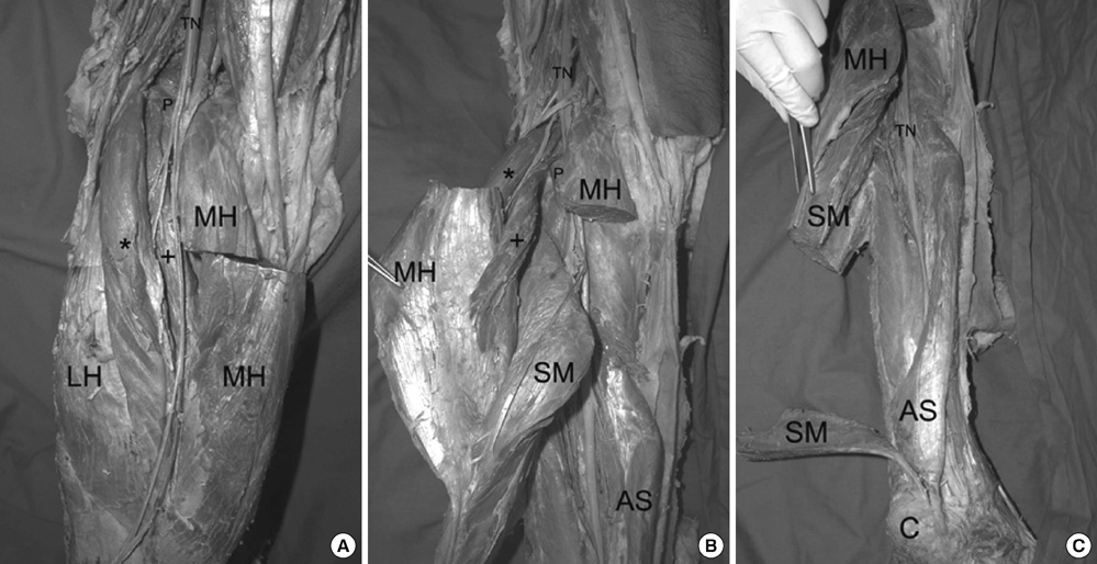
Fig. 1 Photograph of the left side of the czase. (A) Arrangement of the superficial and deep heads of the GCT, (B) Insertion of the deep head of the GCT onto the medial head of the gastrocnemius muscle, (C) Accessory soleus muscle. AS, accessory soleus muscle; C, calcaneus; LH, lateral head of the gastrocnemius muscle; MH, Medial head of the gastrocnemius muscle; P, plantaris muscle; SM, soleus muscle; TN, tibial nerve. *, superficial belly of the gastrocnemius tertius; +, deep belly of the gastrocnemius tertius.

Fig. 2 Appearance of the gastrocnemius tertius muscle in the right side of the case. LH, lateral head of the gastrocnemius muscle; MH, Medial head of the gastrocnemius muscle; GCT, gastrocnemius tertius.
Reference
-
1. Bergman RA, Walker CW, el-Khour GY. The third head of gastrocnemius in CT images. Ann Anat. 1995. 177:291–294.2. Iwai T, Sato S, Yamada T, Muraoka Y, Sakurazawa K, Kinoshita H, Inoue Y, Endo M, Yoshida T, Suzuki S. Popliteal vein entrapment caused by the third head of the gastrocnemius muscle. Br J Surg. 1987. 74:1006–1008.3. Assoun J, Railhac JJ, Richardi G, Fajadet P, Fourcade D, Sans N. CT and MR of accessory soleus muscle. J Comput Assist Tomogr. 1995. 19:333–335.4. Baran G, Sundaram M. Radiologic case study. Accessory soleus muscle. Orthopedics. 1991. 14:499. 501.5. Bergman RA, Thompson SA, Afifi AK, Saadeh FA. Compendium of human anatomical variation. 1988. Baltimore: Urban and Schwarzenberg;7.6. Brodie JT, Dormans JP, Gregg JR, Davidson RS. Accessory soleus muscle. A report of 4 cases and review of literature. Clin Orthop Relat Res. 1997. (337):180–186.7. Tochihara J, Onozawa T. M. gastrocnemius tertius in Japanesae. Jpn J Anat. 1932. 5:589–600.8. Rosset E, Hartung O, Brunet C, Roche PH, Magnan PE, Mathieu JP, Branchereau A, Farisse J. Popliteal artery entrapment syndrome. Anatomic and embryologic bases, diagnostic and therapeutic considerations following a series of 15 cases with a review of the literature. Surg Radiol Anat. 1995. 17:161–169. 23–27.9. Bouhoutsos J, Daskalakis E. Muscular abnormalities affecting the popliteal vessels. Br J Surg. 1981. 68:501–506.10. Frey H. Musculus gastrocnemius tertius. Gegenbaurs Morphol Jahrb. 1919. 50:517–530.11. Gerkin TM, Beebe HG, Williams DM, Bloom JR, Wakefield TW. Popliteal vein entrapment presenting as deep venous thrombosis and chronic venous insufficiency. J Vasc Surg. 1993. 18:760–766.12. Miles S, Roediger W, Cooke P, Mieny CJ. Doppler ultrasound in the diagnosis of the popliteal artery entrapment syndrome. Br J Surg. 1977. 64:883–884.13. Boisgard S, Peronne E, Kalfon P, Levai JP, Michel JL. Accessory soleus muscle. Two case-reports, with a review of the literature. Rev Rhum Engl Ed. 1996. 63:862–865.14. Lorentzon R, Wirell S. Anatomic variations of the accessory soleus muscle. Acta Radiol. 1987. 28:627–629.15. Yu JS, Resnick D. MR imaging of the accessory soleus muscle appearance in six patients and a review of the literature. Skeletal Radiol. 1994. 23:525–528.16. Bonnell J, Cruess RL. Anomalous insertion of the soleus muscle as a cause of fixed equinus deformity. A case report. J Bone Joint Surg Am. 1969. 51:999–1000.17. Chotigavanichaya C, Scaduto AA, Jadhav A, Otsuka NY. Accessory soleus muscle as a cause of resistance to correction in congenital club foot: a case report. Foot Ankle Int. 2000. 21:948–950.18. Peterson DA, Stinson W, Carter J. Bilateral accessory soleus: a report on four patients with partial fasciectomy. Foot Ankle. 1993. 14:284–288.19. Ekstrom JE, Shuman WP, Mack LA. MR imaging of accessory soleus muscle. J Comput Assist Tomogr. 1990. 14:239–242.20. Wu KK. Accessory soleus muscle simulating a soft tissue tumor of the posteromedial ankle region. J Foot Surg. 1991. 30:470–471.
- Full Text Links
-
- Actions
-
Cited
- CITED
-
- Close
- Share
-
- Similar articles
-
- Morphological variations and accessory ossicles in the peroneal and tibialis muscles
- Surface Mapping of Motor Points of Gastrocnemius and Soleus Muscles
- A bilateral gastrocnemius tertius coexisting with a unilateral two-headed plantaris muscle
- Glycogen depletion - repletion Characteristics of Different Types of Skeletal Muscle Fiber in Streptozotocin - diabetic Rats
- Histochemical Muscle Fiber Types of Autopsied Human Gastrocnemius, Soleus, Peroneus longus and Tibialis anterior Muscles
