Optimal microcatheter shaping method customized for a patient-specific vessel using a three-dimensional printer
- Affiliations
-
- 1Department of Neurosurgery, CHA Bundang Medical Center, School of Medicine, CHA University, Seongnam, Korea
- KMID: 2514334
- DOI: http://doi.org/10.7461/jcen.2021.E2020.08.005
Abstract
Objective
In coil embolization of cerebral aneurysms, it is very important to guide microcatheters to the appropriate location in the aneurysm and stabilize them during procedures. To do this, microcatheters need to be properly shaped. In this study, we aim to use a computer application program and a three-dimensional (3D) printer to make a patientspecific shaped microcatheter.
Methods
We simplified, skeletalized, and oversized the existing 3D vascular imaging structures and created the central line structure of the blood vessels. These processes were performed using a computer application program developed by our team. The microcatheters were shaped according to the skeletalized data shape, and the catheterization procedures were simulated using the 3D hollow model of the blood vessel region of interest; the number of hollow models was 10. The compatibility of the microcatheters shaped according to the skeletalized data shape was validated if the microcatheter tip was positioned into the aneurysm.
Results
In all 10 hollow models, the positioning of the microcatheter into the aneurysms was successful following one or two attempts.
Conclusions
When shaping microcatheters during endovascular coil embolization, it may be useful to use central line structures with some expansions customized for a patientspecific vessel using a computer application program and a 3D printer. In the future, it may be necessary to apply this technique to actual patients.
Figure
-
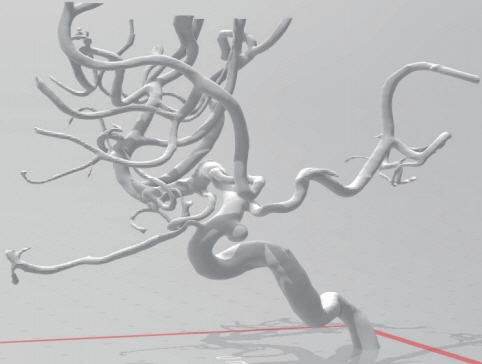
Fig. 1. Three-dimensional image data obtained using conventional cerebral angiography
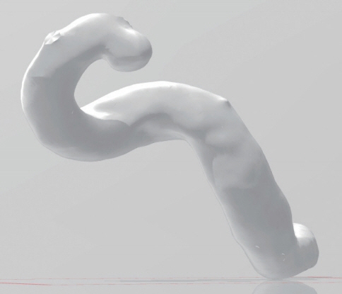
Fig. 2. The blood vessel regions of interest, extracted from the 3D image data from entire blood vessels.
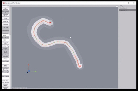
Fig. 3. The central line value of a blood vessel obtained by calculation using the computer application program we developed. BVPF, Blood Vessel Path Finder.
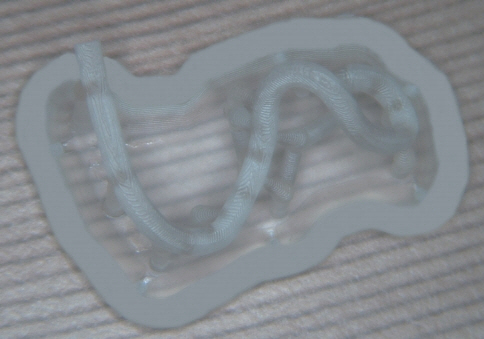
Fig. 4. A 3D model of the central line values of the blood vessel region of interest, which is a 3D printer output of the central line value of a blood vessel obtained in Fig. 3.
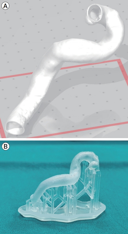
Fig. 5. (A, B) A 3D hollow model of a patient’s blood vessel regions of interest, created using a 3D printer.

Fig. 6. The simulation procedure of catheterization into the aneurysm using the 3D hollow model of the blood vessel regions of interest. These procedures could be visually observed because the 3D hollow model was semi-transparent due to the use of clear resins.
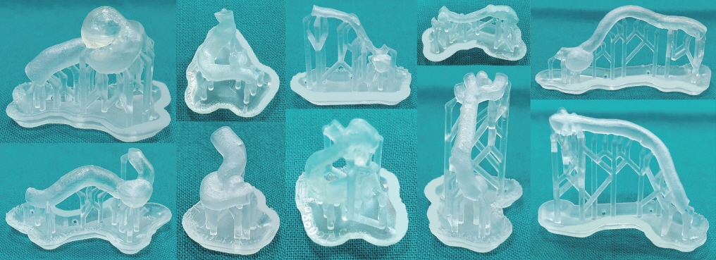
Fig. 7. Ten 3D hollow model, including (according to the sequence from left to right) two internal cerebral artery-paraclinoid aneurysms, two internal cerebral artery-posterior communicating artery aneurysms, two anterior cerebral artery aneurysms, two middle cerebral artery aneurysms, and two posterior circulation aneurysms.
Reference
-
1. Anderson JH, Brody WR, Chui CK, Cai Y, Wang Y, Nowinski WL. Simulation method for designing customized medical devices. United State patent US 7371067B2. 2019 May 13.2. Babiker MH, Frakes DH, Chong BW. Device specific finite element models for simulating endovascular treatment. United State patent US 10290230B2. 2019 May 14.3. Hwang JH, Roh HG, Chun YI, Kang HS, Choi JW, Moon WJ, et al. Endovascular coil embolization of very small intracranial aneurysms. Neuroradiology. 2011; May. 53(5):349–57.
Article4. Ishibashi T, Takao H, Suzuki T, Yuki I, Kaku S, Kan I, et al. Tailor-made shaping of microcatheters using three-dimensional printed vessel models for endovascular coil embolization. Comput Biol Med. 2016; Oct. 77:59–63.
Article5. Kono K, Shintani A, Okada H, Terada T. Preoperative simulations of endovascular treatment for a cerebral aneurysm using a patient-specific vascular silicone model. Neurol Med Chir (Tokyo). 2013; 53(5):347–51.
Article6. Kwon BJ, Im SH, Park JC, Cho YD, Kang HS, Kim JE, et al. Shaping and navigating methods of microcatheters for endovascular treatment of paraclinoid aneurysms. Neurosurgery. 2010; Jul. 67(1):34–40. discussion 40.
Article7. Lee JY, Seo JH, Cho YD, Kang HS, Han MH. Endovascular treatment of 429 anterior communicating artery aneurysms using bare-platinum coils: clinical and radiologic outcomes at the long-term follow-up. J Korean Neurosurg Soc. 2015; Mar. 57(3):159–66.8. Namba K, Higaki A, Kaneko N, Mashiko T, Nemoto S, Watanabe E. Microcatheter shaping for intracranial aneurysm coiling using the 3-dimensional printing rapid prototyping technology: preliminary result in the first 10 consecutive cases. World Neurosurg. 2015; Jul. 84(1):178–86.
Article9. Ohshima T, Imai T, Goto S, Yamamoto T, Nishizawa T, Shimato S, et al. A novel technique of microcatheter shaping with cerebral aneurysmal coil embolization: In vivo printing method. J Neuroendovasc Ther. 2016; 11(1):48–52.
Article10. Yamaguchi S, Ito O, Koyanagi Y, Iwaki K, Matsukado K. Microcatheter shaping using intravascular placement during intracranial aneurysm coiling. Interv Neuroradiol. 2017; Jun. 23(3):249–54.
Article
- Full Text Links
-
- Actions
-
Cited
- CITED
-
- Close
- Share
-
- Similar articles
-
- Fabrication of a Patient-Customized Helmet with a Three-Dimensional Printer for Radiation Therapy of Scalp
- Suction thrombectomy of distal medium vessel occlusion using microcatheter during mechanical thrombectomy for acute ischemic stroke: A case series
- Z-Shaped Microcatheter Tip Shaping for Embolization of Aneurysms at the Proximal A1 Segment of the Anterior Cerebral Artery: A Technical Note
- Nasoethmoid orbital fracture reconstruction using a three-dimensional printing-based craniofacial plate
- Endovascular Rescue Method for Undesirably Stretched Coil
