Triggered Electrooculography for Identification of Oculomotor and Abducens Nerves during Skull Base Surgery
- Affiliations
-
- 1Department of Neurology, Yonsei University College of Medicine, Seoul, Korea
- 2Department of Neurology, Myongji Hospital, Goyang, Korea
- 3Department of Neurosurgery, Yonsei University College of Medicine, Seoul, Korea
- 4Brain Tumor Center, Severance Hospital, Seoul, Korea
- KMID: 2513859
- DOI: http://doi.org/10.3340/jkns.2020.0179
Abstract
Objective
: Electrooculography (EOG) records eyeball movements as changes in the potential difference between the negatively charged retina and the positively charged cornea. We aimed to investigate whether reliable EOG waveforms can be evoked by electrical stimulation of the oculomotor and abducens nerves during skull base surgery.
Methods
: We retrospectively reviewed the records of 18 patients who had undergone a skull base tumor surgery using EOG (11 craniotomies and seven endonasal endoscopic surgeries). Stimulation was performed at 5 Hz with a stimulus duration of 200 μs and an intensity of 0.1–5 mA using a concentric bipolar probe. Recording electrodes were placed on the upper (active) and lower (reference) eyelids, and on the outer corners of both eyes; the active electrode was placed on the contralateral side.
Results
: Reproducibly triggered EOG waveforms were observed in all cases. Electrical stimulation of cranial nerves (CNs) III and VI elicited positive waveforms and negative waveforms, respectively, in the horizontal recording. The median latencies were 3.1 and 0.5 ms for craniotomies and endonasal endoscopic surgeries, respectively (p=0.007). Additionally, the median amplitudes were 33.7 and 46.4 μV for craniotomies and endonasal endoscopic surgeries, respectively (p=0.40).
Conclusion
: This study showed reliably triggered EOG waveforms with stimulation of CNs III and VI during skull base surgery. The latency was different according to the point of stimulation and thus predictable. As EOG is noninvasive and relatively easy to perform, it can be used to identify the ocular motor nerves during surgeries as an alternative of electromyography.
Figure
-
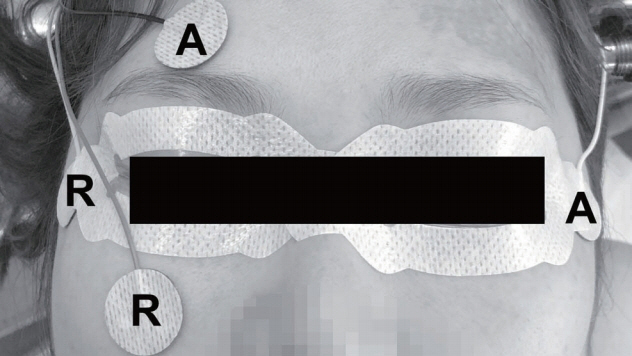
Fig. 1. A schematic design showing the placement of electrodes for the intraoperative electrooculography. R : reference electrode, A : active electrode.
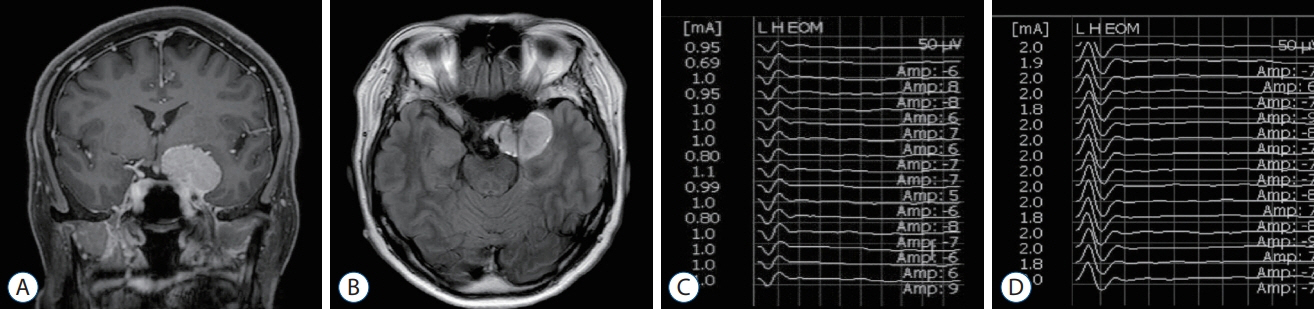
Fig. 2. T1-weighted coronal (A) and fluid attenuated inversion recovery axial (B) magnetic resonance images with enhancement showed left anterior clinoid process meningioma. The triggered electrooculographic responses of left eye in horizontal recording showed the biphasic positive waveforms with a latency of 0.5 ms and an amplitude of 50 μV when stimulating left oculomotor nerve at a stimulation intensity of 1 mA (C) and the biphasic negative waveforms with a latency of 0.5 ms and an amplitude of 75 μV when stimulating left abducens nerve at a stimulation intensity of 2 mA (D).
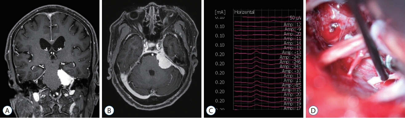
Fig. 3. T1-weighted coronal (A) and axial (B) magnetic resonance images with enhancement showed left petrous meningioma. The triggered electrooculographic responses of left eye in horizontal recording showed the monophasic negative waveforms with a latency of 3.2 ms and an amplitude of 27 μV (C) when stimulating left abducens nerve at a stimulation intensity of 0.2 mA (D).
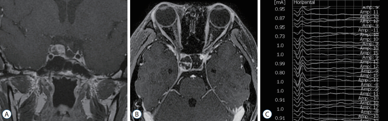
Fig. 4. T1-weighted coronal (A) and axial (B) magnetic resonance images with enhancement showed recurrent craniopharyngioma in right sellar area. The triggered electrooculographic responses of right eye in horizontal recording showed the biphasic positive waveforms with a latency of 0.6 ms and an amplitude of 61.1 μV (C) when stimulating right oculomotor nerve indirectly over the dural layer at a stimulation intensity of 1 mA.
Reference
-
References
1. Cornelius JF, Schipper J, Tortora A, Krause-Molle Z, Smuga M, Petridis AK, et al. Continuous and dynamic facial nerve mapping during surgery of cerebellopontine angle tumors: clinical pilot series. World Neurosurg. 119:e855–e863. 2018.
Article2. Fukaya C, Katayama Y, Kasai M, Kurihara J, Yamamoto T. Intraoperative electrooculographic monitoring of oculomotor nerve function during skull base surgery. Technical note. J Neurosurg. 91:157–159. 1999.
Article3. Hariharan P, Balzer JR, Anetakis K, Crammond DJ, Thirumala PD. Electrophysiology of extraocular cranial nerves: oculomotor, trochlear, and abducens nerve. J Clin Neurophysiol. 35:11–15. 2018.4. Kaspera W, Adamczyk P, Ślaska-Kaspera A, Ładziński P. Usefulness of intraoperative monitoring of oculomotor and abducens nerves during surgical treatment of the cavernous sinus meningiomas. Adv Med Sci. 60:25–30. 2015.
Article5. Kassam AB, Prevedello DM, Carrau RL, Snyderman CH, Thomas A, Gardner P, et al. Endoscopic endonasal skull base surgery: analysis of complications in the authors' initial 800 patients. J Neurosurg. 114:1544–1568. 2011.
Article6. Kawaguchi M, Ohnishi H, Sakamoto T, Shimizu K, Touho H, Monobe T, et al. Intraoperative electrophysiologic monitoring of cranial motor nerves in skull base surgery. Surg Neurol. 43:177–181. 1995.
Article7. Kawamata T, Ishii N, Amano K, Namioka T, Hori T, Okada Y. A novel simple real-time electrooculographic monitoring system during transsphenoidal surgeries to prevent postoperative extraocular motor nerve dysfunction. Neurosurg Rev. 36:371–376. 2013.
Article8. López JR. Neurophysiologic intraoperative monitoring of the oculomotor, trochlear, and abducens nerves. J Clin Neurophysiol. 28:543–550. 2011.
Article9. Schlake HP, Goldbrunner R, Siebert M, Behr R, Roosen K. Intra-operative electromyographic monitoring of extra-ocular motor nerves (Nn. III, VI) in skull base surgery. Acta Neurochir (Wien). 143:251–261. 2001.
Article10. Sekiya T, Hatayama T, Iwabuchi T, Maeda S. Intraoperative recordings of evoked extraocular muscle activities to monitor ocular motor nerve function. Neurosurgery. 32:227–235. discussion 235. 1993.
Article11. Sheshadri V, Bharadwaj S, Chandramouli BA. Intra-operative electrooculographic monitoring to prevent post-operative extraocular motor nerve dysfunction during skull base surgeries. Indian J Anaesth. 60:560–565. 2016.
Article12. Singh H, Vogel RW, Lober RM, Doan AT, Matsumoto CI, Kenning TJ, et al. Intraoperative neurophysiological monitoring for endoscopic endonasal approaches to the skull base: a technical guide. Scientifica (Cairo). 2016:1751245. 2016.
Article13. Thirumala PD, Mohanraj SK, Habeych M, Wichman K, Chang YF, Gardner P, et al. Value of free-run electromyographic monitoring of extraocular cranial nerves during expanded endonasal surgery (EES) of the skull base. J Neurol Surg Rep. 74:43–50. 2013.
Article14. Zhou Q, Zhang M, Jiang Y. Intraoperative oculomotor nerve monitoring predicts outcome following clipping of posterior communicating artery aneurysms. J Clin Neurosci. 19:706–711. 2012.
Article
- Full Text Links
-
- Actions
-
Cited
- CITED
-
- Close
- Share
-
- Similar articles
-
- A Case of Traumatic Bilateral Abducens Nerve Palsy Associated with Skull Base Fracture
- A Case of Isolated Unilateral Abducens Nerve Palsy Caused by Clival Metastasis from Rectal Cancer
- Central Skull Base Osteomyelitis Presenting with Bilateral Abducens Nerve Palsy
- Oculomotor and Abducens Nerve Palsy Complicated by Cavernous Sinus Thrombophlebitis Resulting from Acute Sinusitis
- Iatrogenic Skull Base Defect Accompanied by Brain Injury After Endoscopic Sinus Surgery: A Report of Two Cases
