Iatrogenic Skull Base Defect Accompanied by Brain Injury After Endoscopic Sinus Surgery: A Report of Two Cases
- Affiliations
-
- 1Department of Otorhinolaryngology-Head and Neck Surgery, Asan Medical Center, University of Ulsan College of Medicine, Seoul, Republic of Korea
- KMID: 2544621
- DOI: http://doi.org/10.18787/jr.2023.00021
Abstract
- Although iatrogenic skull base injuries after endoscopic sinus surgery (ESS) are rare (overall complication rate, 0.5%), they can be fatal or cause significant morbidity. Conventionally, skull base injuries were repaired using an external approach. However, in recent years, most skull base injuries after ESS have been repaired using an endoscopic transnasal approach due to its lower morbidity, lower risk of postoperative complications, and shorter hospital stay. We report two cases of iatrogenic skull base injury accompanied by brain injury following ESS and describe the skull base repair techniques employed for each case. In both cases, the skull base defects were successfully repaired using an endoscopic transnasal approach, although craniotomy was also performed in the first case to remove bone fragments from the right frontal base and lateral ventricle. Both patients recovered without residual neurologic deficits.
Figure
-

Fig. 1. Preoperative computed tomography findings. Intracranial pneumocephalus (A). Subdural hemorrhage in the right frontal base (arrowhead) (A, B). Multifocal bone fragments in the right frontal base and lateral ventricle (arrow) (B, C). A 1×2.5 cm right anterior skull base defect (circle) (A, C).
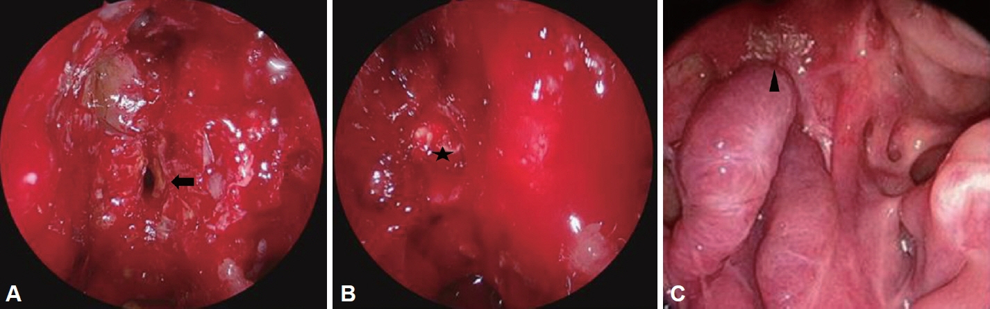
Fig. 2. Endoscopic intranasal images. Intraoperative findings (A, B): right anterior ethmoid roof defect (arrow). Pericranial flap reconstruction (★). Endoscopic view of well-healed reconstruction site at a 23-month follow-up (arrowhead) (C).
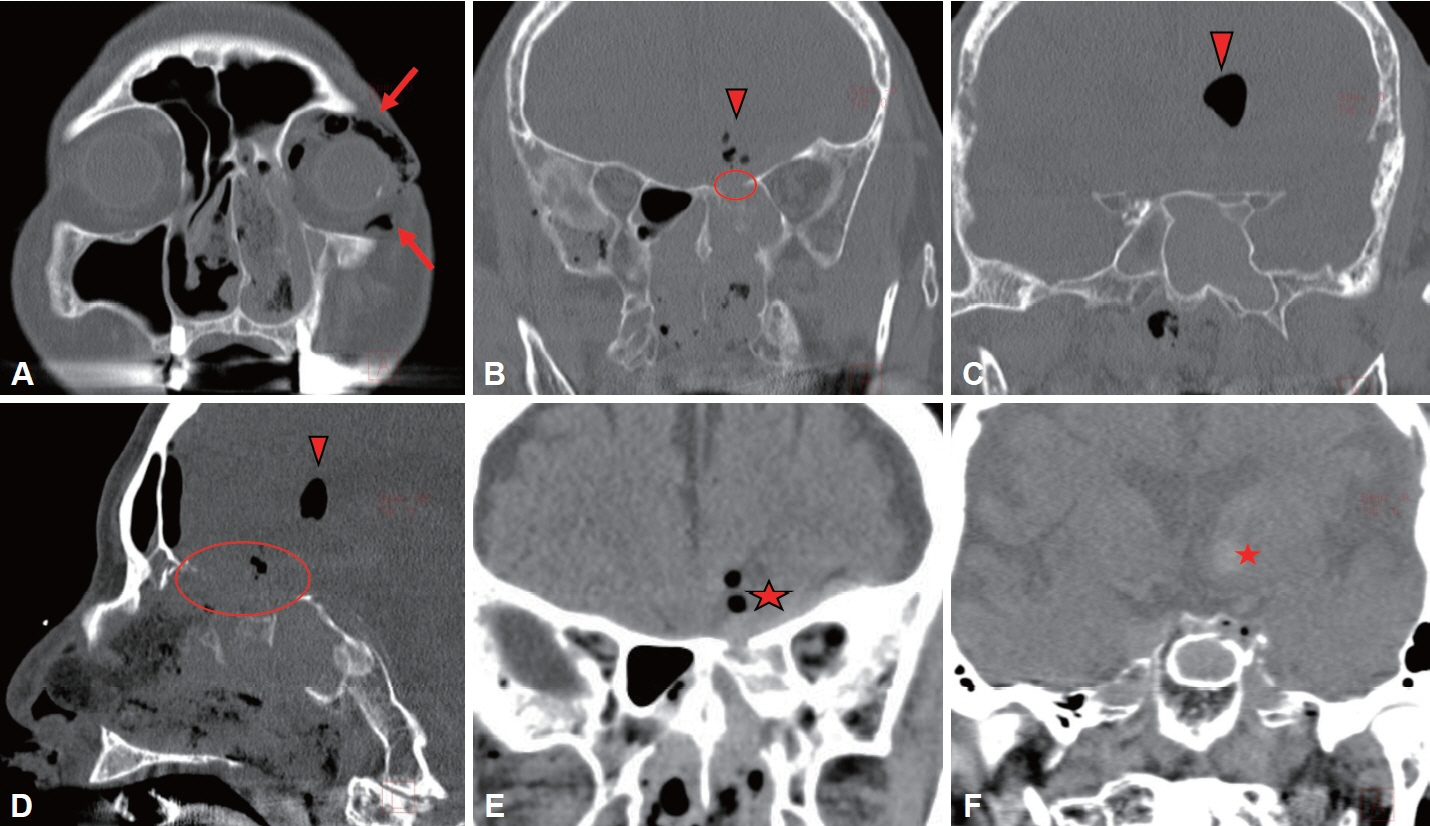
Fig. 3. Preoperative computed tomography findings. Intraorbital emphysema in the left orbit (arrows) (A). Multifocal intracranial pneumocephalus (arrowhead) (B-D). Intracranial hemorrhage in the left inferomedial frontal lobe and basal ganglia (★) (E, F). A 4×1.5 cm right anterior skull base defect (circle) (B, D).

Fig. 4. Endoscopic intranasal images. Intraoperative findings (A, B): left anterior ethmoid roof defect (arrow). Nasoseptal flap reconstruction (★). Endoscopic view of the well-healed reconstruction site at a 9-month follow-up (arrowhead) (C).
Reference
-
References
1. Stevens WW, Schleimer RP, Kern RC. Chronic rhinosinusitis with nasal polyps. J Allergy Clin Immunol Pract. 2016; 4(4):565–72.2. Kubik M, Lee S, Snyderman C, Wang E. Neurologic sequelae associated with delayed identification of iatrogenic skull base injury during endoscopic sinus surgery (ESS). Rhinology. 2017; 55(1):53–8.3. Abdullah B, Chew SC, Aziz ME, Shukri NM, Husain S, Joshua SW, et al. A new radiological classification for the risk assessment of anterior skull base injury in endoscopic sinus surgery. Sci Rep. 2020; 10(1):4600.4. Ibrahim AA, Okasha M, Elwany S. Endoscopic endonasal multilayer repair of traumatic CSF rhinorrhea. Eur Arch Otorhinolaryngol. 2016; 273(4):921–6.5. Bly RA, Morton RP, Kim LJ, Moe KS. Tension pneumocephalus after endoscopic sinus surgery: a technical report of multiportal endoscopic skull base repair. Otolaryngol Head Neck Surg. 2014; 151(6):1081–3.6. Ramakrishnan VR, Kingdom TT, Nayak JV, Hwang PH, Orlandi RR. Nationwide incidence of major complications in endoscopic sinus surgery. Int Forum Allergy Rhinol. 2012; 2(1):34–9.7. Siedek V, Pilzweger E, Betz C, Berghaus A, Leunig A. Complications in endonasal sinus surgery: a 5-year retrospective study of 2,596 patients. Eur Arch Otorhinolaryngol. 2013; 270(1):141–8.8. Suzuki S, Yasunaga H, Matsui H, Fushimi K, Kondo K, Yamasoba T. Complication rates after functional endoscopic sinus surgery: analysis of 50,734 Japanese patients. Laryngoscope. 2015; 125(8):1785–91.9. Fadda GL, Petrelli A, Martino F, Succo G, Castelnuovo P, Bignami M, et al. Anatomic variations of ethmoid roof and risk of skull base injury in endoscopic sinus surgery: statistical correlations. Am J Rhinol Allergy. 2021; 35(6):871–8.10. Keros P. [On the practical value of differences in the level of the lamina cribrosa of the ethmoid]. Z Laryngol Rhinol Otol. 1962; 41:809–13. German.11. Gera R, Mozzanica F, Karligkiotis A, Preti A, Bandi F, Gallo S, et al. Lateral lamella of the cribriform plate, a keystone landmark: proposal for a novel classification system. Rhinology. 2018; 56(1):65–72.12. Ramakrishnan VR, Orlandi RR, Citardi MJ, Smith TL, Fried MP, Kingdom TT. The use of image-guided surgery in endoscopic sinus surgery: an evidence-based review with recommendations. Int Forum Allergy Rhinol. 2013; 3(3):236–41.
- Full Text Links
-
- Actions
-
Cited
- CITED
-
- Close
- Share
-
- Similar articles
-
- A Case of Endoscopic Reconstruction of Skull Base Defect Combined with Meningoencephalocele in the Sphenoid Sinus
- Endoscopic Repair With Contralateral Septal Flap and Fascia Lata for Iatrogenic Skull Base Defect
- Ethmoid Sinus Mucocele Penetrating the Anterior Skull Base: A Case Report
- Endoscopic Transethmoidal Drainage of a Brain Abscess
- Endoscopic Reconstruction of a Total Sella Defect with Nasoseptalflap
