Endoscopic Ultrasound-Guided Vascular Therapy for Portoduodenal Fistula
- Affiliations
-
- 1Department of Internal Medicine, Rajavithi Hospital, Bangkok, Thailand
- 2Department of Internal Medicine, Bangkok Hospital, Bangkok, Thailand
- 3Department of Surgery, Rajavithi Hospital, Bangkok, Thailand
- KMID: 2511237
- DOI: http://doi.org/10.5946/ce.2019.167
Abstract
- Portoenteric fistula is a rare cause of massive upper gastrointestinal bleeding. Most cases can be treated with radiointervention or surgery, but portoenteric fistula is associated with a high mortality. We reported a case of intermittent massive upper gastrointestinal bleeding in a 33-year-old man with cholangiocarcinoma who underwent surgical resection followed by chemoradiation. A portoduodenal fistula due to chronic duodenal ulceration was identified. The bleeding was successfully controlled by endoscopic ultrasound-guided coil placement through the duodenal bulb using the anchoring technique. Follow-up endoscopy and computed tomography scan showed multiple coil placements between a part of the portal vein and the duodenal bulb without any evidence of portal vein thrombosis. There were no complications, and bleeding did not recur during the 8-month follow-up period.
Figure
-

Fig. 1. Endoscopic view showing active bleeding from an ulcer at the duodenal bulb.
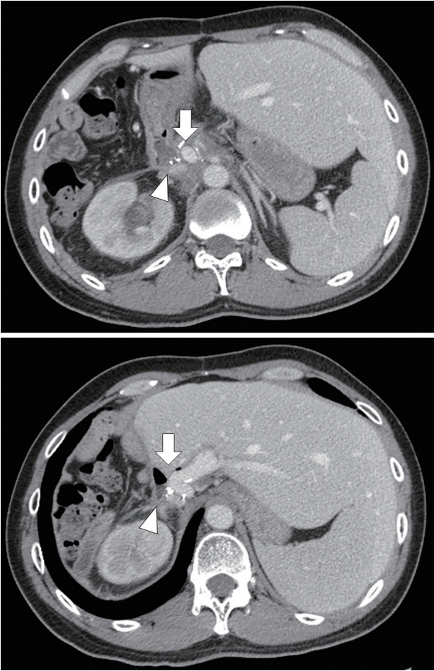
Fig. 2. Computed tomography scan showing portal vein (arrow) attached to about the duodenal bulb (arrowhead).
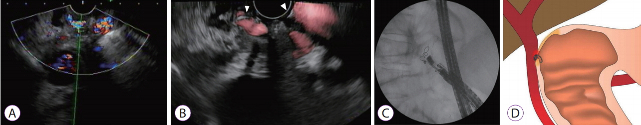
Fig. 3. Endoscopic ultrasound (EUS) images showing the portal vein attached to the base of the ulcer (A). EUS-guided coil embolization was performed (B, coil marked with arrowhead). Fluoroscopic image shows the coil placement without migration (C). The fine aspiration needle was withdrawn during coil deployment, leaving the distal part of the coil anchored against the duodenal wall (D).

Fig. 4. Follow-up endoscopy showing friable ulcer with a visible coil on the ulcer base.
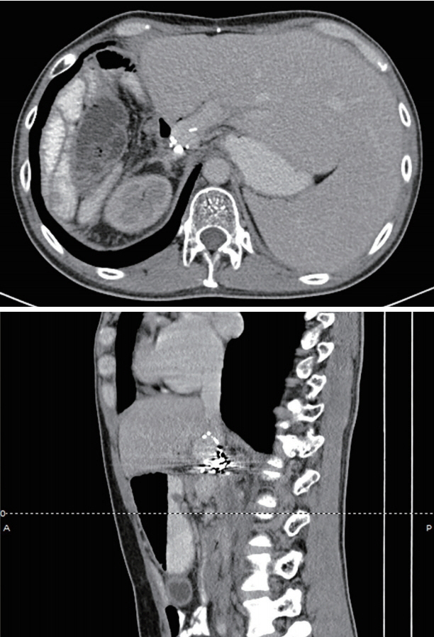
Fig. 5. Follow-up computed tomography scan showing coil embolization at the portoenteric fistula.
Reference
-
1. Lemos DW, Raffetto JD, Moore TC, Menzoian JO. Primary aortoduodenal fistula: a case report and review of the literature. J Vasc Surg. 2003; 37:686–689.
Article2. Fujiki M, Ramirez JR, Aucejo FN. Duodenoportal fistula resulting from peptic ulcer after extended right hepatectomy for cholangiocarcinoma. Am Surg. 2012; 78:E154–E155.
Article3. Kinoshita H, Takifuji K, Nakatani Y, Tani M, Uchiyama K, Yamaue H. Duodenoportal fistula caused by peptic ulcer after extended right hepatectomy for hilar cholangiocarcinoma. World J Surg Oncol. 2006; 4:84.
Article4. Povoski S, Shamma’a J. Fistula involving portal vein and duodenum at the site of a duodenal ulcer in a patient after previous extrahepatic bile duct resection and brachytherapy. Dig Surg. 2003; 20:53–55.
Article5. Masuda T, Yano F, Aoki H, Mitsumori N, Omura N, Yanaga K. A case of a portoenteric fistula due to a duodenal ulcer. Journal of Abdominal Emergency Medicine. 2013; 33:1165–1168.6. Soares MA, Wanless IR, Ambus U, Cameron R. Fistula between duodenum and portal vein caused by peptic ulcer disease and complicated by hemorrhage and portal vein thrombosis. Am J Gastroenterol. 1996; 91:1462–1463.7. Burke CT, Park J. Portal vein pseudoaneurysm with portoenteric fistula: an unusual cause for massive gastrointestinal hemorrhage. Semin Intervent Radiol. 2007; 24:341–345.
Article8. Baron TH, Song LM, Ross A, Tokar JL, Irani S, Kozarek RA. Use of an over-the-scope clipping device: multicenter retrospective results of the first U.S. experience (with videos). Gastrointest Endosc. 2012; 76:202–208.
Article9. Strand DS, Kim D, Peura DA. 25 years of proton pump inhibitors: a comprehensive review. Gut Liver. 2017; 11:27–37.
Article10. Koebbe CJ, Veznedaroglu E, Jabbour P, Rosenwasser RH. Endovascular management of intracranial aneurysms: current experience and future advances. Neurosurgery. 2006; 59:S93–S102. discussion S3-S13.
Article11. Fujii-Lau LL, Law R, Wong Kee Song LM, Levy MJ. Novel techniques for gastric variceal obliteration. Dig Endosc. 2015; 27:189–196.
Article12. Bapaye A, Dubale N, Mahadik M, Bharadwaj T. EUS guided coil embolization of giant gastric varices and modified technique to prevent distant embolization. Gastrointest Endosc. 2017; 85(5 Suppl):AB139.
- Full Text Links
-
- Actions
-
Cited
- CITED
-
- Close
- Share
-
- Similar articles
-
- Endoscopic Ultrasound-Guided Vascular Procedures: A Review
- Endoscopic ultrasound-guided vascular interventions: An overview of current and emerging techniques
- Endoscopic ultrasound-guided vascular intervention for portal hypertension
- Recent development of endoscopic ultrasound-guided biliary drainage
- Present status and perspectives of endosonography 2017 in gastroenterology
