Anat Cell Biol.
2020 Dec;53(4):411-416. 10.5115/acb.20.038.
Sternalis muscle in jordanian population: a prevalence study and level of physicians’ awareness
- Affiliations
-
- 1Department of Medical Laboratory Sciences, Faculty of Allied Medical Sciences, Al-Ahliyya Amman University, Amman, Jordan
- 2Department of Clinical Sciences, Faculty of Medicine, Yarmouk University, Irbid, Jordan
- 3King Abdullah University Hospital (KAUH), Al-Ramtha, Jordan
- KMID: 2509686
- DOI: http://doi.org/10.5115/acb.20.038
Abstract
- Sternalis muscle (SM) is an anatomical variant that lies parallel to the sternum. It is present in (8%) of human population. Awareness about its presence during thoracic imaging is important, since it might be misdiagnosed as a tumor. This study is the first that discusses the prevalence of SM in the Jordanian population and document the level of awareness about SM among intern doctors and surgery and radiology residents. Our aims are to know the prevalence of SM in the Jordanian population, using thoracic multi-detector computerized tomography (CT) images, and to assess the awareness about SM among a sample of intern and resident Jordanian physicians. Random anonymous axial thoracic multi-detector CT images of 1,709 (801 females and 908 males) Jordanian patients, were examined for the presence or absence of unilateral and/ or bilateral SM. A questionnaire aiming to identify SM was distributed among 175 intern doctors, 26 surgery resident and 28 radiology resident doctors, their answers were summarized. The prevalence of SM among Jordanians is 5.9%. The prevalence of unilateral SM is 2.1% on the right side of the thorax and 1.9% on the left side, bilateral prevalence was 1.8%. While 35.7% of the radiology residents could identify SM using CT and/or anatomy images, only 3.9% of surgery residents and none of the intern doctors could. We concluded that SM is present in the Jordanian population, with a prevalence of 5.9% which falls within the global average. Intern doctors and surgery and radiology residents are almost unaware and unfamiliar about SM.
Keyword
Figure
-
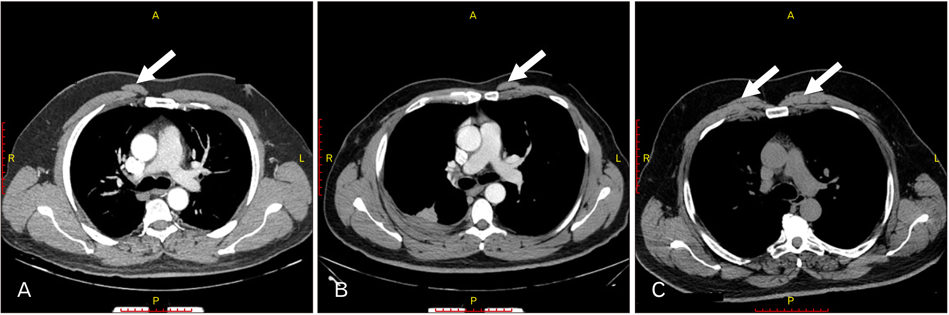
Fig. 1 Multi-detector computerized tomography images showing sternalis muscle (white arrows) on (A) the right of thorax, (B) the left side of thorax and (C) both sides of the thorax. A, anterior; L, left; P, posterior; R, right.
Reference
-
References
1. Pinhal-Enfield G, Varricchio P, DeFouw D, Vasan NS. 2011; Sternalis muscle: importance of its awareness in chest imaging and clinical significance. Int J Anat Var. 4:106–8.2. Yap SE. 1921; Musculus sternalis in Filipinos. Anat Rec. 21:353–72. DOI: 10.1002/ar.1090210402.
Article3. Taniguchi T, Tochihara J. 1932; Studies of the sternalis muscle in Japanese (No. 2), with special references to fetus and anencephalon of Japanese. Acta Anat Jpn. 7:1232–49.4. Barlow RN. 1935; The sternalis muscle in American whites and Negroes. Anat Rec. 61:413–26. DOI: 10.1002/ar.1090610405.
Article5. Kacker GN. 1960; Sternalis muscle in UP Indian subjects. J Anat Soc India. 9:101–3.6. Ge Z, Tong Y, Zhu S, Fang X, Zhuo L, Gong X. 2014; Prevalence and variance of the sternalis muscle: a study in the Chinese population using multi-detector CT. Surg Radiol Anat. 36:219–24. DOI: 10.1007/s00276-013-1175-4. PMID: 23912561.
Article7. Shoumin YU. 1954; Observations on the musculus sternalis in Chinese. Acta Anat Sin. (2):8. Kim WS, Kim SI, Han SR. 2000; Three cases of sternalis muscle in Korean. Korean J Phys Anthropol. 13:337–44. DOI: 10.11637/kjpa.2000.13.4.337.
Article9. Jelev L, Georgiev G, Surchev L. 2001; The sternalis muscle in the Bulgarian population: classification of sternales. J Anat. 199(Pt 3):359–63. DOI: 10.1046/j.1469-7580.2001.19930359.x. PMID: 11554516. PMCID: PMC1468341.
Article10. Chaijaroonkhanarak W, Amarttayakong P, Pannangron W, Umka J, Namking M, Chaisiwamongkol K, Kondo H, Prachaney P. 2013; Incidence of the sternalis muscle in Northeastern Thais. Srinagarind Med J. 28:62–5. DOI: 10.1007/s12565-013-0200-3. PMID: 23990382.11. Kirirat P, Thaweethamsewee P, Subhadhirasakul P, Puriwathanakul K, Tanomkiat W. 2005; The sternalis muscle in the Thai population. J Health Sci Med Res. 23:255–9.12. Plakornkul V, Viravud Y. 2012; Sternalis muscle: anatomical variations in Thais. Siriraj Med J. 64(suppl):19–21.13. Molina CR, Pinochet JA, Heras AA, Taunton MJ, Letelier RG, Letelier RF. 2017; Prevalence of the sternalis muscle in Chilean population: a computed tomography study. Ital J Anat Embryol. 122:173–8.14. Bradley FM, Hoover HC Jr, Hulka CA, Whitman GJ, McCarthy KA, Hall DA, Moore R, Kopans DB. 1996; The sternalis muscle: an unusual normal finding seen on mammography. AJR Am J Roentgenol. 166:33–6. DOI: 10.2214/ajr.166.1.8571900. PMID: 8571900.
Article15. Nuthakki S, Gross M, Fessell D. 2007; Sonography and helical computed tomography of the sternalis muscle. J Ultrasound Med. 26:247–50. DOI: 10.7863/jum.2007.26.2.247. PMID: 17255189.
Article16. Kobayashi S, Tomizawa H, Tomizawa Y, Manaka Y. 2013; Incidental finding of the sternalis muscle on chest multidetector-row computed tomography (MDCT): the diagnostic value of additional postprocessed MDCT images for an uncommon muscular variant. Intern Med. 52:1137–9. DOI: 10.2169/internalmedicine.52.9392. PMID: 23676605.
Article17. Gruber L, Martinoli C, Tagliafico AS, Gruber J, Klauser AS. 2016; A rare case of a symptomatic sternalis muscle: ultrasonograpy and MRI correlation. Ultrasound Int Open. 2:E140–1. DOI: 10.1055/s-0042-113607. PMID: 27896335. PMCID: PMC5120977.
Article18. Georgiev GP, Jelev L, Ovtscharoff VA. 2009; On the clinical significance of the sternalis muscle. Folia Med (Plovdiv). 51:53–6. PMID: 19957564.19. Saxena AK, Alalayet YF. Saxena AK, editor. 2017. Surgical anatomy of the chest wall. Chest wall deformities. Springer;Berlin: p. 37–53. DOI: 10.1007/978-3-662-53088-7_3. PMCID: PMC5438414.
Article20. Arráez-Aybar LA, Sobrado-Perez J, Merida-Velasco JR. 2003; Left musculus sternalis. Clin Anat. 16:350–4. DOI: 10.1002/ca.10120. PMID: 12794922.
Article21. Bailey PM, Tzarnas CD. 1999; The sternalis muscle: a normal finding encountered during breast surgery. Plast Reconstr Surg. 103:1189–90. DOI: 10.1097/00006534-199904010-00013. PMID: 10088505.
Article22. Jeng H, Su SJ. 1998; The sternalis muscle: an uncommon anatomical variant among Taiwanese. J Anat. 193(Pt 2):287–8. DOI: 10.1046/j.1469-7580.1998.19320287.x. PMID: 9827644. PMCID: PMC1467848.
Article23. Snosek M, Tubbs RS, Loukas M. 2014; Sternalis muscle, what every anatomist and clinician should know. Clin Anat. 27:866–84. DOI: 10.1002/ca.22361. PMID: 24431029.
Article24. Duque-Parra JE, Barco-Ríos J, Vélez-García JF. 2019; Incidence of sternalis muscle in the Caldas population (Colombia): anatomical variations. Int J Morphol. 37:1342–6. DOI: 10.4067/S0717-95022019000401342.
Article25. Davimes JG, Bacci N, Mazengenya P. 2018; Evidence of the sternalis muscle in two South African cadavers. Surg Radiol Anat. 40:1313–7. DOI: 10.1007/s00276-018-2058-5. PMID: 29931531.
Article26. Puthuraj MP, Shanmugam S. 2016; Prevalence of rectus sternalis: a clinical enigma. Indian J Basic Appl Med Res. 5:5–10.27. Parmar ND, Gupta DS. 2016; A study of morphological variation of pectoral region muscle 'Rectus Sternalis' in south Gujarat region. Int J Anat Res. 4:2423–28. DOI: 10.16965/ijar.2016.226.
Article28. Hung L, Lucaciu OC, Wong JJ. 2012; Back to the debate: sternalis muscle. Int J Morphol. 30:330–6. DOI: 10.4067/S0717-95022012000100058.
Article29. Khan UD. 2008; Use of the rectus sternalis in augmentation mammoplasty: case report and literature search. Aesthetic Plast Surg. 32:21–4. DOI: 10.1007/s00266-007-9046-1. PMID: 17965818.
Article
- Full Text Links
-
- Actions
-
Cited
- CITED
-
- Close
- Share
-
- Similar articles
-
- Implication of Sternalis Muscle on Staged Breast Reconstruction with Implant
- Bilateral Sternalis Muscle in Korean
- A Bilateral Double Sternalis Muscle in a Korean Cadaver
- Sternalis Muscle Encountered during Immediate Breast Reconstruction: 2 Case Reports
- Bilateral Sternalis with Unusual Left-Sided Presentation: A Clinical Perspective
