Thyroid Metastasis from Breast and Lung Cancer in Patients with Underlying Hashimoto’s Thyroiditis
- Affiliations
-
- 1Departments of Internal Medicine, Seoul National University Hospital, Seoul, Korea
- 2Departments of Nuclear Medicine, Seoul National University Hospital, Seoul, Korea
- KMID: 2509205
- DOI: http://doi.org/10.11106/ijt.2020.13.2.175
Abstract
- Metastatic disease involving the thyroid gland is uncommon. Thyroid metastases has been previously described from several primary cancers of lung, breast, and kidney. Because of the lower incidence and ambiguous clinical significance, it is not easy to consider thyroid metastasis and decide the optimal time for performing diagnostic examination. Here, we reported two cases of metastatic diseases of thyroid in patients who had underlying Hashimoto’s thyroiditis: a 39-year-old woman who had thyroid metastasis of breast cancer with underlying Hashimoto’s thyroiditis, and a 44-year-old woman with metastatic lung cancer.
Keyword
Figure
-
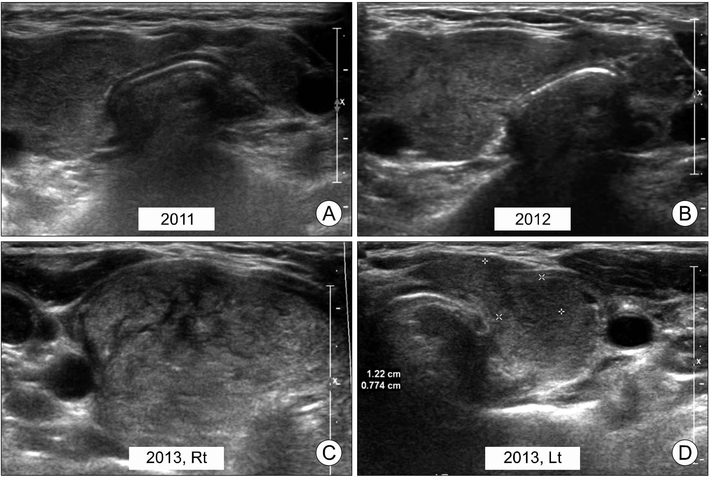
Fig. 1 Ultrasonographic findings of thyroid gland of Case 1 (2011-2013). Enlargement of right thyroid lobe with heterogeneous echogenicity at sonogram obtained in 2011 (A) and 2012 (B). In 2013, markedly enlarged right (C) and left (D) lobes of thyroid gland is observed with progressive changes of heterogeneous echogenicity.
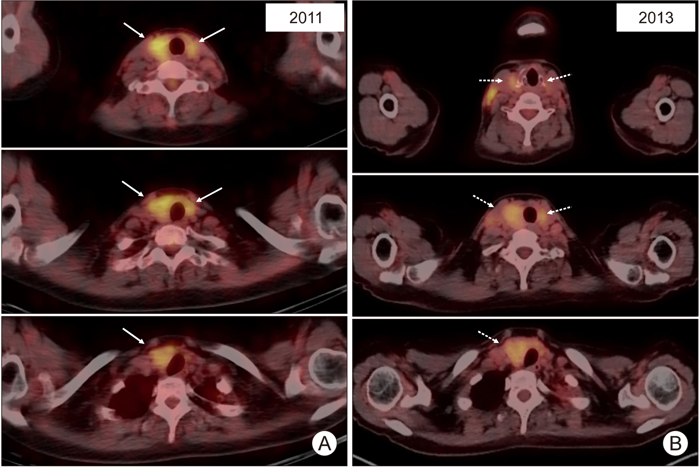
Fig. 2 Whole body [18F] FDG PET/CT images of Case 1. Diffuse [18F]FDG uptake is observed in both thyroid glands (arrows) in 2011 (A) and 2013 (B). The maximum standardized uptake value (SUVmax) of thyroid glands is increased from 3.7 in 2011 to 4.7 in 2013. Abnormal [18F] FDG uptakes is observed at both neck in 2013 (dotted arrows). The SUVmax of right and left neck lymph nodes are 6.0 and 4.0, respectively.
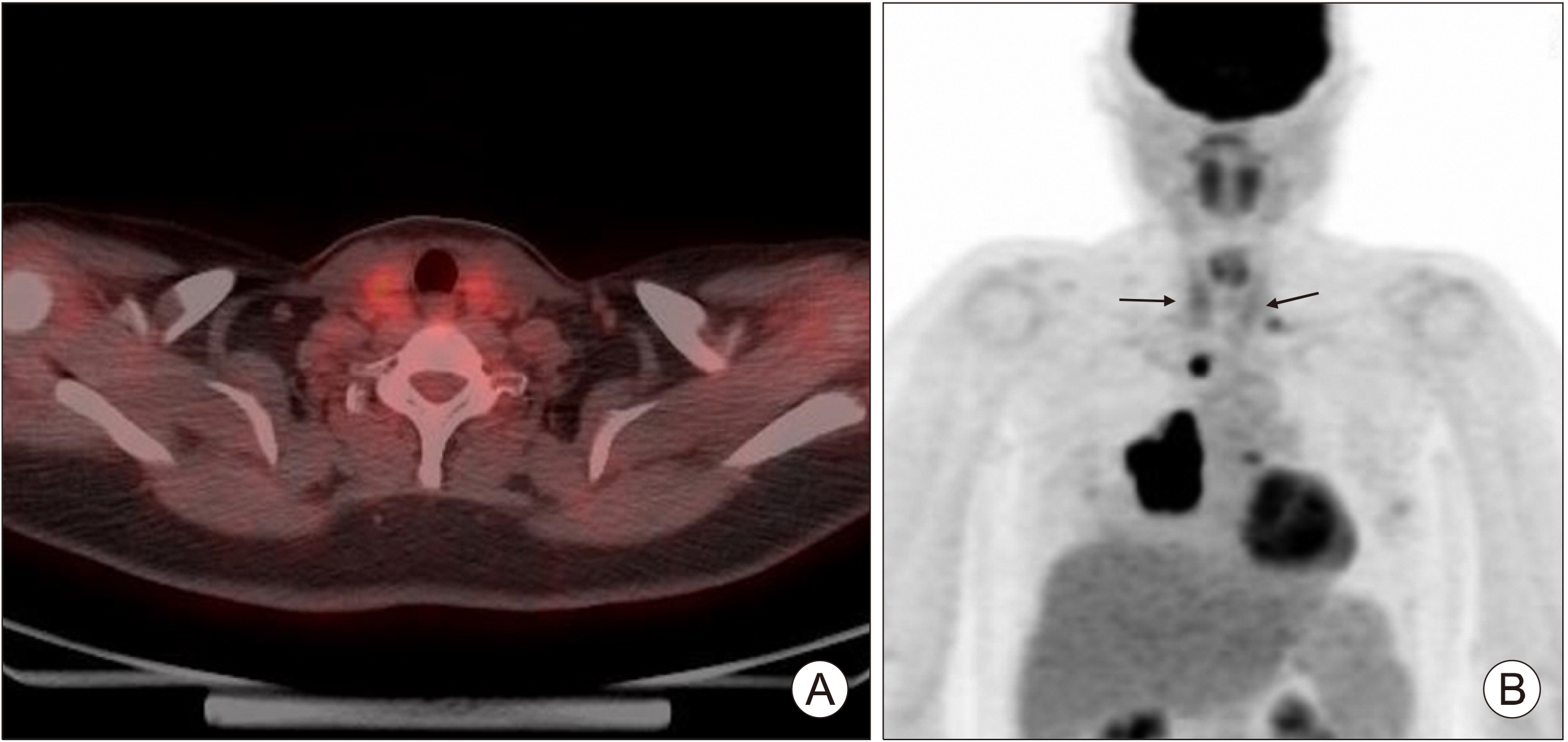
Fig. 3 Whole body [18F] FDG PET/CT images of Case 2. (A, B) Diffuse [18F]FDG uptake is observed in both thyroid glands on [18F]FDG PET scan as an initial evaluation for lung cancer (arrows). A lung cancer lesion and mediastinal lymph node metastasis shows intense [18F]FDG uptake.
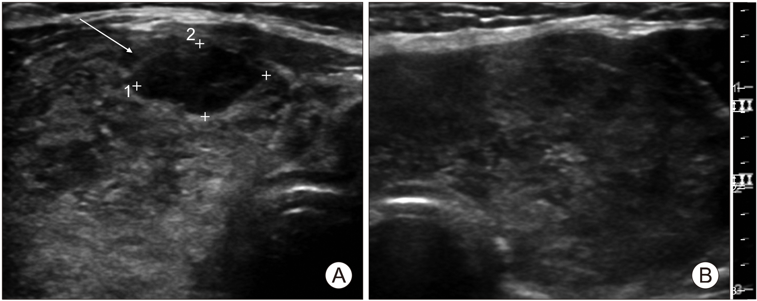
Fig. 4 Ultrasonographic findings of thyroid gland of Case 2. Enlargement of right (A) and left (B) thyroid lobes with heterogeneous echogenicity. Focal hypoechoic lesion is observed in right upper lobe of thyroid gland (A, arrow).
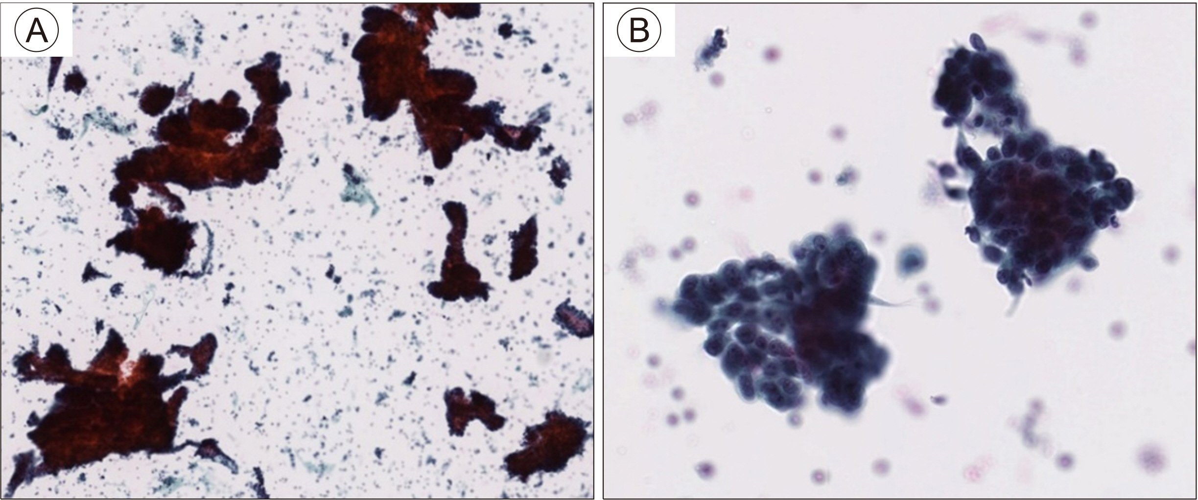
Fig. 5 Cytologic examination of focal thyroid lesion in Case 2. Increased cellularity (A, ×100) and atypical cells with high N/C ratio, pleomorphic nuclei and glandular pattern (B, ×400) are shown.
Reference
-
References
1. Wood K, Vini L, Harmer C. 2004; Metastases to the thyroid gland: the Royal Marsden experience. Eur J Surg Oncol. 30(6):583–8. DOI: 10.1016/j.ejso.2004.03.012. PMID: 15256229.
Article2. Nakhjavani MK, Gharib H, Goellner JR, van Heerden JA. 1997; Metastasis to the thyroid gland. A report of 43 cases. Cancer. 79(3):574–8. DOI: 10.1002/(SICI)1097-0142(19970201)79:3<574::AID-CNCR21>3.0.CO;2-#. PMID: 9028370.3. Barczynski M, Jamski J, Cichon S, Barczynski M, Sulowicz W. 2000; Diagnosis, treatment and prognosis in cases of renal clear cell carcinoma metastases into the thyroid gland. Przegl Lek. 57(3):157–9. PMID: 10909286.4. Chung AY, Tran TB, Brumund KT, Weisman RA, Bouvet M. 2012; Metastases to the thyroid: a review of the literature from the last decade. Thyroid. 22(3):258–68. DOI: 10.1089/thy.2010.0154. PMID: 22313412.
Article5. Lam KY, Lo CY. 1998; Metastatic tumors of the thyroid gland: a study of 79 cases in Chinese patients. Arch Pathol Lab Med. 122(1):37–41. PMID: 9448014.6. Liu Y. 2009; Clinical significance of thyroid uptake on F18-fluorodeoxyglucose positron emission tomography. Ann Nucl Med. 23(1):17–23. DOI: 10.1007/s12149-008-0198-0. PMID: 19205834.
Article7. Ho TY, Liou MJ, Lin KJ, Yen TC. 2011; Prevalence and significance of thyroid uptake detected by 18F-FDG PET. Endocrine. 40(2):297–302. DOI: 10.1007/s12020-011-9470-5. PMID: 21505891.8. McLeod DS, Cooper DS. 2012; The incidence and prevalence of thyroid autoimmunity. Endocrine. 42(2):252–65. DOI: 10.1007/s12020-012-9703-2. PMID: 22644837.
Article9. Vanderpump MP. 2011; The epidemiology of thyroid disease. Br Med Bull. 99:39–51. DOI: 10.1093/bmb/ldr030. PMID: 21893493.
Article10. Caturegli P, De Remigis A, Rose NR. 2014; Hashimoto thyroiditis: clinical and diagnostic criteria. Autoimmun Rev. 13(4-5):391–7. DOI: 10.1016/j.autrev.2014.01.007. PMID: 24434360.
Article11. Czepczynski R. 2012; Nuclear medicine in the diagnosis of benign thyroid diseases. Nucl Med Rev Cent East Eur. 15(2):113–9. PMID: 22936504.12. Bertagna F, Treglia G, Piccardo A, Giubbini R. 2012; Diagnostic and clinical significance of F-18-FDG-PET/CT thyroid incidentalomas. J Clin Endocrinol Metab. 97(11):3866–75. DOI: 10.1210/jc.2012-2390. PMID: 22904176.
Article13. Chen W, Parsons M, Torigian DA, Zhuang H, Alavi A. 2009; Evaluation of thyroid FDG uptake incidentally identified on FDG-PET/CT imaging. Nucl Med Commun. 30(3):240–4. DOI: 10.1097/MNM.0b013e328324b431. PMID: 19262287.
Article14. Kurata S, Ishibashi M, Hiromatsu Y, Kaida H, Miyake I, Uchida M, et al. 2007; Diffuse and diffuse-plus-focal uptake in the thyroid gland identified by using FDG-PET: prevalence of thyroid cancer and Hashimoto's thyroiditis. Ann Nucl Med. 21(6):325–30. DOI: 10.1007/s12149-007-0030-2. PMID: 17705011.
Article15. Bae JS, Chae BJ, Park WC, Kim JS, Kim SH, Jung SS, et al. 2009; Incidental thyroid lesions detected by FDG-PET/CT: prevalence and risk of thyroid cancer. World J Surg Oncol. 7:63. DOI: 10.1186/1477-7819-7-63. PMID: 19664272. PMCID: PMC2732624.
Article16. Bonabi S, Schmidt F, Broglie MA, Haile SR, Stoeckli SJ. 2012; Thyroid incidentalomas in FDG-PET/CT: prevalence and clinical impact. Eur Arch Otorhinolaryngol. 269(12):2555–60. DOI: 10.1007/s00405-012-1941-7. PMID: 22298251.
Article17. Are C, Hsu JF, Schoder H, Shah JP, Larson SM, Shaha AR. 2007; FDG-PET detected thyroid incidentalomas: need for further investigation? Ann Surg Oncol. 14(1):239–47. DOI: 10.1245/s10434-006-9181-y. PMID: 17024553.
Article
- Full Text Links
-
- Actions
-
Cited
- CITED
-
- Close
- Share
-
- Similar articles
-
- Ultrasonographic Findings of Papillary Thyroid Cancer with or without Hashimoto's Thyroiditis
- Thyroid MALT Lymphoma Associated with Thyroid Papillary Cancer
- A Case of Aggravation of Thyroid Goiter after Treatment with PD-1 Inhibitor for Breast Cancer in Patients with Underlying Hashimoto's Thyroiditis
- Progression of Hashimoto’s Thyroiditis to Graves’ Disease: a Report of Two Pediatric Cases
- A Case of Painful Hashimoto's Thyroiditis Successfully Treated with Total Thyroidectomy
