Multimodality Imaging of Large Left Ventricular Apical Pseudoaneurysm after Thoracic Surgery
- Affiliations
-
- 1Department of Cardiology, Kartal Kosuyolu Research and Education Hospital, İstanbul, Turkey
- KMID: 2505742
- DOI: http://doi.org/10.4070/kcj.2020.0074
Figure
-
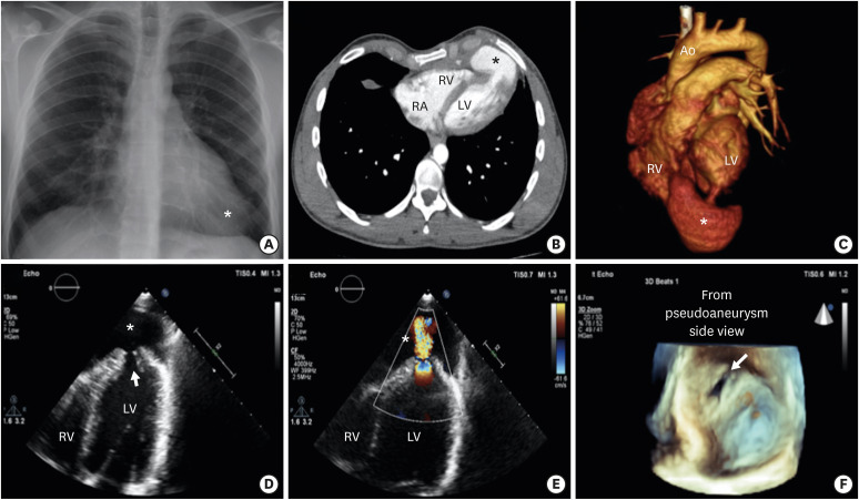
Figure 1 (A) Chest X-ray showed enlarged apical cardiac silhouette (asterisk). (B) CT angiography transvers view showing large apical pseudoaneurysm (asterisk). (C) Three-dimensional CT angiography showing pseudoaneurysm(asterisk) and connection to the LV cavity through a neck. (D) Transtorasic echocardiography apical 4 chamber view showing left ventricle apical pseudoaneurysm(asterisk) and entrance of pseudoaneurysm (arrow). (E) Transtorasic echocardiography apical 4 chamber colour doppler view showing pseudoaneurysm (asterisk) was connected to the LV cavity. (F) Three-dimensional echocardiography view from pseudoaneurysm side showing pseudoaneurysm entrance orifice of 2.5×1.5 cm.Ao = aortic; CT = computed tomography; LV = left ventricular; RV = right ventricular.
- Full Text Links
-
- Actions
-
Cited
- CITED
-
- Close
- Share
-
- Similar articles
-
- Polycythemia Vera Presenting as Left Ventricular Pseudoaneurysm: The Role of Multimodality Imaging
- Erratum: Polycythemia Vera Presenting as Left Ventricular Pseudoaneurysm: The Role of Multimodality Imaging
- Huge Multilobulated Left Ventricular Outflow Tract Pseudoaneurysm Presenting with Ventricular Tachycardia
- Left Ventricular Apical Pseudoaneurysm with Cardiac Tamponade
- Surgery for a Muscular Type Ventricular Septal Defect via Right Apical Ventriculotomy: A case report
