The effect of two phosphodiesterase inhibitors on bone healing in mandibular fractures (animal study in rats)
- Affiliations
-
- 1Department of Oral and Maxillofacial Surgery, Dental School, Islamic Azad University, Isfahan (Khorasgan) Branch, Isfahan, Iran
- 2Torabinejad Dental Research Center and Department of Oral and Maxillofacial Pathology, School of Dentistry, Isfahan University of Medical Sciences, Isfahan, Iran
- KMID: 2505445
- DOI: http://doi.org/10.5125/jkaoms.2020.46.4.258
Abstract
Objectives
Despite advances in maxillofacial surgery, impaired bone healing remains a concern for surgical teams. Many studies have evaluated the effects of sildenafil and pentoxifylline on bone healing. However, their effects on healing of bone fractures have not been well investigated. This study aimed to assess the effects of the phosphodiesterase inhibitors sildenafil and pentoxifylline on healing of mandibular fractures in rats.
Materials and Methods
A total of 60 rats were randomly divided into six groups of 10. Mandibular fracture was induced in all rats. After the surgical procedure, group C1 received saline, group S1 received 10 mg/kg sildenafil and group P1 received 50 mg/kg pentoxifylline. The rats were sacrificed after 1 week. Groups C4, S4, and P4 received pharmaceutical therapy as in groups C1, S1, and P1 but were sacrificed after 4 weeks. The samples then underwent histological analysis.
Results
The mean rate of bone healing of mandibular fractures in groups S1 and P1 was significantly higher than in group C1 at 1 week (p<0.001). The mean rate of bone healing of mandibular fractures in group P1 was higher than in group S1 at 1 week (p=0.04). The mean rate of bone healing of mandibular fractures in groups S4 (p=0.001) and P4 (p=0.004) was significantly higher than in group C4 at 4 weeks, but no significant difference was noted in the rate of healing between groups P4 and S4 (p=0.53).
Conclusion
Sildenafil and pentoxifylline can be used as adjuncts to enhance bone healing in rats.
Figure
-

Fig. 1 Surgical steps: ① shaving of surgical site, ② submandibular cutaneous incision, ③ exposure of masseter muscle, ④ dissection of the masseter muscle and exposure of the body of mandible, ⑤ fracture line, ⑥ placement of microplate, ⑦ fixation with screw, and ⑧ suturing.
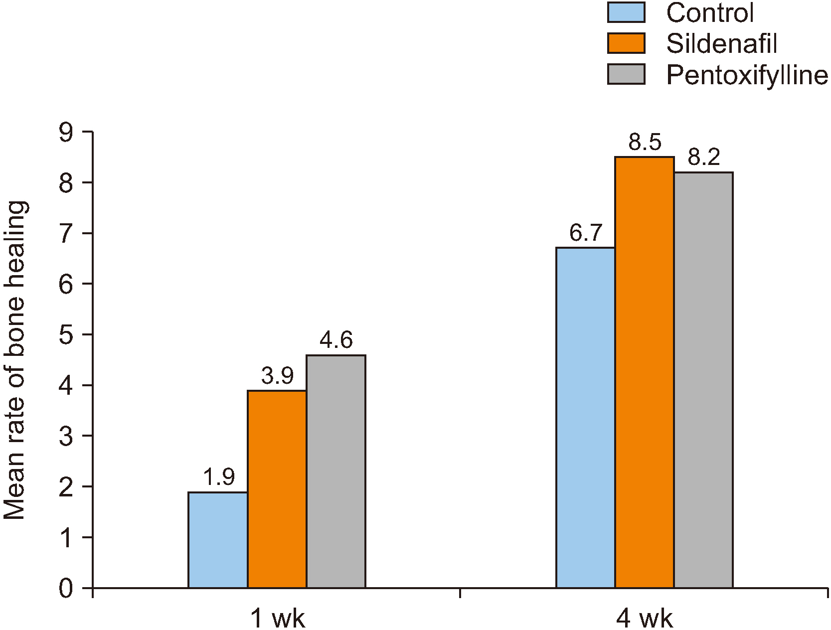
Fig. 2 Mean rate of bone healing in mandibular fractures in the study groups.

Fig. 3 Histological images of study groups (H&E staining, ×100). A. A specimen from group C1 with bone healing score 1 (healing with fibrous tissue). B. A specimen from group S1 with bone healing score 3 (healing with equal amounts of fibrous and cartilage tissue). C. A specimen from group P1 with bone healing score 5 (healing with mainly cartilage tissue and small amount of immature [woven] bone). D. A specimen from group C4 with bone healing score 6 (healing with equal amounts of cartilage tissue and immature bone). E. A specimen from group S4 with bone healing score 7 (healing with mainly immature bone and small amount of cartilage). F. A specimen from group P4 with bone healing score 9 (healing with immature bone and small amount of mature bone).

Fig. 4 Histological image of group P1 with bone healing score 5 (H&E staining, ×100). (arrows: cartilage tissue, x: cartilage calcification and conversion to bone, asterisks: lamellar bone, circle: immature bone, square: interface of the newly formed bone and older bone)
Cited by 1 articles
-
Comparative effects of systemic administration of levofloxacin and cephalexin on fracture healing in rats
Shayan Golestani, Arash Golestaneh, Atousa Aminzadeh Gohari
J Korean Assoc Oral Maxillofac Surg. 2022;48(2):94-100. doi: 10.5125/jkaoms.2022.48.2.94.
Reference
-
References
1. Glória JCR, Fernandes IA, Silveira EMD, Souza GM, Rocha RL, Galvão EL, et al. 2018; Comparison of bite force with locking plates versus non-locking plates in the treatment of mandibular fractures: a meta-analysis. Int Arch Otorhinolaryngol. 22:181–9. https://doi.org/10.1055/s-0037-1604056 . DOI: 10.1055/s-0037-1604056. PMID: 29619110. PMCID: PMC5882372.
Article2. Uçan MC, Koparal M, Ağaçayak S, Gunay A, Ozgoz M, Atilgan S, et al. 2013; Influence of caffeic acid phenethyl ester on bone healing in a rat model. J Int Med Res. 41:1648–54. https://doi.org/10.1177/0300060513490613 . DOI: 10.1177/0300060513490613. PMID: 24065455.
Article3. Fernández JR, Gallas M, Burguera M, Viaño JM. 2003; A three-dimensional numerical simulation of mandible fracture reduction with screwed miniplates. J Biomech. 36:329–37. https://doi.org/10.1016/s0021-9290(02)00416-5 . DOI: 10.1016/S0021-9290(02)00416-5. PMID: 12594981.
Article4. Durmuş K, Turgut NH, Doğan M, Tuncer E, Özer H, Altuntaş EE, et al. 2017; Histopathological evaluation of the effect of locally administered strontium on healing time in mandibular fractures: an experimental study. Adv Clin Exp Med. 26:1063–7. https://doi.org/10.17219/acem/65477 . DOI: 10.17219/acem/65477. PMID: 29211352.
Article5. Hausman MR, Schaffler MB, Majeska RJ. 2001; Prevention of fracture healing in rats by an inhibitor of angiogenesis. Bone. 29:560–4. https://doi.org/10.1016/s8756-3282(01)00608-1 . DOI: 10.1016/S8756-3282(01)00608-1. PMID: 11728927.
Article6. Dincel YM, Alagoz E, Arikan Y, Caglar AK, Dogru SC, Ortes F, et al. 2018; Biomechanical, histological, and radiological effects of different phosphodiesterase inhibitors on femoral fracture healing in rats. J Orthop Surg (Hong Kong). 26:2309499018777885. https://doi.org/10.1177/2309499018777885 . DOI: 10.1177/2309499018777885. PMID: 29848169.
Article7. Hankenson KD, Zimmerman G, Marcucio R. 2014; Biological perspectives of delayed fracture healing. Injury. 45(Suppl 2):S8–15. https://doi.org/10.1016/j.injury.2014.04.003 . DOI: 10.1016/j.injury.2014.04.003. PMID: 24857030. PMCID: PMC4406220.
Article8. Cook JJ, Summers NJ, Cook EA. 2015; Healing in the new millennium: bone stimulators: an overview of where we've been and where we may be heading. Clin Podiatr Med Surg. 32:45–59. https://doi.org/10.1016/j.cpm.2014.09.003 . DOI: 10.1016/j.cpm.2014.09.003. PMID: 25440417.
Article9. Chao EY, Inoue N. 2003; Biophysical stimulation of bone fracture repair, regeneration and remodelling. Eur Cell Mater. 6:72–84. discussion 84-5. https://doi.org/10.22203/ecm.v006a07 . DOI: 10.22203/eCM.v006a07. PMID: 14722904.
Article10. Massari L, Caruso G, Sollazzo V, Setti S. 2009; Pulsed electromagnetic fields and low intensity pulsed ultrasound in bone tissue. Clin Cases Miner Bone Metab. 6:149–54. PMID: 22461165. PMCID: PMC2781224.11. Perry AC, Prpa B, Rouse MS, Piper KE, Hanssen AD, Steckelberg JM, et al. 2003; Levofloxacin and trovafloxacin inhibition of experimental fracture-healing. Clin Orthop Relat Res. (414):95–100. https://doi.org/10.1097/01.blo.0000087322.60612.14 . DOI: 10.1097/01.blo.0000087322.60612.14. PMID: 12966282.
Article12. Zandi M, Dehghan A, Amini P, Rezaeian L, Doulati S. 2017; Evaluation of mandibular fracture healing in rats under zoledronate therapy: a histologic study. Injury. 48:2683–7. https://doi.org/10.1016/j.injury.2017.10.026 . DOI: 10.1016/j.injury.2017.10.026. PMID: 29042034.
Article13. Colnot C, Lu C, Hu D, Helms JA. 2004; Distinguishing the contributions of the perichondrium, cartilage, and vascular endothelium to skeletal development. Dev Biol. 269:55–69. https://doi.org/10.1016/j.ydbio.2004.01.011 . DOI: 10.1016/j.ydbio.2004.01.011. PMID: 15081357.
Article14. Lienau J, Schell H, Epari DR, Schütze N, Jakob F, Duda GN, et al. 2006; CYR61 (CCN1) protein expression during fracture healing in an ovine tibial model and its relation to the mechanical fixation stability. J Orthop Res. 24:254–62. https://doi.org/10.1002/jor.20035 . DOI: 10.1002/jor.20035. PMID: 16435358.
Article15. Keramaris NC, Calori GM, Nikolaou VS, Schemitsch EH, Giannoudis PV. 2008; Fracture vascularity and bone healing: a systematic review of the role of VEGF. Injury. 39(Suppl 2):S45–57. https://doi.org/10.1016/S0020-1383(08)70015-9 . DOI: 10.1016/S0020-1383(08)70015-9.
Article16. Diwan AD, Wang MX, Jang D, Zhu W, Murrell GA. 2000; Nitric oxide modulates fracture healing. J Bone Miner Res. 15:342–51. https://doi.org/10.1359/jbmr.2000.15.2.342 . DOI: 10.1359/jbmr.2000.15.2.342. PMID: 10703937.
Article17. Baldik Y, Talu U, Altinel L, Bilge H, Demiryont M, Aykac-Toker G. 2002; Bone healing regulated by nitric oxide: an experimental study in rats. Clin Orthop Relat Res. (404):343–52. https://doi.org/10.1097/00003086-200211000-00051 . DOI: 10.1097/00003086-200211000-00051. PMID: 12439279.
Article18. Derici H, Kamer E, Unalp HR, Diniz G, Bozdag AD, Tansug T, et al. 2010; Effect of sildenafil on wound healing: an experimental study. Langenbecks Arch Surg. 395:713–8. https://doi.org/10.1007/s00423-009-0471-2 . DOI: 10.1007/s00423-009-0471-2. PMID: 19224243.
Article19. Histing T, Marciniak K, Scheuer C, Garcia P, Holstein JH, Klein M, et al. 2011; Sildenafil accelerates fracture healing in mice. J Orthop Res. 29:867–73. https://doi.org/10.1002/jor.21324 . DOI: 10.1002/jor.21324. PMID: 21246617.
Article20. Vidavalur R, Penumathsa SV, Zhan L, Thirunavukkarasu M, Maulik N. 2006; Sildenafil induces angiogenic response in human coronary arteriolar endothelial cells through the expression of thioredoxin, hemeoxygenase and vascular endothelial growth factor. Vascul Pharmacol. 45:91–5. https://doi.org/10.1016/j.vph.2006.03.010 . DOI: 10.1016/j.vph.2006.03.010. PMID: 16716755.
Article21. Hart K, Baur D, Hodam J, Lesoon-Wood L, Parham M, Keith K, et al. 2006; Short- and long-term effects of sildenafil on skin flap survival in rats. Laryngoscope. 116:522–8. https://doi.org/10.1097/01.mlg.0000200792.67802.3b . DOI: 10.1097/01.mlg.0000200792.67802.3b. PMID: 16585853.
Article22. Das A, Xi L, Kukreja RC. 2005; Phosphodiesterase-5 inhibitor sildenafil preconditions adult cardiac myocytes against necrosis and apoptosis. Essential role of nitric oxide signaling. J Biol Chem. 280:12944–55. https://doi.org/10.1074/jbc.M404706200 . DOI: 10.1074/jbc.M404706200. PMID: 15668244.
Article23. Essayan DM. 2001; Cyclic nucleotide phosphodiesterases. J Allergy Clin Immunol. 108:671–80. https://doi.org/10.1067/mai.2001.119555 . DOI: 10.1067/mai.2001.119555. PMID: 11692087.
Article24. Delanian S, Porcher R, Rudant J, Lefaix JL. 2005; Kinetics of response to long-term treatment combining pentoxifylline and tocopherol in patients with superficial radiation-induced fibrosis. J Clin Oncol. 23:8570–9. https://doi.org/10.1200/JCO.2005.02.4729 . DOI: 10.1200/JCO.2005.02.4729. PMID: 16260695.
Article25. Ward A, Clissold SP. 1987; Effect of pentoxifylline administration on an experimental rat model of femur fracture healing with intramedullary fixation. Drugs. 34:50–97. https://doi.org/10.2165/00003495-198734010-00003 . DOI: 10.2165/00003495-198734010-00003. PMID: 3308412.
Article26. Bayat M, Amini A, Rezaie F, Bayat S. 2014; Patents of pentoxifylline administration on some diseases and chronic wounds. Recent Pat Regen Med. 4:137–43. https://doi.org/10.2174/2210296504666140813194744 . DOI: 10.2174/2210296504666140813194744.
Article27. Vashghani Farahani MM, Masteri Farahani R, Mostafavinia A, Abbasian MR, Pouriran R, Noruzian M, et al. 2015; Effect of pentoxifylline administration on an experimental rat model of femur fracture healing with intramedullary fixation. Iran Red Crescent Med J. 17:e29513. https://doi.org/10.5812/ircmj.29513 . DOI: 10.5812/ircmj.29513. PMID: 26756019. PMCID: PMC4707237.
Article28. Ghofrani HA, Osterloh IH, Grimminger F. 2006; Sildenafil: from angina to erectile dysfunction to pulmonary hypertension and beyond. Nat Rev Drug Discov. 5:689–702. https://doi.org/10.1038/nrd2030 . DOI: 10.1038/nrd2030. PMID: 16883306. PMCID: PMC7097805.
Article29. Koneru S, Varma Penumathsa S, Thirunavukkarasu M, Vidavalur R, Zhan L, Singal PK, et al. 2008; Sildenafil-mediated neovascularization and protection against myocardial ischaemia reperfusion injury in rats: role of VEGF/angiopoietin-1. J Cell Mol Med. 12:2651–64. https://doi.org/10.1111/j.1582-4934.2008.00319.x . DOI: 10.1111/j.1582-4934.2008.00319.x. PMID: 18373738. PMCID: PMC3828881.
Article30. Kinoshita T, Kobayashi S, Ebara S, Yoshimura Y, Horiuchi H, Tsutsumimoto T, et al. 2000; Phosphodiesterase inhibitors, pentoxifylline and rolipram, increase bone mass mainly by promoting bone formation in normal mice. Bone. 27:811–7. https://doi.org/10.1016/s8756-3282(00)00395-1 . DOI: 10.1016/S8756-3282(00)00395-1. PMID: 11113392.
Article31. Labib GS, Farid RM. 2015; Osteogenic effect of locally applied Pentoxyfilline gel: in vitro and in vivo evaluations. Drug Deliv. 22:1094–102. https://doi.org/10.3109/10717544.2014.884193 . DOI: 10.3109/10717544.2014.884193. PMID: 24555662.
Article32. Kahenasa N, Sung EC, Nabili V, Kelly J, Garrett N, Nishimura I. 2012; Resolution of pain and complete healing of mandibular osteoradionecrosis using pentoxifylline and tocopherol: a case report. Oral Surg Oral Med Oral Pathol Oral Radiol. 113:e18–23. https://doi.org/10.1016/j.oooo.2011.10.014 . DOI: 10.1016/j.oooo.2011.10.014. PMID: 22668439.
Article33. Delanian S, Depondt J, Lefaix JL. 2005; Major healing of refractory mandible osteoradionecrosis after treatment combining pentoxifylline and tocopherol: a phase II trial. Head Neck. 27:114–23. https://doi.org/10.1002/hed.20121 . DOI: 10.1002/hed.20121. PMID: 15641107.
Article34. Aydin K, Sahin V, Gürsu S, Mercan AS, Demir B, Yildirim T. 2011; Effect of pentoxifylline on fracture healing: an experimental study. Eklem Hastalik Cerrahisi. 22:160–5. PMID: 22085352.35. Clark JD, Gebhart GF, Gonder JC, Keeling ME, Kohn DF. 1997; The 1996 guide for the care and use of laboratory animals. ILAR J. 38:41–8. https://doi.org/10.1093/ilar.38.1.41 . DOI: 10.1093/ilar.38.1.41. PMID: 11528046.
Article36. Schlundt C, El Khassawna T, Serra A, Dienelt A, Wendler S, Schell H, et al. 2018; Macrophages in bone fracture healing: their essential role in endochondral ossification. Bone. 106:78–89. https://doi.org/10.1016/j.bone.2015.10.019 . DOI: 10.1016/j.bone.2015.10.019. PMID: 26529389.
Article37. Rotter N, Haisch A, Bücheler M. 2005; Cartilage and bone tissue engineering for reconstructive head and neck surgery. Eur Arch Otorhinolaryngol. 262:539–45. https://doi.org/10.1007/s00405-004-0866-1 . DOI: 10.1007/s00405-004-0866-1. PMID: 16091977.
Article38. Vega LG. 2011; Reoperative mandibular trauma: management of posttraumatic mandibular deformities. Oral Maxillofac Surg Clin North Am. 23:47–61. v-vi. https://doi.org/10.1016/j.coms.2010.12.003 . DOI: 10.1016/j.coms.2010.12.003. PMID: 21272766.
Article39. Irkorucu O, Taşcilar O, Cakmak GK, Karakaya K, Emre AU, Ucan BH, et al. 2008; The effect of sildenafil on an animal model for ischemic colitis. Dig Dis Sci. 53:1618–23. https://doi.org/10.1007/s10620-007-0033-9 . DOI: 10.1007/s10620-007-0033-9. PMID: 17932755.
Article40. Atalay Y, Bozkurt MF, Gonul Y, Cakmak O, Agacayak KS, Köse I, et al. 2015; The effects of amlodipine and platelet rich plasma on bone healing in rats. Drug Des Devel Ther. 9:1973–81. https://doi.org/10.2147/DDDT.S80778 . DOI: 10.2147/DDDT.S80778. PMID: 25897207. PMCID: PMC4396585.
Article41. Anitua E, Sánchez M, Orive G, Andía I. 2007; The potential impact of the preparation rich in growth factors (PRGF) in different medical fields. Biomaterials. 28:4551–60. https://doi.org/10.1016/j.biomaterials.2007.06.037 . DOI: 10.1016/j.biomaterials.2007.06.037. PMID: 17659771.
Article42. Yaman F, Atilgan S, Günes N, Agacayak S, Günay A, Ucan MC, et al. 2011; Phosphodiesterase-5 inhibitors may facilitate bone defect recovery. Eur Rev Med Pharmacol Sci. 15:1301–5. PMID: 22195363.43. Rajkumar DS, Faitelson AV, Gudyrev OS, Dubrovin GM, Pokrovski MV, Ivanov AV. 2013; Comparative evaluation of enalapril and losartan in pharmacological correction of experimental osteoporosis and fractures of its background. J Osteoporos. 2013:325693. https://doi.org/10.1155/2013/325693 . DOI: 10.1155/2013/325693. PMID: 23401845. PMCID: PMC3562670.
Article44. Corbett SA, Hukkanen M, Batten J, McCarthy ID, Polak JM, Hughes SP. 1999; Phosphodiesterase inhibitors stimulate osteoclast formation via TRANCE/RANKL expression in osteoblasts: possible involvement of ERK and p38 MAPK pathways. J Bone Joint Surg Br. 81:531–7. https://doi.org/10.1302/0301-620x.81b3.8852 . DOI: 10.1302/0301-620X.81B3.8852. PMID: 10872379.
Article45. Si W, Kang Q, Luu HH, Park JK, Luo Q, Song WX, et al. 2006; CCN1/Cyr61 is regulated by the canonical Wnt signal and plays an important role in Wnt3A-induced osteoblast differentiation of mesenchymal stem cells. Mol Cell Biol. 26:2955–64. https://doi.org/10.1128/MCB.26.8.2955-2964.2006 . DOI: 10.1128/MCB.26.8.2955-2964.2006. PMID: 16581771. PMCID: PMC1446962.
Article46. Takami M, Cho ES, Lee SY, Kamijo R, Yim M. 2005; Phosphodiesterase inhibitors stimulate osteoclast formation via TRANCE/RANKL expression in osteoblasts: possible involvement of ERK and p38 MAPK pathways. FEBS Lett. 579:832–8. https://doi.org/10.1016/j.febslet.2004.12.066 . DOI: 10.1016/j.febslet.2004.12.066. PMID: 15670856.
Article47. Horiuchi H, Saito N, Kinoshita T, Wakabayashi S, Tsutsumimoto T, Otsuru S, et al. 2004; Enhancement of recombinant human bone morphogenetic protein-2 (rhBMP-2)-induced new bone formation by concurrent treatment with parathyroid hormone and a phosphodiesterase inhibitor, pentoxifylline. J Bone Miner Metab. 22:329–34. https://doi.org/10.1007/s00774-003-0490-y . DOI: 10.1007/s00774-003-0490-y.
Article48. Horiuchi H, Saito N, Kinoshita T, Wakabayashi S, Tsutsumimoto T, Takaoka K. 2001; Enhancement of bone morphogenetic protein-2-induced new bone formation in mice by the phosphodiesterase inhibitor pentoxifylline. Bone. 28:290–4. https://doi.org/10.1016/s8756-3282(00)00450-6 . DOI: 10.1016/S8756-3282(00)00450-6.
Article49. Tsutsumimoto T, Wakabayashi S, Kinoshita T, Horiuchi H, Takaoka K. 2002; A phosphodiesterase inhibitor, pentoxifylline, enhances the bone morphogenetic protein-4 (BMP-4)-dependent differentiation of osteoprogenitor cells. Bone. 31:396–401. https://doi.org/10.1016/s8756-3282(02)00839-6 . DOI: 10.1016/S8756-3282(02)00839-6.
Article50. Gong Y, Xu CY, Wang JR, Hu XH, Hong D, Ji X, et al. 2014; Inhibition of phosphodiesterase 5 reduces bone mass by suppression of canonical Wnt signaling. Cell Death Dis. 5:e1544. https://doi.org/10.1038/cddis.2014.510 . DOI: 10.1038/cddis.2014.510. PMID: 25429621. PMCID: PMC4260761.
Article
- Full Text Links
-
- Actions
-
Cited
- CITED
-
- Close
- Share
-
- Similar articles
-
- Comparative effects of systemic administration of levofloxacin and cephalexin on fracture healing in rats
- The histomorphometric study on the healing process of a mandibular fracture in the streptozotocin-induced diabetic rats
- Effect of rhBMP-2 on the healing of bone defect in the low calcium diet rat
- Intermittent Parathyroid Hormone Treatment for Stimulation of Callus Formation on Distal Femoral Fracture in Elderly Patients: Case Report
- Radiologic and Histological Study of Healing Process on Malunion Rat Model after Zygomatic Arch Fracture
