Three-dimensional assessment of nasal changes after maxillary advancement with impaction using stereophotogrammetry
- Affiliations
-
- 1Department of Orthodontics, Faculty of Dentistry, Erciyes University, Kayseri, Turkey
- 2Department of Oral and Maxillofacial Surgery, Faculty of Dentistry, Erciyes University, Kayseri, Turkey
- KMID: 2504142
- DOI: http://doi.org/10.4041/kjod.2020.50.4.249
Abstract
Objective
To evaluate the changes in the nose in three dimensions after Le Fort I osteotomy in patients with skeletal Class III malocclusion.
Methods
The subjects were 40 adult patients (20 females and 20 males; mean age, 20.3 ± 3.0 years; range, 17.0 to 31.1 years) who underwent one-piece Le Fort I osteotomy with maxillary advancement and impaction treatment for maxillary hypoplasia. The mean maxillary advancement was 4.56 ± 1.34 mm, and the mean maxillary impaction was 2.03 ± 1.04 mm. Stereophotogrammetry was used to acquire three-dimensional images before and at least 6 months after surgery.
Results
Alare (Al) and alare curvature (Ac) points had moved vertically and anterolaterally postoperatively. A significant increase was observed in the nasal ala width and alar base width, and no changes were noted in the columellar length, nasolabial angle, and nasal area. There was a significant relationship between maxillary impaction and nasal ala width and horizontal and sagittal positions of the bilateral Al and Ac. The only relationship found was between maxillary advancement and postoperative sagittal location of the subnasale and pronasale.
Conclusions
Nasal soft tissues were highly affected by the vertical movement of the maxilla; however, the soft tissue responses were individual-dependent.
Figure
-
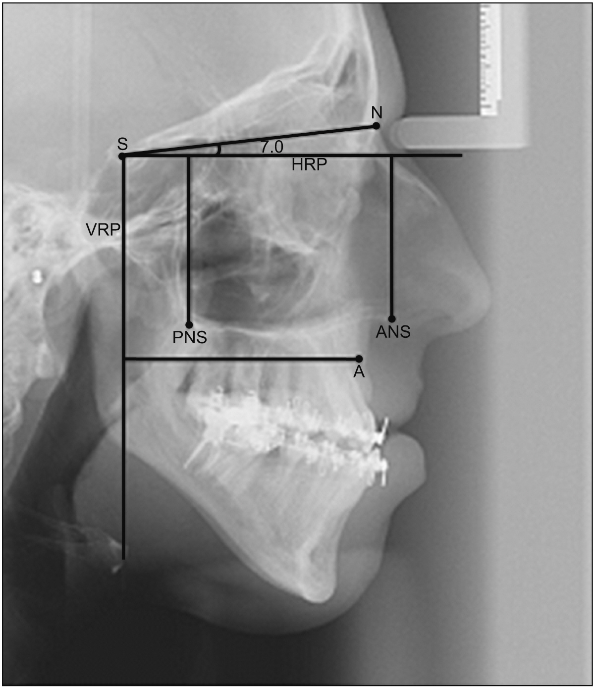
Figure 1 Evaluation of surgical quantities of hard tissues. N, Nasion; S, sella; A, A point; ANS, anterior nasal spina; PNS, posterior nasal spina; HRP, horizontal reference plane; VRP, vertical reference plane.
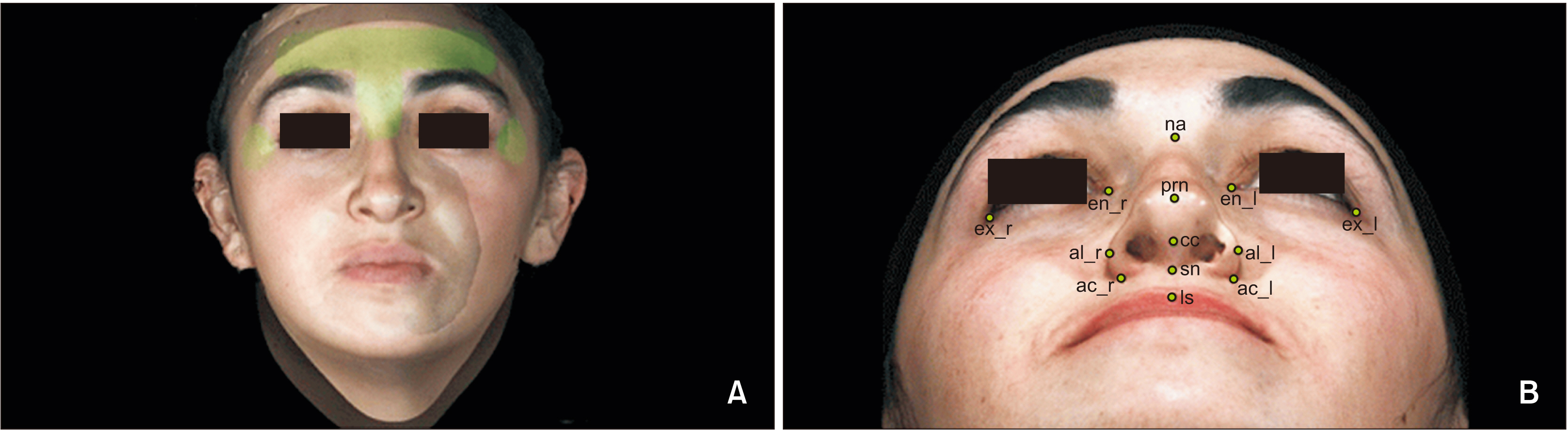
Figure 2 A, Reference markers used for superimposition: forehead area (the wide surface over the eyebrows), nasion area (the corresponding soft tissue point from the nasal root to the nasal hump), and exocanthion (the 3/4 lateral regions of the exocanthion). B, Soft tissue landmarks. na, Soft tissue nasion; en, endocanthion; ex, exocanthion; prn, pronasale; cc, columella constructed point; al, alare; ac, alare curvature; sn, subnasale; ls, labiale superior; r, right; l, left.
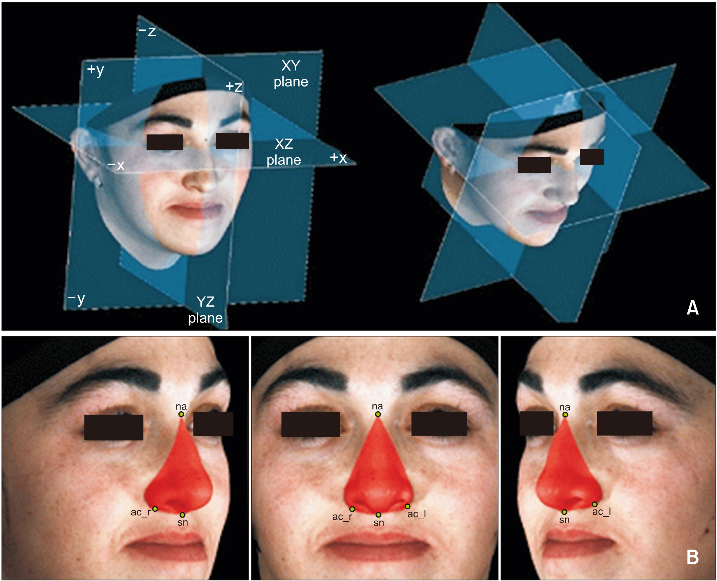
Figure 3 A, Axial reference plane passing through both endocanthions when the patients had their natural head positions. Sagittal reference plane passing through na, perpendicular to the axial and coronal reference planes; coronal reference plane passing through both exocanthions, perpendicular to the axial and sagittal planes. B, External nasal surface area measurement was computed following soft tissue nasal borders. na, Soft tissue nasion; ac, alare curvature; sn, subnasale; r, right; l, left.
Reference
-
1. Arnett GW, McLaughlin RP. 2004. Facial and dental planning for orthodontists and oral surgeons. Mosby;London: p. 4.2. Schendel SA, Carlotti AE Jr. 1991; Nasal considerations in orthognathic surgery. Am J Orthod Dentofacial Orthop. 100:197–208. DOI: 10.1016/0889-5406(91)70056-3. PMID: 1877544.
Article3. Naini FB, Gill DS. 2017. Orthognathic surgery: principles, planning and practice. John Wiley & Sons;Chichester: p. 417. DOI: 10.1002/9781119004370.4. Larrabee WF Jr. 1987; Facial analysis for rhinoplasty. Otolaryngol Clin North Am. 20:653–74. PMID: 3320863.
Article5. van Loon B Jr, van Heerbeek N, Bierenbroodspot F, Verhamme L, Xi T, de Koning MJ, et al. 2015; Three-dimensional changes in nose and upper lip volume after orthognathic surgery. Int J Oral Maxillofac Surg. 44:83–9. DOI: 10.1016/j.ijom.2014.08.001. PMID: 25218802.
Article6. Baik HS, Kim SY. 2010; Facial soft-tissue changes in skeletal Class III orthognathic surgery patients analyzed with 3-dimensional laser scanning. Am J Orthod Dentofacial Orthop. 138:167–78. DOI: 10.1016/j.ajodo.2010.02.022. PMID: 20691358.
Article7. Ferrario VF, Sforza C, Schmitz JH, Santoro F. 1999; Three-dimensional facial morphometric assessment of soft tissue changes after orthognathic surgery. Oral Surg Oral Med Oral Pathol Oral Radiol Endod. 88:549–56. DOI: 10.1016/S1079-2104(99)70084-3. PMID: 10556748.
Article8. Honrado CP, Lee S, Bloomquist DS, Larrabee WF Jr. 2006; Quantitative assessment of nasal changes after maxillomandibular surgery using a 3-dimensional digital imaging system. Arch Facial Plast Surg. 8:26–35. DOI: 10.1001/archfaci.8.1.26. PMID: 16415444.
Article9. Schendel SA, Williamson LW. 1983; Muscle reorientation following superior repositioning of the maxilla. J Oral Maxillofac Surg. 41:235–40. DOI: 10.1016/0278-2391(83)90265-3.
Article10. Mitchell C, Oeltjen J, Panthaki Z, Thaller SR. 2007; Nasolabial aesthetics. J Craniofac Surg. 18:756–65. DOI: 10.1097/scs.0b013e3180684360. PMID: 17667661.
Article11. Guenthner TA, Sather AH, Kern EB. 1984; The effect of Le Fort I maxillary impaction on nasal airway resistance. Am J Orthod. 85:308–15. DOI: 10.1016/0002-9416(84)90188-X.
Article12. Chung C, Lee Y, Park KH, Park SH, Park YC, Kim KH. 2008; Nasal changes after surgical correction of skeletal Class III malocclusion in Koreans. Angle Orthod. 78:427–32. DOI: 10.2319/041207-186.1. PMID: 18416623.
Article13. Choi JW, Lee JY, Oh TS, Kwon SM, Yang SJ, Koh KS. 2014; Frontal soft tissue analysis using a 3 dimensional camera following two-jaw rotational orthognathic surgery in skeletal class III patients. J Craniomaxillofac Surg. 42:220–6. DOI: 10.1016/j.jcms.2013.05.004. PMID: 23870714.
Article14. Solow B, Tallgren A. 1971; Natural head position in standing subjects. Acta Odontol Scand. 29:591–607. DOI: 10.3109/00016357109026337. PMID: 5290983.
Article15. Lim YK, Chu EH, Lee DY, Yang IH, Baek SH. 2010; Three-dimensional evaluation of soft tissue change gradients after mandibular setback surgery in skeletal Class III malocclusion. Angle Orthod. 80:896–903. DOI: 10.2319/021210-90.1. PMID: 20578861.
Article16. Lin HH, Chiang WC, Lo LJ, Sheng-Pin Hsu S, Wang CH, Wan SY. 2013; Artifact-resistant superimposition of digital dental models and cone-beam computed tomography images. J Oral Maxillofac Surg. 71:1933–47. DOI: 10.1016/j.joms.2013.06.199. PMID: 23911142.
Article17. Kim YK, Lee NK, Moon SW, Jang MJ, Kim HS, Yun PY. 2015; Evaluation of soft tissue changes around the lips after bracket debonding using three-dimensional stereophotogrammetry. Angle Orthod. 85:833–40. DOI: 10.2319/090414.622.1. PMID: 26308106.
Article18. Liu ZY, Yu J, Dai FF, Jiang RP, Xu TM. 2019; Three-dimensional changes in lip vermilion morphology of adult female patients after extraction and non-extraction orthodontic treatment. Korean J Orthod. 49:222–34. DOI: 10.4041/kjod.2019.49.4.222. PMID: 31367577. PMCID: PMC6658897.
Article19. Rasse M, Forkert G, Waldhäusl P. 1991; Stereophotogrammetry of facial soft tissue. Int J Oral Maxillofac Surg. 20:163–6. DOI: 10.1016/S0901-5027(05)80008-6. PMID: 1890324.
Article20. Betts NJ, Vig KW, Vig P, Spalding P, Fonseca RJ. 1993; Changes in the nasal and labial soft tissues after surgical repositioning of the maxilla. Int J Adult Orthodon Orthognath Surg. 8:7–23.21. Sarver DM, Weissman SM. 1991; Long-term soft tissue response to LeFort I maxillary superior repositioning. Angle Orthod. 61:267–76.22. Misir AF, Manisali M, Egrioglu E, Naini FB. 2011; Retrospective analysis of nasal soft tissue profile changes with maxillary surgery. J Oral Maxillofac Surg. 69:e190–4. DOI: 10.1016/j.joms.2010.10.032. PMID: 21367507.23. Gassmann CJ, Nishioka GJ, Van Sickels JE, Thrash WJ. 1989; A lateral cephalometric analysis of nasal morphology following Le Fort I osteotomy applying photometric analysis techniques. J Oral Maxillofac Surg. 47:926–30. DOI: 10.1016/0278-2391(89)90375-3.
Article24. McCance AM, Moss JP, Fright WR, James DR, Linney AD. 1992; A three dimensional analysis of soft and hard tissue changes following bimaxillary orthognathic surgery in skeletal III patients. Br J Oral Maxillofac Surg. 30:305–12. DOI: 10.1016/0266-4356(92)90180-Q. PMID: 1390562.
Article25. Radney LJ, Jacobs JD. 1981; Soft-tissue changes associated with surgical total maxillary intrusion. Am J Orthod. 80:191–212. DOI: 10.1016/0002-9416(81)90218-9. PMID: 6943938.
Article26. Sforza C, Peretta R, Grandi G, Ferronato G, Ferrario VF. 2007; Soft tissue facial volumes and shape in skeletal Class III patients before and after orthognathic surgery treatment. J Plast Reconstr Aesthet Surg. 60:130–8. DOI: 10.1016/j.bjps.2006.06.003. PMID: 17223510.
Article27. Soncul M, Bamber MA. 2004; Evaluation of facial soft tissue changes with optical surface scan after surgical correction of Class III deformities. J Oral Maxillofac Surg. 62:1331–40. DOI: 10.1016/j.joms.2004.04.019. PMID: 15510353.
Article28. Freihofer HP Jr. 1977; Changes in nasal profile after maxillary advancement in cleft and non-cleft patients. J Maxillofac Surg. 5:20–7. DOI: 10.1016/S0301-0503(77)80071-4. PMID: 265348.
Article29. Mansour S Jr, Burstone C, Legan H. 1983; An evaluation of soft-tissue changes resulting from Le Fort I maxillary surgery. Am J Orthod. 84:37–47. DOI: 10.1016/0002-9416(83)90146-X.
Article30. Koh CH, Chew MT. 2004; Predictability of soft tissue profile changes following bimaxillary surgery in skeletal class III Chinese patients. J Oral Maxillofac Surg. 62:1505–9. DOI: 10.1016/j.joms.2004.04.022. PMID: 15573350.
Article
- Full Text Links
-
- Actions
-
Cited
- CITED
-
- Close
- Share
-
- Similar articles
-
- QUANTITATIVE ASSESSMENT OF NASAL AND UPPER LIP CHANGES AFTER LE FORT I OSTEOTOMY SURGERY USING A 3-DIMENSIONAL COMPUTED TOMOGRAPHY
- A clinico-statistical study of soft tissue changes of upper lip & nose following Le Fort I maxillary movement
- Management of Maxillary Impacted Canines
- Nose changes after maxillary advancement surgery in skeletal Class III malocclusion
- Nasal airway function after Le Fort I osteotomy with maxillary impaction: A prospective study using the Nasal Obstruction Symptom Evaluation scale
