Construction reproducibility of a composite tooth model composed of an intraoral-scanned crown and a cone-beam computed tomography-scanned root
- Affiliations
-
- 1Department of Orthodontics, School of Dentistry, Chonnam National University, Gwangju, Korea
- 2Department of Oral Anatomy, School of Dentistry, Chonnam National University, Gwangju, Korea
- 3Department of Orthodontics, School of Dentistry, Seoul National University, Seoul, Korea
- KMID: 2504140
- DOI: http://doi.org/10.4041/kjod.2020.50.4.229
Abstract
Objective
To evaluate the construction reproducibility of a composite tooth model (CTM) composed of an intraoral-scanned crown and a cone-beam computed tomography (CBCT)-scanned root.
Methods
The study assessed 240 teeth (30 central incisors, 30 canines, 30 second premolars, and 30 first molars in the maxillary and mandibular arches) from 15 young adult patients whose pre-treatment intraoral scan and CBCT were available. Examiner-Reference (3 years’ experience in CTM construction) and Examiners-A and Examiner-B (no experience) constructed the individual CTMs independently by performing the following steps: image acquisition and processing into a three-dimensional model, integration of intraoral-scanned crowns and CBCT-scanned teeth, and replacement of the CBCT-scanned crown with the intraoral-scanned crown. The tooth axis angle in terms of mesiodistal angulation and buccolingual inclination of the CTMs constructed by the three examiners were measured. To assess the construction reproducibility of CTMs, intraclass correlation coefficient (ICC) assessments were performed.
Results
The ICC values of mesiodistal angulation and buccolingual inclination among the 3 examiners showed excellent agreement (0.950–0.992 and 0.965–0.993; 0.976–0.994 and 0.973–0.995 in the maxillary and mandibular arches, respectively).
Conclusions
The CTM showed excellent construction reproducibility in mesiodistal angulation and buccolingual inclination regardless of the construction skill and experience levels of the examiners.
Keyword
Figure
-
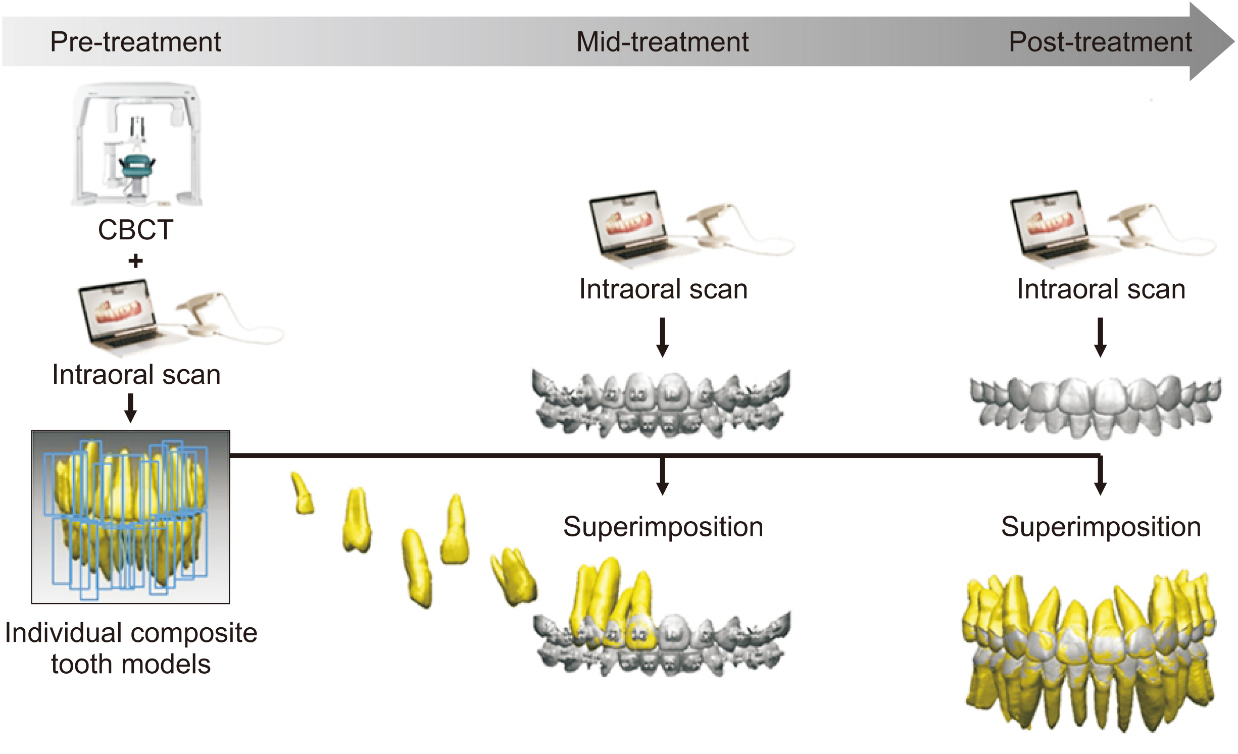
Figure 1 Clinical application of the composite tooth model (CTM). The CTMs constructed at the pre-treatment stage by combining cone-beam computed tomography (CBCT) and intraoral scan data can be used for evaluation of the root position at the mid- and post-treatment stages.
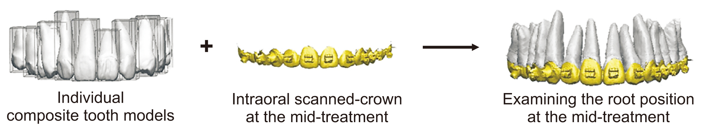
Figure 2 Evaluation of root position at the mid-treatment stage by superimposing individual composite tooth models onto the mid-treatment intraoral-scanned image using the crown as an index.
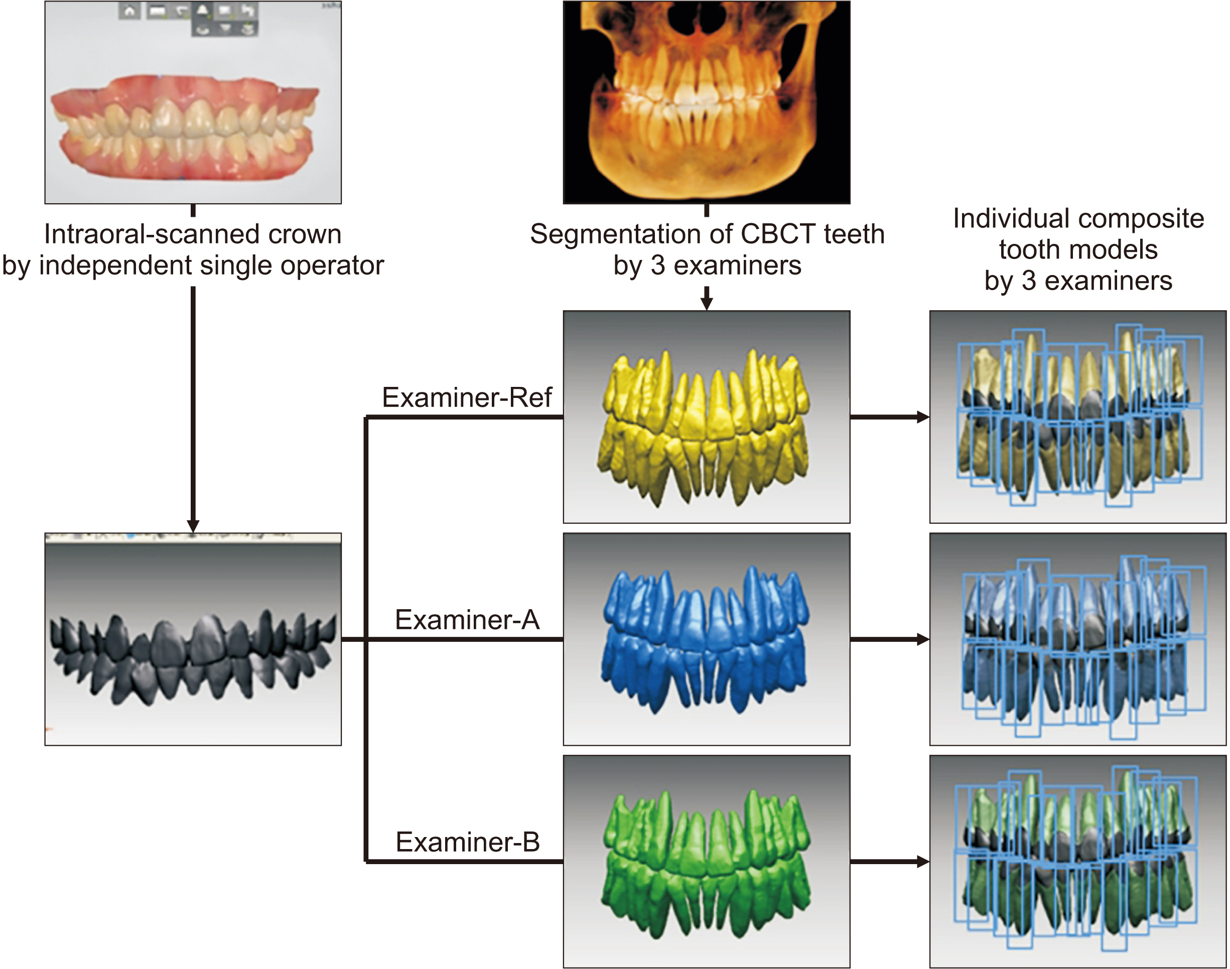
Figure 3 Study design for evaluation of inter-examiner reliability in construction of individual composite tooth models (CTMs). Examiner-Reference (Examiner-Ref), Examiner-A, and Examiner-B independently constructed the individual CTMs using the intraoral-scanned crown and the cone-beam computed tomography (CBCT)-segmented teeth.
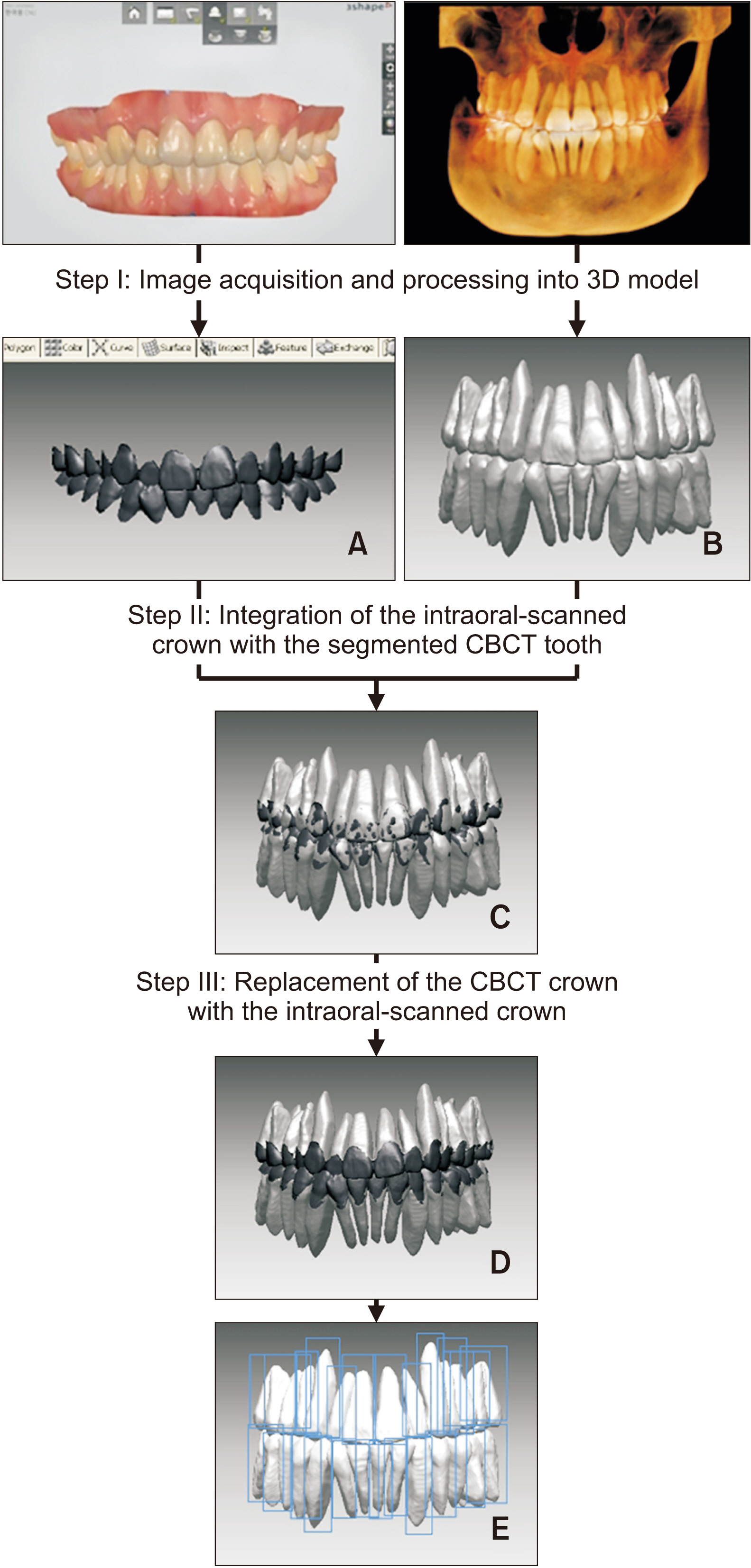
Figure 4 Construction procedure of the individual composite tooth models. A, Intraoral-scanned crown. B, Segmented cone-beam computed tomography (CBCT)-scanned teeth. C, Integration of A and B by crown registration. D, Intraoral-scanned crown with CBCT-scanned root. E, Individual composite tooth models. 3D, Three-dimensional.
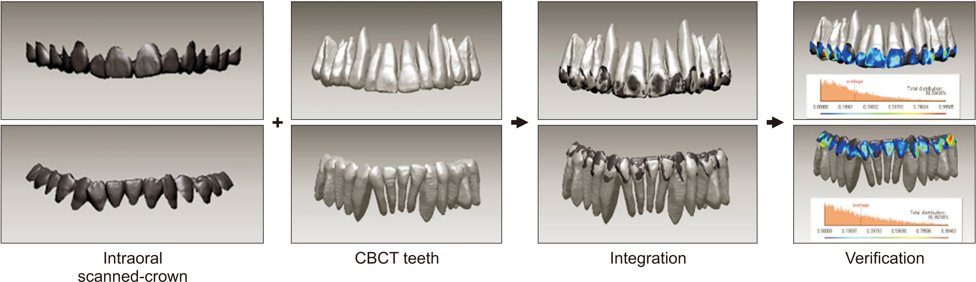
Figure 5 Verification of the integration accuracy. Registration errors were evaluated by measuring the absolute values of the three-dimensional Euclidean distances between the surface points on the two images. CBCT, Cone-beam computed tomography.
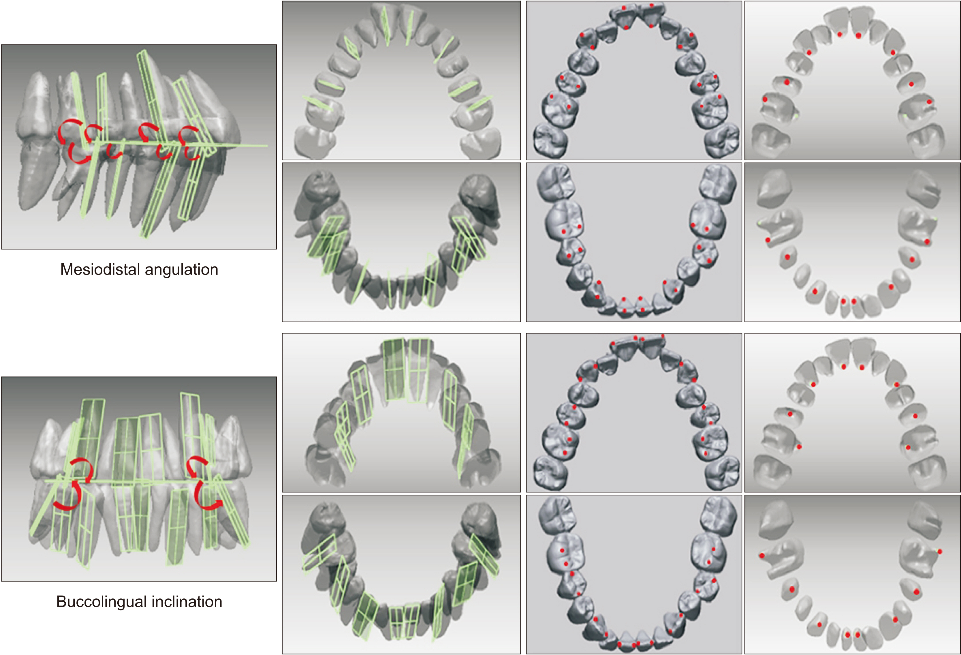
Figure 6 Measurements of tooth axis angle. The mesiodistal angulation was measured at the distal angle in conjunction with the occlusal plane and the tooth axis plane (upper row). The buccolingual inclination was measured at the lingual angle in conjunction with the occlusal plane and the tooth axis plane (lower row). The green square-shaped plane presents tooth axis planes constructed by two points in the crown and one point in the root apex.
Cited by 1 articles
-
Consideration of root position in virtual tooth setup for extraction treatment: A comparative study of simulated and actual treatment results
Mirinae Park, Veerasathpurush Allareddy, Phimon Atsawasuwan, Min Kyeong Lee, Kyungmin Clara Lee
Korean J Orthod. 2023;53(1):26-34. doi: 10.4041/kjod22.105.
Reference
-
1. Garcia-Figueroa MA, Raboud DW, Lam EW, Heo G, Major PW. 2008; Effect of buccolingual root angulation on the mesiodistal angulation shown on panoramic radiographs. Am J Orthod Dentofacial Orthop. 134:93–9. DOI: 10.1016/j.ajodo.2006.07.034. PMID: 18617108.
Article2. Mckee IW, Glover KE, Williamson PC, Lam EW, Heo G, Major PW. 2001; The effect of vertical and horizontal head positioning in panoramic radiography on mesiodistal tooth angulations. Angle Orthod. 71:442–51.3. Song IT, Cho JH, Chae JM, Chang NY. 2011; Evaluation of mesiodistal tooth axis using a CBCT-generated panoramic view. Korean J Orthod. 41:255–67. DOI: 10.4041/kjod.2011.41.4.255.
Article4. Lascala CA, Panella J, Marques MM. 2004; Analysis of the accuracy of linear measurements obtained by cone beam computed tomography (CBCT-NewTom). Dentomaxillofac Radiol. 33:291–4. DOI: 10.1259/dmfr/25500850. PMID: 15585804.
Article5. Tong H, Enciso R, Van Elslande D, Major PW, Sameshima GT. 2012; A new method to measure mesiodistal angulation and faciolingual inclination of each whole tooth with volumetric cone-beam computed tomography images. Am J Orthod Dentofacial Orthop. 142:133–43. DOI: 10.1016/j.ajodo.2011.12.027. PMID: 22748999.
Article6. Ender A, Mehl A. 2011; Full arch scans: conventional versus digital impressions--an in-vitro study. Int J Comput Dent. 14:11–21.7. Hayashi K, Sachdeva AU, Saitoh S, Lee SP, Kubota T, Mizoguchi I. 2013; Assessment of the accuracy and reliability of new 3-dimensional scanning devices. Am J Orthod Dentofacial Orthop. 144:619–25. DOI: 10.1016/j.ajodo.2013.04.021. PMID: 24075671.
Article8. Sun L, Lee JS, Choo HH, Hwang HS, Lee KM. 2018; Reproducibility of an intraoral scanner: a comparison between in-vivo and ex-vivo scans. Am J Orthod Dentofacial Orthop. 154:305–10. DOI: 10.1016/j.ajodo.2017.09.022. PMID: 30075932.9. Lim SW, Hwang HS, Cho IS, Baek SH, Cho JH. 2020; Registration accuracy between intraoral-scanned and cone-beam computed tomography-scanned crowns in various registration methods. Am J Orthod Dentofacial Orthop. 157:348–56. DOI: 10.1016/j.ajodo.2019.04.031. PMID: 32115113.
Article10. Macchi A, Carrafiello G, Cacciafesta V, Norcini A. 2006; Three-dimensional digital modeling and setup. Am J Orthod Dentofacial Orthop. 129:605–10. DOI: 10.1016/j.ajodo.2006.01.010. PMID: 16679200.
Article11. Guo H, Zhou J, Bai Y, Li S. 2011; A three-dimensional setup model with dental roots. J Clin Orthod. 45:209–16. quiz 235–6. PMID: 21785205.12. Kihara T, Tanimoto K, Michida M, Yoshimi Y, Nagasaki T, Murayama T, et al. 2012; Construction of orthodontic setup models on a computer. Am J Orthod Dentofacial Orthop. 141:806–13. DOI: 10.1016/j.ajodo.2011.10.027. PMID: 22640682.
Article13. Lee RJ, Pham J, Choy M, Weissheimer A, Dougherty HL Jr, Sameshima GT, et al. 2014; Monitoring of typodont root movement via crown superimposition of single cone-beam computed tomography and consecutive intraoral scans. Am J Orthod Dentofacial Orthop. 145:399–409. DOI: 10.1016/j.ajodo.2013.12.011. PMID: 24582031.
Article14. Lee RJ, Weissheimer A, Pham J, Go L, de Menezes LM, Redmond WR, et al. 2015; Three-dimensional monitoring of root movement during orthodontic treatment. Am J Orthod Dentofacial Orthop. 147:132–42. DOI: 10.1016/j.ajodo.2014.10.010. PMID: 25533080.
Article15. Lee RJ, Pi S, Park J, Devgon D, Nelson G, Hatcher D, et al. 2018; Accuracy and reliability of the expected root position setup methodology to evaluate root position during orthodontic treatment. Am J Orthod Dentofacial Orthop. 154:583–95. DOI: 10.1016/j.ajodo.2018.05.010. PMID: 30268268.
Article16. Walter SD, Eliasziw M, Donner A. 1998; Sample size and optimal designs for reliability studies. Stat Med. 17:101–10. DOI: 10.1002/(SICI)1097-0258(19980115)17:1<101::AID-SIM727>3.0.CO;2-E. PMID: 9463853.
Article17. Schunk DH. 2012. Learning theories: an educational perspective. 6th ed. Pearson Education;Boston: p. 38–40.18. Wang C, Liu Y, Wang S, Wang Y, Zhao Y. 2018; Evaluation of in vivo digital root reconstruction based on anatomical characteristics of the periodontal ligament using cone beam computed tomography. Sci Rep. 8:269. DOI: 10.1038/s41598-017-18592-4. PMID: 29321613. PMCID: PMC5762886.
Article19. Ye N, Long H, Xue J, Wang S, Yang X, Lai W. 2014; Integration accuracy of laser-scanned dental models into maxillofacial cone beam computed tomography images of different voxel sizes with different segmentation threshold settings. Oral Surg Oral Med Oral Pathol Oral Radiol. 117:780–6. DOI: 10.1016/j.oooo.2014.02.022. PMID: 24767699.
Article20. Marmulla R, Wörtche R, Mühling J, Hassfeld S. 2005; Geometric accuracy of the NewTom 9000 Cone Beam CT. Dentomaxillofac Radiol. 34:28–31. DOI: 10.1259/dmfr/31342245. PMID: 15709102.
Article21. Sang YH, Hu HC, Lu SH, Wu YW, Li WR, Tang ZH. 2016; Accuracy assessment of three-dimensional surface reconstructions of in vivo teeth from cone-beam computed tomography. Chin Med J (Engl). 129:1464–70. DOI: 10.4103/0366-6999.183430. PMID: 27270544. PMCID: PMC4910372.
Article22. European Commission. 2012. Mar. Cone Beam CT for dental and maxillofacial radiology: evidence-based guidelines [Internet]. SEDENTEXCT;Luxembourg: Available from: http://www.sedentexct.eu/files/radiation_protection_172.pdf. cited 2020 Jan 30.
- Full Text Links
-
- Actions
-
Cited
- CITED
-
- Close
- Share
-
- Similar articles
-
- Integrated three-dimensional digital assessment of accuracy of anterior tooth movement using clear aligners
- Cone-beam computed tomography for the assessment of root–crown ratios of the maxillary and mandibular incisors in a Korean population
- Crown and root lengths of incisors, canines, and premolars measured by cone-beam computed tomography in patients with malocclusions
- Comparison of interference from eccentric movements of dental crowns fabricated via dynamic jaw motion tracking and conventional methods: a double-blind clinical study
- Evaluation of imaging reformation for root and pulp canal shapes of permanent teeth using a cone beam computed tomography
