Crown and root lengths of incisors, canines, and premolars measured by cone-beam computed tomography in patients with malocclusions
- Affiliations
-
- 1Department of Orthodontics, School of Dentistry, Chosun University, Gwangju, Korea. shlim@chosun.ac.kr
- 2Division in Anatomy and Developmental Biology, College of Dentistry, Yonsei University, Seoul, Korea.
- KMID: 2273420
- DOI: http://doi.org/10.4041/kjod.2013.43.6.271
Abstract
OBJECTIVE
The purposes of this study were to determine the accuracy of crown and root length measurements of premolars using cone-beam computed tomography (CBCT) and to generate reference CBCT-based data on incisor, canine, and premolar lengths in patients with malocclusions.
METHODS
Imaging was performed using a CBCT scanner with a 0.292-mm voxel size and 12-bit grayscale. The CBCT-based length measurements were compared with direct measurements of 94 subsequently extracted premolars without metal restorations using the paired t-test. Furthermore, the crown and root lengths of incisors, canines, and premolars in 62 Korean patients with malocclusions were measured using CBCT, and Pearson's correlation coefficients were calculated to examine the relationship between the crown and root length measurements of each tooth type.
RESULTS
The differences between the CBCT-based and direct measurements of the extracted premolars were not significant, with 95% limits of agreement of -0.90 to 0.90 mm for crown length and -1.23 to 1.18 mm for root length. Weak positive correlations between the crown and root length measurements were observed for the mandibular canine and premolars.
CONCLUSIONS
The CBCT-based measurements showed a wider range of limits of agreements for root length than for crown length. The CBCT-based data can be used as a reference for evaluating root length and resorption of teeth without metal restorations in patients with malocclusions.
Keyword
MeSH Terms
Figure
-

Figure 1 Schematic of the landmarks used for measuring the total tooth, crown, and root lengths of the extracted premolars.
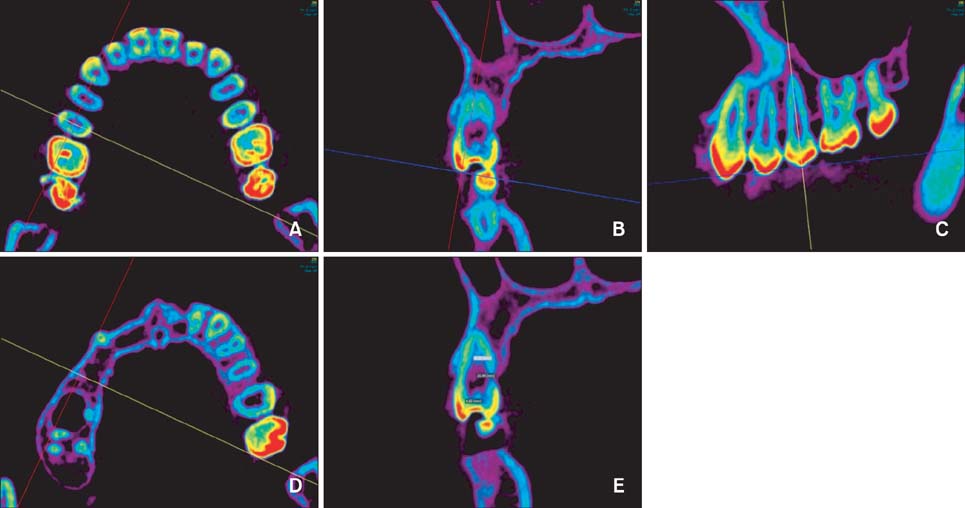
Figure 2 Alignment procedure of multiplanar reconstruction windows for the cone-beam computed tomography-based length measurements. The red, yellow, and blue lines indicate the sagittal, coronal, and axial planes, respectively. A, The intersection of the sagittal and coronal planes is aligned with the center of the pulp chamber in the axial view of the tooth to be measured. B, The sagittal plane is rotated in the coronal view of the tooth such that it passes through the buccal cusp and root tips. C, The coronal plane in the sagittal view of the tooth is rotated such that it passes through the buccal cusp and root tips, while ensuring that the sagittal plane also passes through the buccal cusp and root tips. D, Verification that the sagittal and coronal planes pass through the buccal cusp and root tips by moving the axial slice until the cusp or root tip disappears. E, Measurement of crown, root, and total tooth lengths in the sagittal view of the tooth. These measurements were performed similarly to the corresponding direct measurements.
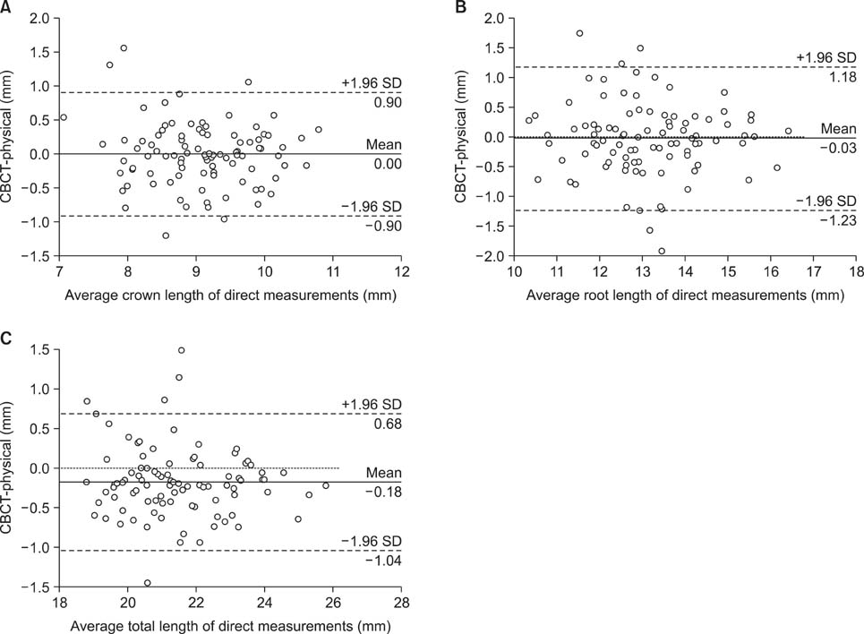
Figure 3 Bland-Altman plots of the cone-beam computed tomography (CBCT)-based and direct length measurements of the extracted premolars. The CBCT-physical difference (dotted line), mean difference (thick solid line) and 95% limits of agreement (dashed line) are shown. A, Crown length. B, Root length. C, Total tooth length. SD, Standard deviation; CBCT-physical indicates difference of CBCT-based and direct length measurements.
Cited by 2 articles
-
Cone-beam computed tomography for the assessment of root–crown ratios of the maxillary and mandibular incisors in a Korean population
Sung-Hwan Choi, Jung-Suk Kim, Cheol-Soon Kim, Hyung-Seog Yu, Chung-Ju Hwang
Korean J Orthod. 2017;47(1):39-49. doi: 10.4041/kjod.2017.47.1.39.Root proximity of the anchoring miniscrews of orthodontic miniplates in the mandibular incisal area: Cone-beam computed tomographic analysis
Do-Min Jeong, Song Hee Oh, HyeRan Choo, Yong-Suk Choi, Seong-Hun Kim, Jin-Suk Lee, Eui-Hwan Hwang
Korean J Orthod. 2021;51(4):231-240. doi: 10.4041/kjod.2021.51.4.231.
Reference
-
1. Brezniak N, Wasserstein A. Orthodontically induced inflammatory root resorption. Part I: The basic science aspects. Angle Orthod. 2002; 72:175–179.2. Sameshima GT, Sinclair PM. Predicting and preventing root resorption: Part I. Diagnostic factors. Am J Orthod Dentofacial Orthop. 2001; 119:505–510.
Article3. Lupi JE, Handelman CS, Sadowsky C. Prevalence and severity of apical root resorption and alveolar bone loss in orthodontically treated adults. Am J Orthod Dentofacial Orthop. 1996; 109:28–37.
Article4. Plets JH, Isaacson RJ, Speidel TM, Worms FW. Maxillary central incisor root length in orthodontically treated and untreated patients. Angle Orthod. 1974; 44:43–47.5. Black GV. Descriptive anatomy of the human teeth. Philadelphia: White manufacturing;1902.6. Verhoeven JW, van Aken J, van der Weerdt GP. The length of teeth. A statistical analysis of the differences in length of human teeth for radiologic purposes. Oral Surg Oral Med Oral Pathol. 1979; 47:193–199.7. Maina SW, Wagaiyu CK. The average human tooth lengths for black Kenyan population. East Afr Med J. 1990; 67:33–38.8. Bjorndal AM, Henderson WG, Skidmore AE, Kellner FH. Anatomic measurements of human teeth extracted from males between the ages of 17 and 21 years. Oral Surg Oral Med Oral Pathol. 1974; 38:791–803.
Article9. Jayawardena CK, Abesundara AP, Nanayakkara DC, Chandrasekara MS. Age-related changes in crown and root length in Sri Lankan Sinhalese. J Oral Sci. 2009; 51:587–592.
Article10. Ozaki T, Satake T, Kanazawa E. Morphological significance of root length variability in comparison with other crown dimensions. I. Basic statistics and sex difference. J Nihon Univ Sch Dent. 1987; 29:233–240.
Article11. Ozaki T, Satake T, Kanazawa E. Morphological significance of root length variability in comparison with other crown dimensions. II. Correlation between crown and root measurements. J Nihon Univ Sch Dent. 1988; 30:11–21.
Article12. Brezniak N, Goren S, Zoizner R, Shochat T, Dinbar A, Wasserstein A, et al. The accuracy of the cementoenamel junction identification on periapical films. Angle Orthod. 2004; 74:496–500.13. Jakobsson R, Lind V. Variation in root length of the permanent maxillary central incisor. Scand J Dent Res. 1973; 81:335–338.
Article14. Brezniak N, Goren S, Zoizner R, Dinbar A, Arad A, Wasserstein A, et al. A comparison of three methods to accurately measure root length. Angle Orthod. 2004; 74:786–791.15. Thanyakarn C, Hansen K, Rohlin M, Akesson L. Measurements of tooth length in panoramic radiographs. 1. The use of indicators. Dentomaxillofac Radiol. 1992; 21:26–30.
Article16. Yassaei S, Ezoddini-Ardakani F, Ostovar N. Predicting the actual length of premolar teeth on the basis of panoramic radiology. Indian J Dent Res. 2010; 21:468–473.
Article17. Lien LC, Soh G. Accuracy of the orthopantomogram in assessment of tooth length in orthodontic patients. Singapore Dent J. 2000; 23:1 Suppl. 68–71.18. Hölttä P, Nyström M, Evälahti M, Alaluusua S. Root-crown ratios of permanent teeth in a healthy Finnish population assessed from panoramic radiographs. Eur J Orthod. 2004; 26:491–497.
Article19. Stramotas S, Geenty JP, Darendeliler MA, Byloff F, Berger J, Petocz P. The reliability of crown-root ratio, linear and angular measurements on panoramic radiographs. Clin Orthod Res. 2000; 3:182–191.
Article20. Lund H, Gröndahl K, Gröndahl HG. Cone beam computed tomography for assessment of root length and marginal bone level during orthodontic treatment. Angle Orthod. 2010; 80:466–473.
Article21. Sherrard JF, Rossouw PE, Benson BW, Carrillo R, Buschang PH. Accuracy and reliability of tooth and root lengths measured on cone-beam computed tomographs. Am J Orthod Dentofacial Orthop. 2010; 137:4 Suppl. S100–S108.
Article22. Stratemann SA, Huang JC, Maki K, Miller AJ, Hatcher DC. Comparison of cone beam computed tomography imaging with physical measures. Dentomaxillofac Radiol. 2008; 37:80–93.
Article23. Pinsky HM, Dyda S, Pinsky RW, Misch KA, Sarment DP. Accuracy of three-dimensional measurements using cone-beam CT. Dentomaxillofac Radiol. 2006; 35:410–416.
Article24. Baumgaertel S, Palomo JM, Palomo L, Hans MG. Reliability and accuracy of cone-beam computed tomography dental measurements. Am J Orthod Dentofacial Orthop. 2009; 136:19–25. discussion 25-8.
Article25. Ponder SN, Benavides E, Kapila S, Hatch NE. Quantification of external root resorption by low- vs high-resolution cone-beam computed tomography and periapical radiography: A volumetric and linear analysis. Am J Orthod Dentofacial Orthop. 2013; 143:77–91.
Article26. Kim MS, Kim SH, Kim HJ, Kim HJ, Park , KP , Park BG, et al. Dental anatomy and morphology. Seoul, Korea: Jeesung Publishing;2005. p. 27.27. Hwang CJ, Song YY. A radiographic study on root resorption in the malocclusion patients before orthodontic treatment. Korean J Orthod. 1999; 29:219–236.28. Nackaerts O, Maes F, Yan H, Couto Souza P, Pauwels R, Jacobs R. Analysis of intensity variability in multislice and cone beam computed tomography. Clin Oral Implants Res. 2011; 22:873–879.
Article29. Pauwels R, Nackaerts O, Bellaiche N, Stamatakis H, Tsiklakis K, Walker A, et al. SEDENTEXCT Project Consortium. Variability of dental cone beam CT grey values for density estimations. Br J Radiol. 2013; 86:20120135.
Article
- Full Text Links
-
- Actions
-
Cited
- CITED
-
- Close
- Share
-
- Similar articles
-
- Cone-beam computed tomography for the assessment of root–crown ratios of the maxillary and mandibular incisors in a Korean population
- Retrospective Analysis of Incisor Root Resorption Associated with Impacted Maxillary Canines
- Analysis of the root position and angulation of maxillary premolars in alveolar bone using cone-beam computed tomography
- Three-dimensional analysis of dental decompensation for skeletal Class III malocclusion on the basis of vertical skeletal patterns obtained using cone-beam computed tomography
- Factors affecting external apical root resorption of maxillary incisors associated with microimplantassisted rapid palatal expansion
