Neurolymphomatosis in patients withmantle cell lymphoma diagnosed byFDG PET-CT
- Affiliations
-
- 1Department of Neurology, Chungnam National University Hospital, Deajeon, Korea
- 2Department of Hematology, Chungnam National University Hospital, Deajeon, Korea
- KMID: 2500303
- DOI: http://doi.org/10.14253/acn.2020.22.1.37
Abstract
- Neurolymphomatosis (NL) is characterized by the infiltration of malignant lymphoma cells into peripheral nerves, nerve roots, plexuses, or cranial nerves. This is a very rare complication of mantle-cell lymphoma. Diagnosing NL is made difficult by cerebrospinal fluid cytology and bone-marrow biopsy results often being negative. NL can appear as the only sign of recurrence in a patient with a previous diagnosis of lymphoma. Here we present two cases of NL in patients with mantle-cell lymphoma diagnosed by positron emission tomography with deoxy-fluoro-D-glucose integrated with computed tomography.
Keyword
Figure
-
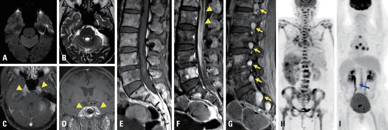
Fig. 1. Brain magnetic resonance imaging (MRI) (A-D), spine MRI (E-G), and positron emission tomography with deoxy-fluoro-D-glucose integrated with computed tomography (FDG PET-CT) for diagnosing mantle-cell lymphoma (H) and neurolymphomatosis (I). There were no abnormal signal intensities in diffusion weighted image (A) and T2-weighted (B) images. Diffuse thickening and bilateral enhancement (arrowheads) of the trigeminal nerve were observed in T1-weighted gadolinium-enhanced images (C, D). Diffuse leptomeningeal enhancement (arrowheads) was observed on a T1-weighted gadolinium-enhanced image (F) compared to a nonenhanced T1-weighted image (E). Multiple spinal nerve enhancements (arrows) were observed in a T1-weighted gadolinium-enhanced image (G). Multiple regions exhibiting FDG uptake were observed in the lymph nodes and bone marrow in initial FDG PET-CT (H). Complete remission of the previous lesion with new linear area (blue arrow) of high uptake in the lower spinal cord and cauda equine were observed in subsequent FDG PET-CT (I).
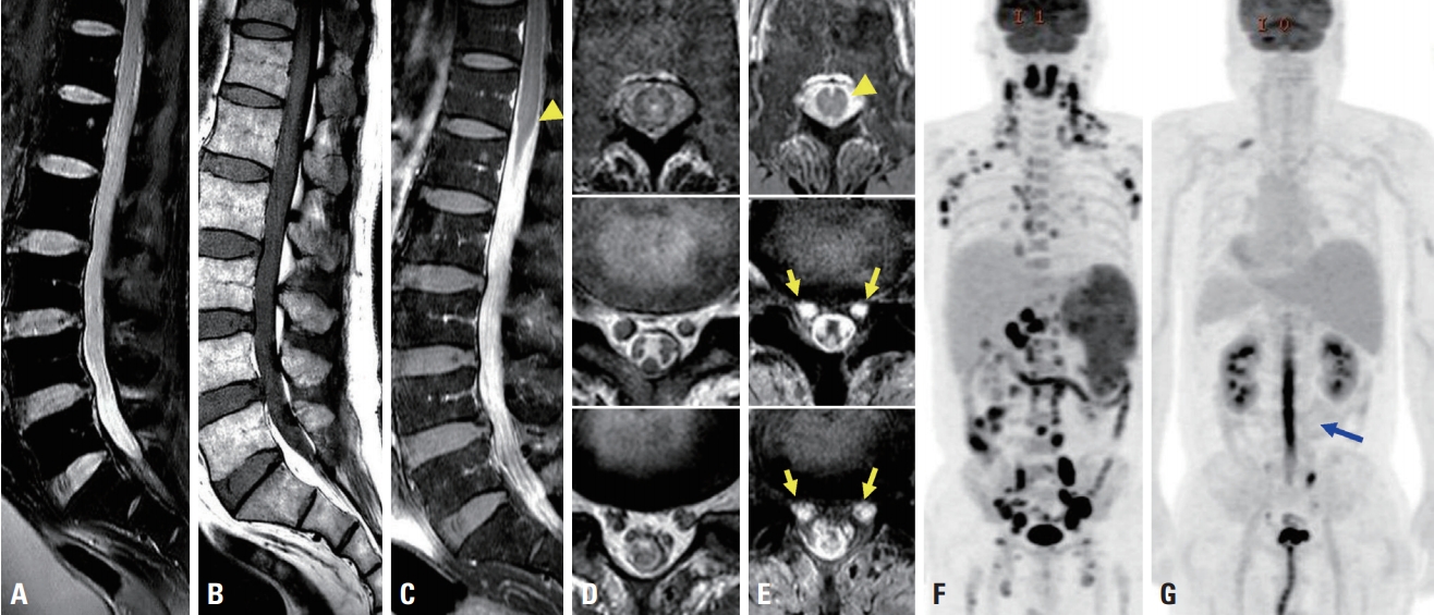
Fig. 2. Spine magnetic resonance imaging (A-E) and positron emission tomography with deoxy-fluoro-D-glucose integrated with computed tomography for diagnosing mantle-cell lymphoma (F) and neurolymphomatosis (G). Enlargement of the spinal nerves was observed in T2-weighted (A) and T1-weighted (B) image. Diffuse leptomeningeal enhancement (arrowheads) was observed in T1-weighted gadolinium-enhanced images (C, E). Multiple spinal nerve roots were enhanced (arrows) after gadolinium injection (E) compared to a T2-weighted axial image (D). Multiple lymph nodes showed hypermetabolism in initial FDG PET-CT (F). Thoracolumbar spinal cord hypermetabolism (blue arrow) with improvement of the previous lesion was evident in subsequent FDG PET-CT (G).
Reference
-
1. Davidson T, Kedmi M, Avigdor A, Komisar O, Chikman B, Lidar M, et al. FDG PET-CT evaluation in neurolymphomatosis: imaging characteristics and clinical outcomes. Leuk Lymphoma. 2018; 59:348–356.
Article2. Pham M, Awad M. Lymphoma relapse presenting as neurolymphomatosis. Asian J Neurosurg. 2016; 11:73.
Article3. DeVries AH, Howe BM, Spinner RJ, Broski SM. B-cell peripheral neurolymphomatosis: MRI and (18)F-FDG PET/CT imaging characteristics. Skeletal Radiol. 2019; 48:1043–1050.
Article4. Vargas TC, Thomas RL, Erickson JC. Leptomeningeal enhancement in a patient with progressive cranial neuropathies and lumbosacral radiculopathies. JAMA Neurol. 2016; 73:345–346.
Article5. Lin M, Kilanowska J, Taper J, Chu J. Neurolymphomatosis–diagnosis and assessment of treatment response by FDG PET-CT. Hematol Oncol. 2008; 26:43–45.
Article6. Ferrer A, Bosch F, Villamor N, Rozman M, Graus F, Gutiérrez G, et al. Central nervous system involvement in mantle cell lymphoma. Ann Oncol. 2008; 19:135–141.
Article7. Faivre G, Lagarde J, Choquet S, Maillart E, Lubetzki C. CNS involvement at diagnosis in mantle cell lymphoma with atypical MRI features. J Neurol. 2014; 261:1018–1020.
Article8. Baehring JM, Damek D, Martin EC, Betensky RA, Hochberg FH. Neurolymphomatosis. Neuro Oncol. 2003; 5:104–115.
Article9. Gan HK, Azad A, Cher L, Mitchell PL. Neurolymphomatosis: diagnosis, management, and outcomes in patients treated with rituximab. Neuro Oncol. 2010; 12:212–215.
Article10. Grisariu S, Avni B, Batchelor TT, van den Bent MJ, Bokstein F, Schiff D, et al. Neurolymphomatosis: an International Primary CNS Lymphoma Collaborative Group report. Blood. 2010; 115:5005–5011.
Article
- Full Text Links
-
- Actions
-
Cited
- CITED
-
- Close
- Share
-
- Similar articles
-
- When We Consider Neurolymphomatosis in Patient with Lumbosacral Plexopathy with an Extreme Leg Pain?
- A Case of Neurolymphomatosis Involving Cranial Nerve Diagnosed by PET-CT Imaging
- Nasal-Type Extranodal Natural Killer/T-cell Neurolymphomatosis Confined to the Lumbar Nerve Roots: A Case Report
- Cervical Nerve Root Neurolymphomatosis Detected on F-18 FDG PET/CT
- Neurolymphomatosis Involving Antebrachial Cutaneous Nerve
