Imaging Sci Dent.
2020 Mar;50(1):45-52. 10.5624/isd.2020.50.1.45.
Development of an evidence-based clinical imaging diagnostic guideline for implant planning: Joint recommendations of the Korean Academy of Oral and Maxillofacial Radiology and National Evidence-based Healthcare Collaborating Agency
- Affiliations
-
- 1Department of Oral and Maxillofacial Radiology and Dental Research Institute, School of Dentistry, Seoul National University, Seoul, Korea. raylee@snu.ac.kr
- 2Division for Healthcare Technology Assessment Research, National Evidence-based Healthcare Collaborating Agency, Seoul, Korea.
- 3Department of Radiology, Ajou University School of Medicine, Suwon, Korea.
- 4Department of Oral and Maxillofacial Radiology, Yonsei University College of Dentistry, Seoul, Korea.
- KMID: 2471845
- DOI: http://doi.org/10.5624/isd.2020.50.1.45
Abstract
- PURPOSE
This study was conducted to develop an evidence-based clinical imaging diagnostic guideline for implant planning, taking into account efficacy, benefits, and risks.
MATERIALS AND METHODS
The guideline development process employed the adaptation methodology used for Korean clinical imaging guidelines(K-CIG). Core databases(Ovid-Medline, Ovid-Embase, National Guideline Clearinghouse, Guideline International Network) and domestic databases (KoreaMed, KMbase, and KoMGI) were searched for guidelines. The retrieved articles were analyzed by 2 reviewers, and articles were selected using well-established inclusion criteria.
RESULTS
The search identified 294 articles, of which 3 were selected as relevant guidelines. Based on those 3 guidelines, 3 recommendations for implant planning were derived.
CONCLUSION
We recommend radiography or cone-beam computed tomography (CBCT) scanning for individual patients judged to require a cross-sectional image after reading of a panoramic X-ray image and a conventional intraoral radiological image. Various steps should be taken to raise awareness of these recommendations among clinicians and the public, and K-CIG should be regularly reviewed and revised.
Keyword
Figure
-
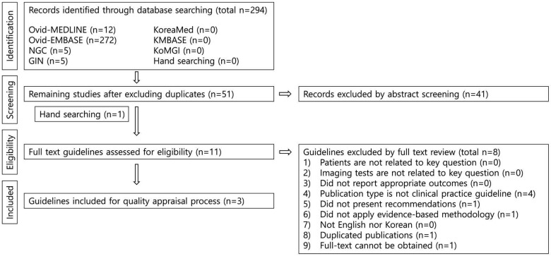
Fig. 1 The guidelines identified by searching were selected by step-by-step screening. This is the flow diagram of guideline selection.
Reference
-
1. Harris D, Horner K, Gröndahl K, Jacobs R, Helmrot E, Benic GI, et al. E.A.O. guidelines for the use of diagnostic imaging in implant dentistry 2011. A consensus workshop organized by the European Association for Osseointegration at the Medical University of Warsaw. Clin Oral Implants Res. 2012; 23:1243–1253. PMID: 22432473.
Article2. Fienitz T, Schwarz F, Ritter L, Dreiseidler T, Becker J, Rothamel D. Accuracy of cone beam computed tomography in assessing peri-implant bone defect regeneration: a histologically controlled study in dogs. Clin Oral Implants Res. 2012; 23:882–887. PMID: 21707753.
Article3. Bornstein MM, Horner K, Jacobs R. Use of cone beam computed tomography in implant dentistry: current concepts, indications and limitations for clinical practice and research. Periodontol 2000. 2017; 73:51–72. PMID: 28000270.
Article4. Choi SJ, Jeong WK, Jo AJ, Choi JA, Kim MJ, Lee M, et al. Methodology for developing evidence-based clinical imaging guidelines: joint recommendations by Korean Society of Radiology and National Evidence-Based Healthcare Collaborating Agency. Korean J Radiol. 2017; 18:208–216. PMID: 28096730.
Article5. Brouwers MC, Kho ME, Browman GP, Burgers JS, Cluzeau F, Feder G, et al. AGREE II: advancing guideline development, reporting and evaluation in health care. CMAJ. 2010; 182:E839–E842. PMID: 20603348.
Article6. Bornstein MM, Scarfe WC, Vaughn VM, Jacobs R. Cone beam computed tomography in implant dentistry: a systematic review focusing on guidelines, indications, and radiation dose risks. Int J Oral Maxillofac Implants. 2014; 29 Suppl:55–77. PMID: 24660190.
Article7. Bornstein MM, Al Nawas B, Kuchler U, Tahmaseb A. Consensus statements and recommended clinical procedures regarding contemporary surgical and radiographic techniques in implant dentistry. Int J Oral Maxillofac Implants. 2014; 29 Suppl:78–82. PMID: 24660191.
Article8. SEDENTEXCT Guideline Development Panel. Radiation protection No 172. Cone beam CT for dental and maxillofacial radiology. Evidence based guidelines. Luxembourg: European Comminssion Directorate-General for Energy;2012.9. Jacobs R, Salmon B, Codari M, Hassan B, Bornstein MM. Cone beam computed tomography in implant dentistry: recommendations for clinical use. BMC Oral Health. 2018; 18:88. PMID: 29764458.
Article10. Arisan V, Karabuda ZC, Avsever H, Özdemir T. Conventional multi-slice computed tomography (CT) and cone-beam CT (CBCT) for computer-assisted implant placement. Part I: relationship of radiographic gray density and implant stability. Clin Implant Dent Relat Res. 2013; 15:893–906. PMID: 22251553.
Article11. Lee C, Lee SS, Kim JE, Symkhampha K, Lee WJ, Huh KH, et al. A dose monitoring system for dental radiography. Imaging Sci Dent. 2016; 46:103–108. PMID: 27358817.
Article
- Full Text Links
-
- Actions
-
Cited
- CITED
-
- Close
- Share
-
- Similar articles
-
- Methodology for Developing Evidence-Based Clinical Imaging Guidelines: Joint Recommendations by Korean Society of Radiology and National Evidence-Based Healthcare Collaborating Agency
- Evidence-based Healthcare in Korea
- Guidelines for Primary Imaging Test and Biopsy Methods in the Diagnosis of Thyroid Nodules: Joint Report by the Korean Society of Radiology and National Evidence-Based Healthcare Collaborating Agency
- Primary Imaging Test for Suspected Traumatic Thoracolumbar Spine Injury: 2017 Guidelines by the Korean Society of Radiology and National Evidence-Based Healthcare Collaborating Agency
- Korean Clinical Imaging Guideline for Hemoptysis
