Contrast-Enhanced Spectral Mammography Versus Ultrasonography: Diagnostic Performance in Symptomatic Patients with Dense Breasts
- Affiliations
-
- 1Department of Radiology, Yantai Yuhuangding Hospital, Yantai, China.
- 2Department of Ultrasound, Yantai Yuhuangding Hospital, Yantai, China. 2189425576@qq.com
- KMID: 2471810
- DOI: http://doi.org/10.3348/kjr.2019.0393
Abstract
OBJECTIVE
To compare the diagnostic performance of contrast-enhanced spectral mammography (CESM) versus ultrasonography (US) in symptomatic patients with dense breasts, while using histology as the gold standard.
MATERIALS AND METHODS
After obtaining approval from the local ethics board, this prospective study collected data from patients with symptomatic breasts who underwent CESM and US examinations from May 1, 2017 to September 30, 2017. We then selected those with dense breasts and pathological results as our sample population. Both CESM and US results were classified by a radiologist through the Breast Imaging Reporting and Data System, and the results were compared with their corresponding histological results. The chi-square test was conducted to compare the diagnostic performance of CESM and US, and the receiver operating characteristic curves for the two imaging modalities were obtained.
RESULTS
A total of 131 lesions from 115 patients with dense breasts were included in this study. Sensitivity, specificity, positive predictive value (PPV), negative predictive value (NPV), and accuracy were 93.8%, 88.1%, 88.2%, 93.7%, and 90.8% for CESM, and 90.6%, 82.1%, 82.9%, 90.2%, and 86.3% for US, respectively. The p values for sensitivity, specificity, PPV, NPV, and accuracy were 0.687, 0.388, 0.370, 0.702, and 0.238, respectively. The area under the curve of CESM (0.917) was comparable with that of US (0.884); however, the differences between CESM and US were not statistically significant (p = 0.225). Eight false-positive cases and 4 false-negative cases for breast cancer were found in CESM, while 12 false-positive cases and 6 false-negative cases were found in US.
CONCLUSION
The diagnostic performances of CESM and US are comparable in symptomatic women with dense breasts; however, the routine use of additional US imaging is questionable for lesions that can be detected by CESM.
MeSH Terms
Figure
-
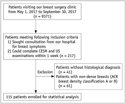
Fig. 1 Flow chart of patient inclusion and exclusion criteria. ACR = American College of Radiology, CESM = contrast-enhanced spectral mammography, US = ultrasonography
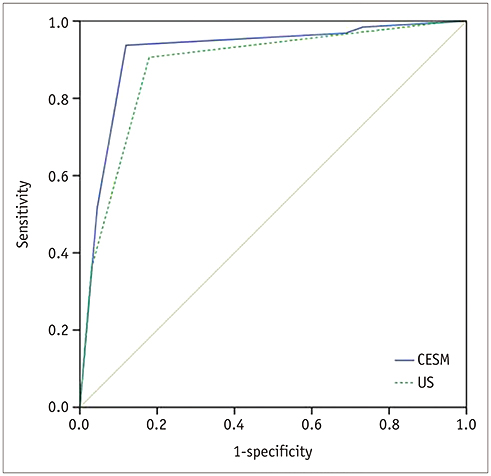
Fig. 2 Receiver operating characteristic curves to illustrate diagnostic performances of CESM and US. AUC for CESM in diagnosis of breast lesions was 0.917 (95% CI, 0.863–0.970). AUC for US in diagnosis of breast lesions was 0.884 (95% CI, 0.823–0.944). Difference of AUC between CESM and US was not significant (p = 0.225). AUC = area under curve, CI = confidence interval
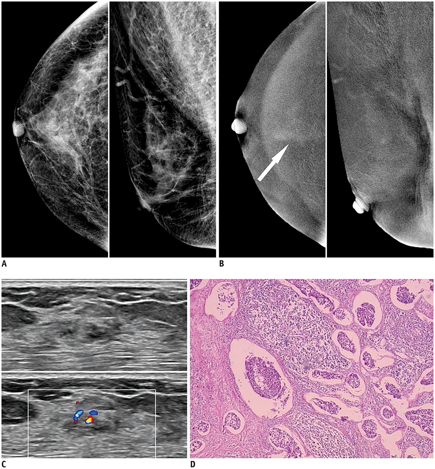
Fig. 3 59-year-old woman presented to her doctor with dull pain in right breast. A. Low-energy images of CESM showed heterogeneously dense breast with no abnormal findings. B. Recombined images of CESM showed slight cottony enhancement in craniocaudal view (arrow). C. US showed irregular hypoechoic mass with significant internal blood flow. D. Histological result from surgical specimen: invasive ductal carcinoma, no special type, grade II; nuclear grade II; tumor size: 10 × 5 mm (hematoxylin-eosin staining, original magnification × 40).
Reference
-
1. Løberg M, Lousdal ML, Bretthauer M, Kalager M. Benefits and harms of mammography screening. Breast Cancer Res. 2015; 17:63.
Article2. Ursin G, Ma H, Wu AH, Bernstein L, Salane M, Parisky YR, et al. Mammographic density and breast cancer in three ethnic groups. Cancer Epidemiol Biomarkers Prev. 2003; 12:332–338.3. Hooley RJ, Greenberg KL, Stackhouse RM, Geisel JL, Butler RS, Philpotts LE. Screening US in patients with mammographically dense breasts: initial experience with Connecticut Public Act 09-41. Radiology. 2012; 265:59–69.
Article4. Abdullah N, Mesurolle B, El-Khoury M, Kao E. Breast imaging reporting and data system lexicon for US: interobserver agreement for assessment of breast masses. Radiology. 2009; 252:665–672.
Article5. Hobbs MM, Taylor DB, Buzynski S, Peake RE. Contrast-enhanced spectral mammography (CESM) and contrast enhanced MRI (CEMRI): patient preferences and tolerance. J Med Imaging Radiat Oncol. 2015; 59:300–305.
Article6. Li L, Roth R, Germaine P, Ren S, Lee M, Hunter K, et al. Contrast-enhanced spectral mammography (CESM) versus breast magnetic resonance imaging (MRI): a retrospective comparison in 66 breast lesions. Diagn Interv Imaging. 2017; 98:113–123.
Article7. Phillips J, Miller MM, Mehta TS, Fein-Zachary V, Nathanson A, Hori W, et al. Contrast-enhanced spectral mammography (CESM) versus MRI in the high-risk screening setting: patient preferences and attitudes. Clin Imaging. 2017; 42:193–197.
Article8. Fallenberg EM, Dromain C, Diekmann F, Engelken F, Krohn M, Singh JM, et al. Contrast-enhanced spectral mammography versus MRI: initial results in the detection of breast cancer and assessment of tumour size. Eur Radiol. 2014; 24:256–264.
Article9. Fallenberg EM, Schmitzberger FF, Amer H, Ingold-Heppner B, Balleyguier C, Diekmann F, et al. Contrast-enhanced spectral mammography vs. mammography and MRI - clinical performance in a multi-reader evaluation. Eur Radiol. 2017; 27:2752–2764.
Article10. Lobbes MB, Lalji UC, Nelemans PJ, Houben I, Smidt ML, Heuts E, et al. The quality of tumor size assessment by contrast-enhanced spectral mammography and the benefit of additional breast MRI. J Cancer. 2015; 6:144–150.
Article11. Kolb TM, Lichy J, Newhouse JH. Comparison of the performance of screening mammography, physical examination, and breast US and evaluation of factors that influence them: an analysis of 27,825 patient evaluations. Radiology. 2002; 225:165–175.
Article12. Emaus MJ, Bakker MF, Peeters PH, Loo CE, Mann RM, de Jong MD, et al. MR Imaging as an additional screening modality for the detection of breast cancer in women aged 50-75 years with extremely dense breasts: the DENSE trial study design. Radiology. 2015; 277:527–537.
Article13. Cheung YC, Tsai HP, Lo YF, Ueng SH, Huang PC, Chen SC. Clinical utility of dual-energy contrast-enhanced spectral mammography for breast microcalcifications without associated mass: a preliminary analysis. Eur Radiol. 2016; 26:1082–1089.
Article14. Lobbes MB, Lalji U, Houwers J, Nijssen EC, Nelemans PJ, van Roozendaal L, et al. Contrast-enhanced spectral mammography in patients referred from the breast cancer screening programme. Eur Radiol. 2014; 24:1668–1676.
Article15. Fallenberg EM, Dromain C, Diekmann F, Renz DM, Amer H, Ingold-Heppner B, et al. Contrast-enhanced spectral mammography: does mammography provide additional clinical benefits or can some radiation exposure be avoided? Breast Cancer Res Treat. 2014; 146:371–381.
Article16. Ohuchi N, Suzuki A, Sobue T, Kawai M, Yamamoto S, Zheng YF, et al. Sensitivity and specificity of mammography and adjunctive ultrasonography to screen for breast cancer in the Japan Strategic Anti-cancer Randomized Trial (J-START): a randomised controlled trial. Lancet. 2016; 387:341–348.
Article17. Luczyńska E, Heinze S, Adamczyk A, Rys J, Mitus JW, Hendrick E. Comparison of the mammography, contrast-enhanced spectral mammography and ultrasonography in a group of 116 patients. Anticancer Res. 2016; 36:4359–4366.18. Klang E, Krosser A, Amitai MM, Sorin V, Halshtok Neiman O, Shalmon A, et al. Utility of routine use of breast ultrasound following contrast-enhanced spectral mammography. Clin Radiol. 2018; 73:908.e11–908.e16.
Article19. Taylor D, O'Hanlon S, Latham B. False-negative contrast-enhanced spectral mammography: use of more than one imaging modality and application of the triple test avoids misdiagnosis. BMJ Case Rep. 2017; 2017:bcr2016218556.
Article20. Dromain C, Balleyguier C, Muller S, Mathieu MC, Rochard F, Opolon P, et al. Evaluation of tumor angiogenesis of breast carcinoma using contrast-enhanced digital mammography. AJR Am J Roentgenol. 2006; 187:W528–W537.
Article21. Luczynska E, Niemiec J, Ambicka A, Adamczyk A, Walasek T, Rys´ J, et al. Correlation between blood and lymphatic vessel density and results of contrast-enhanced spectral mammography. Pol J Pathol. 2015; 66:310–322.
Article
- Full Text Links
-
- Actions
-
Cited
- CITED
-
- Close
- Share
-
- Similar articles
-
- Automated Breast Ultrasound Screening for Dense Breasts
- Medical auditing of whole-breast screening ultrasonography
- Equity in breast cancer screening for Asian women with dense breasts through ultrasonography: lessons learned from Japanese mammography screening and the J-START trial
- Performance of Digital Mammography-Based Artificial Intelligence Computer-Aided Diagnosis on Synthetic Mammography From Digital Breast Tomosynthesis
- Distribution of dense breasts using screening mammography in Korean women: a retrospective observational study
