Rapid Imaging: Recent Advances in Abdominal MRI for Reducing Acquisition Time and Its Clinical Applications
- Affiliations
-
- 1Department of Radiology, Seoul National University Hospital, Seoul National University College of Medicine, Seoul, Korea. jhjhry@gmail.com
- 2MR Application Predevelopment, Siemens Healthcare GmbH, Erlangen, Germany.
- 3MR Clinical Science, Philips Healthcare, Best, The Netherlands.
- 4Institute of Radiation Medicine, Seoul National University Medical Research Center, Seoul, Korea.
- KMID: 2471673
- DOI: http://doi.org/10.3348/kjr.2018.0931
Abstract
- Magnetic resonance imaging (MRI) plays an important role in abdominal imaging. The high contrast resolution offered by MRI provides better lesion detection and its capacity to provide multiparametric images facilitates lesion characterization more effectively than computed tomography. However, the relatively long acquisition time of MRI often detrimentally affects the image quality and limits its accessibility. Recent developments have addressed these drawbacks. Specifically, multiphasic acquisition of contrast-enhanced MRI, free-breathing dynamic MRI using compressed sensing technique, simultaneous multi-slice acquisition for diffusion-weighted imaging, and breath-hold three-dimensional magnetic resonance cholangiopancreatography are recent notable advances in this field. This review explores the aforementioned state-of-the-art techniques by focusing on their clinical applications and potential benefits, as well as their likely future direction.
Keyword
Figure
-
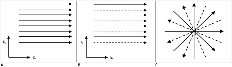
Fig. 1 Different k-space sampling schemes.Cartesian acquisition with fully sampled k-space (A), uniformly undersampled with acceleration factor of 2 (B), or non-Cartesian acquisition with skipping spokes in radial acquisition (C). Solid and dashed lines refer to acquired and skipped k-space data, respectively. Kx = frequency encoding direction, Ky = phase encoding direction
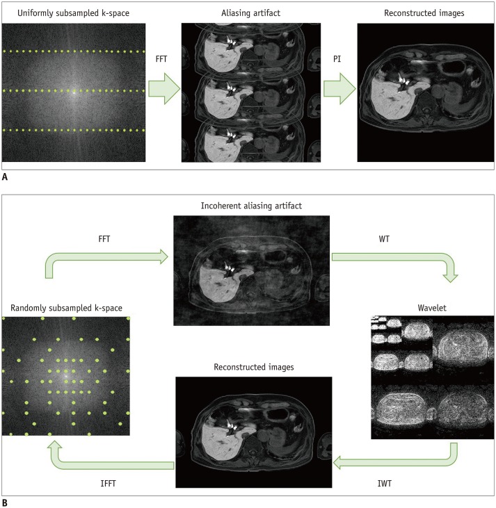
Fig. 2 Graphical representation of principles of parallel imaging and compressed sensing.In parallel imaging (A), uniform subsampling gives typical aliasing artifacts. Parallel imaging reconstruction makes it possible to achieve alias-free image. For SENSE, parallel imaging reconstruction is done in image space, whereas de-aliasing is already done before FFT in generalized autocalibrating partially parallel acquisitions (GRAPPA). In compressed sensing (B), variable-density, pseudo-random subsampling produces incoherent noise-like aliasing artifact after FFT. Sparsity (in this case, wavelet) transform allows setting of sparsity constraints. Image is obtained after IWT into image domain. IFFT back to k-space allows data consistency checking. After several iterations, final image is delivered with optimal balance between data consistency and sparsity constraints. FFT = Fourier transform, IFFT = inverse Fourier transform, IWT = inverse transform, PI = parallel imaging, SENSE = sensitivity encoding, WT = wavelet transform
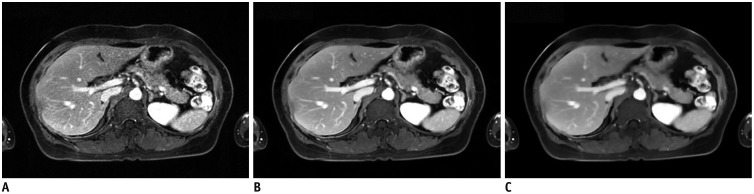
Fig. 3 Effect of regularization parameters.Same dataset was reconstructed using no regularization parameter (A), suggested regularization parameter (B), and 10-fold higher regularization parameter than that suggested (C). Images show different imaging textures and signal-to-noise ratios, according to regularization parameters.
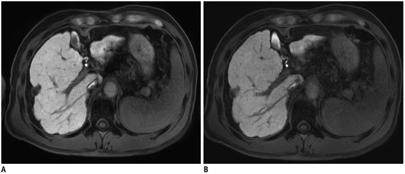
Fig. 4 Hepatobiliary phase of gadoxetic acid-enhanced MRI in 69-year-old man.A. First image was obtained with parallel imaging alone (SENSE) with acceleration factor of 2.8. B. Next image was acquired using compressed sensing and SENSE with acceleration factor of 7.17. Although both images show comparable image quality and spatial resolution (reconstruction voxel size 0.99 × 0.99 × 3 mm), image acquisition time was 15 seconds in (A) and 6 seconds in (B).
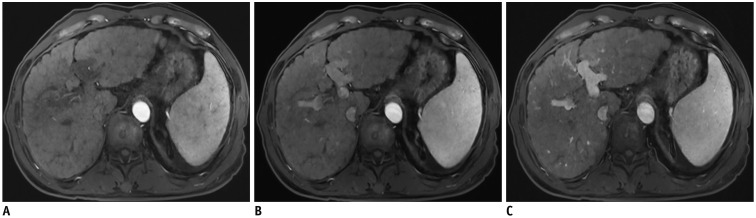
Fig. 5 Multi-arterial phase of gadoxetic acid-enhanced MRI in 63-year-old man obtained using compressed sensing and parallel imaging.First (A), second (B), and third (C) arterial phases clearly captured different timings of contrast-enhancement of liver, with sufficient spatial resolution (reconstruction voxel size of 0.98 × 1.41 × 3 mm), without noticeable temporal blurring in single breath-hold. Temporal resolution of each phase was 5.3 seconds.
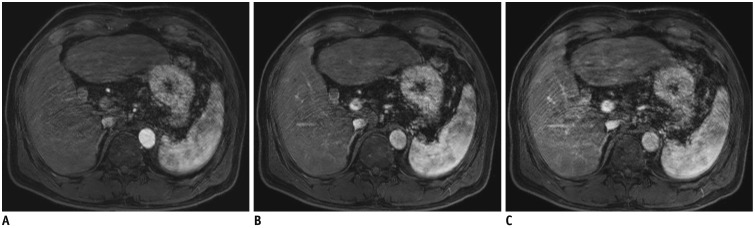
Fig. 6 Multi-arterial phase of gadoxetic acid-enhanced MRI in 66-year-old man obtained using view-sharing technique.All three arterial phases (A–C) show persistent motion artifacts, which decrease image quality.
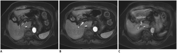
Fig. 7 Multi-arterial phase of gadoxetic acid-enhanced MRI in 88-year-old woman obtained using compressed sensing and parallel imaging.Even though patient failed to hold her breath during scan, first (A) and second (B) phases were saved because motion artifact was limited to last phase (C).
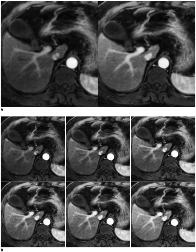
Fig. 8 Flexible temporal resolution of GRASP imaging of liver MRI in 61-year-old man.Images with temporal resolution of 13.3 seconds (A) and 3.3 seconds (B) were retrospectively reconstructed from single free-breathing examination. GRASP = golden-angle radial sparse parallel
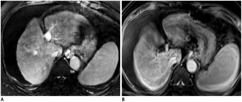
Fig. 9 T1-weighted images of gadoxetic acid-enhanced liver MRI in 56-year-old woman with limited breath-holding capacity.Motion artifacts are significantly less in free-breathing, motion-resolved reconstructed images (extra-dimension-VIBE, A) than in subsequent breath-hold 3D GRE transitional phase images (breath-hold VIBE, B). GRE = gradient-echo, VIBE = volumetric interpolated breath-hold examination, 3D = three-dimensional
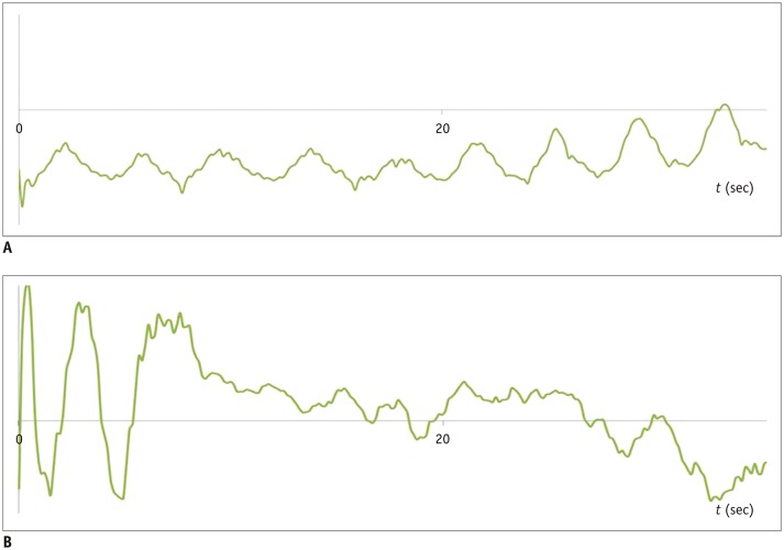
Fig. 10 Self-gated signals extracted from k-space in GRASP imaging sequence.Regular breathing pattern (A) and irregular breathing pattern (B) are seen.
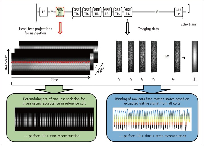
Fig. 11 Fast fat-saturated T1-weighted imaging acquires imaging data in form of echo trains following fat-suppression pulse.For free-breathing acquisitions, additional GRE with same excitation but with selectable readout direction can be inserted (top). Consequently, head-feet projections for each coil element can be obtained along with imaging data (middle). This can either be used for gated reconstruction that only utilizes specified fraction of data with smallest variation (bottom left) or for extracting gating signal to assign each echo train to motion state, followed by motion-resolved reconstruction (bottom right). FS = fat-suppressed, SI = signal intensity, TR = repetition time
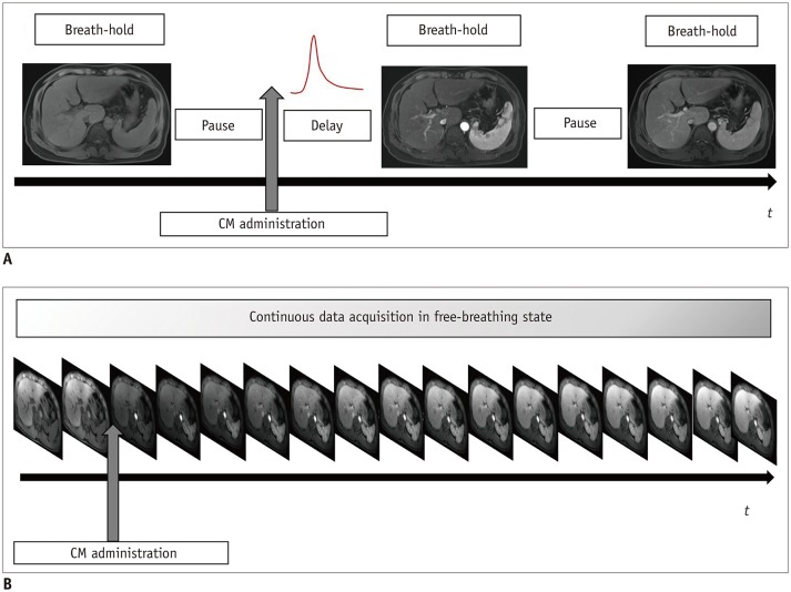
Fig. 12 Shift of acquisition scheme in contrast-enhanced abdominal MRI.Current protocol of dynamic sequence (A) includes several pauses and instances of breath-holding. In dynamic sequences using compressed sensing VIBE or GRASP, continuous data acquisition is possible (B) without breath-holding, because images are retrospectively reconstructed including motion correction. CM = contrast media
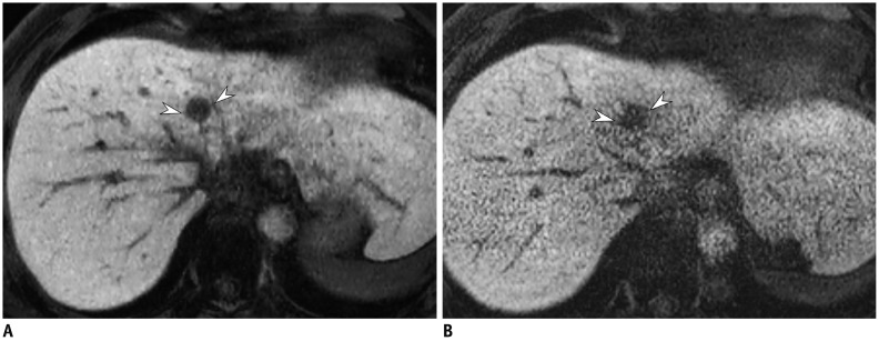
Fig. 13 Hepatobiliary phase of gadoxetic acid-enhanced liver MRI in 51-year-old man.Image obtained with compressed sensing and parallel imaging (A) shows less image noise and better overall image quality than that obtained with parallel imaging only (B). Treated hepatocellular carcinoma (arrowheads) is more visible in image obtained by using both compressed sensing and parallel imaging, than in that obtained with parallel imaging alone. Acquisition time is 15 seconds for both images and spatial resolution is same (reconstruction voxel size: 0.98 × 0.98 × 1.5 mm).
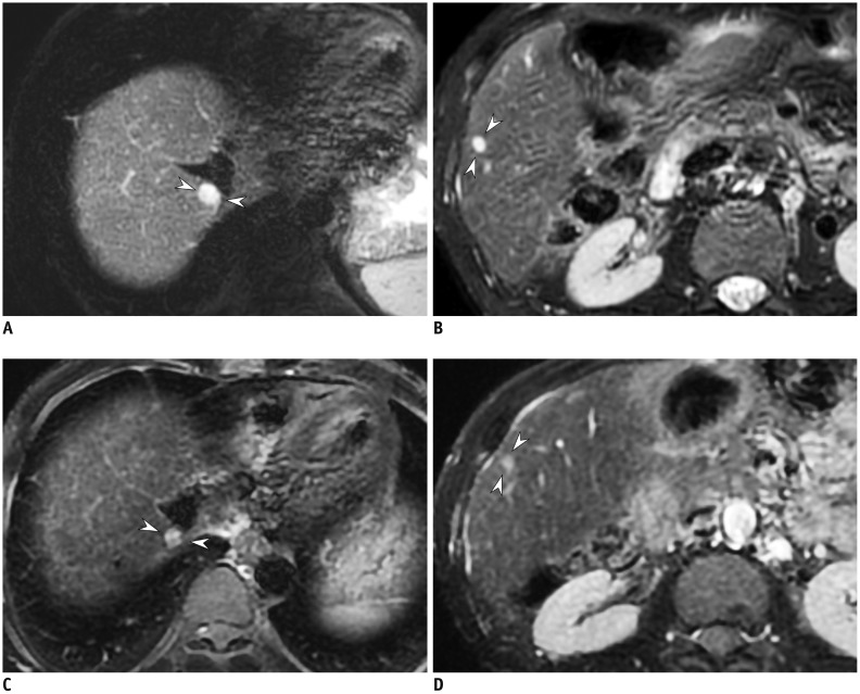
Fig. 14 T2-weighted image of 49-year-old man with hemangiomas.2D T2-weighted images using compressed sensing and parallel imaging with 4-mm slice thickness and 4-mm gap (A, B) provide better conspicuity of small hemangiomas (arrowheads) than that obtained in 2D T2-weighted images with 8-mm slice thickness and 8-mm gap (C, D). TR/TE were 3240/80 ms (A, B) and 2050/83.6 ms (C, D), respectively. TE = echo time, 2D = two-dimensional
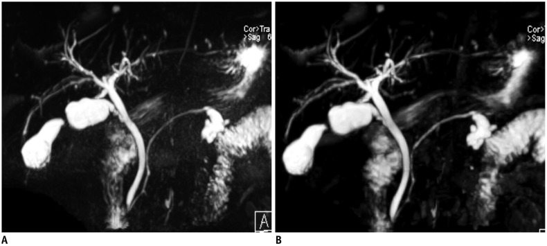
Fig. 15 Respiratory-triggered 3D MRCP in 67-year-old man.Conventional 3D MRCP (A) and compressed sensing 3D MRCP (B) show comparable image quality, with acquisition times of 5 minutes 35 seconds and 2 minutes 4 seconds, respectively. TR/TE was 4172/702 ms for conventional 3D MRCP (A) and 3861/725 ms for compressed sensing 3D MRCP (B). MRCP = magnetic resonance cholangiopancreatography
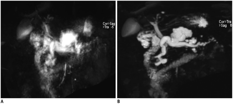
Fig. 16 3D MRCP using compressed sensing in 68-year-old woman.Conventional respiratory-triggered 3D MRCP using parallel imaging (A) shows substantial motion artifacts due to irregular breathing patterns, whereas breath-hold 3D MRCP using compressed sensing and parallel imaging (B) shows acceptable image quality. TR/TE was 4421/699 ms for conventional respiratory-triggered 3D MRCP (A) and 1700/674 ms for breath-hold 3D MRCP (B).
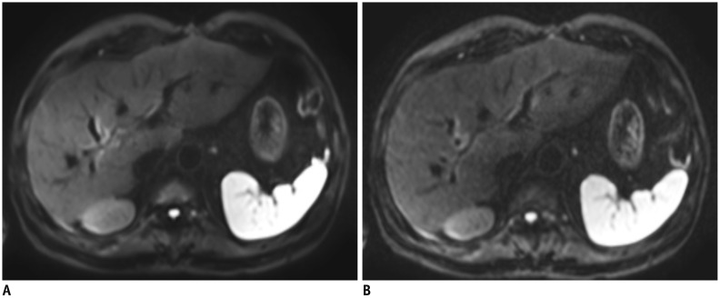
Fig. 17 Respiratory-triggered DWI using b-value of 800 s/mm2 in 64-year-old man.Images of conventional DWI (A) and SMS-DWI (B) show comparable image quality, but scan time was significantly shorter in SMS-DWI than in conventional DWI. TR/TE were 2100/60 ms for conventional DWI (A) and 2200/62 ms for SMS-DWI (B). Field of view (400 × 320 mm2) and matrix (150 × 120) were identical. DWI = diffusion-weighted imaging, SMS = simultaneous multi-slice
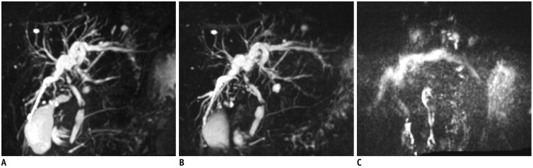
Fig. 18 3D MRCP in 61-year-old woman with limited breath-holding capability.Breath-hold 3D MRCP using gradient and spin-echo (A) shows comparable image quality to that of breath-hold 3D MRCP using compressed sensing (B), and better image quality than that of respiratory-triggered 3D MRCP (C).
Reference
-
1. Bashir MR, Castelli P, Davenport MS, Larson D, Marin D, Hussain HK, et al. Respiratory motion artifact affecting hepatic arterial phase MR imaging with gadoxetate disodium is more common in patients with a prior episode of arterial phase motion associated with gadoxetate disodium. Radiology. 2015; 274:141–148. PMID: 25162310.
Article2. Pietryga JA, Burke LM, Marin D, Jaffe TA, Bashir MR. Respiratory motion artifact affecting hepatic arterial phase imaging with gadoxetate disodium: examination recovery with a multiple arterial phase acquisition. Radiology. 2014; 271:426–434. PMID: 24475864.
Article3. Griswold MA, Jakob PM, Heidemann RM, Nittka M, Jellus V, Wang J, et al. Generalized autocalibrating partially parallel acquisitions (GRAPPA). Magn Reson Med. 2002; 47:1202–1210. PMID: 12111967.
Article4. Pruessmann KP, Weiger M, Scheidegger MB, Boesiger P. SENSE: sensitivity encoding for fast MRI. Magn Reson Med. 1999; 42:952–962. PMID: 10542355.
Article5. Hoogeveen RM, von Falkenhausen M, Gieseke J. Fast dynamic, high resolution contrast-enhanced MR angiography with CENTRA keyhole and SENSE. Proceedings of the 12th scientific meeting and exhibition of the International Society for Magnetic Resonance in Medicine. Kyoto, Japan: 2004. 5. p. 15–21.6. Kazmierczak PM, Theisen D, Thierfelder KM, Sommer WH, Reiser MF, Notohamiprodjo M, et al. Improved detection of hypervascular liver lesions with CAIPIRINHA-Dixon-TWIST-volume-interpolated breath-hold examination. Invest Radiol. 2015; 50:153–160. PMID: 25478742.
Article7. van Vaals JJ, Brummer ME, Dixon WT, Tuithof HH, Engels H, Nelson RC, et al. “Keyhole” method for accelerating imaging of contrast agent uptake. J Magn Reson Imaging. 1993; 3:671–675. PMID: 8347963.
Article8. Willinek WA, Hadizadeh DR, von Falkenhausen M, Urbach H, Hoogeveen R, Schild HH, et al. 4D time-resolved MR angiography with keyhole (4D-TRAK): more than 60 times accelerated MRA using a combination of CENTRA, keyhole, and SENSE at 3.0T. J Magn Reson Imaging. 2008; 27:1455–1460. PMID: 18504736.
Article9. Yoon JH, Lee JM, Yu MH, Kim EJ, Han JK. Triple arterial phase MR imaging with gadoxetic acid using a combination of contrast enhanced time robust angiography, keyhole, and viewsharing techniques and two-dimensional parallel imaging in comparison with conventional single arterial phase. Korean J Radiol. 2016; 17:522–532. PMID: 27390543.
Article10. Hope TA, Saranathan M, Petkovska I, Hargreaves BA, Herfkens RJ, Vasanawala SS. Improvement of gadoxetate arterial phase capture with a high spatio-temporal resolution multiphase three-dimensional SPGR-Dixon sequence. J Magn Reson Imaging. 2013; 38:938–945. PMID: 23371926.
Article11. Saranathan M, Rettmann DW, Hargreaves BA, Clarke SE, Vasanawala SS. DIfferential subsampling with Cartesian ordering (DISCO): a high spatio-temporal resolution Dixon imaging sequence for multiphasic contrast enhanced abdominal imaging. J Magn Reson Imaging. 2012; 35:1484–1492. PMID: 22334505.
Article12. Donoho DL. Compressed sensing. IEEE T Inform Theory. 2006; 52:1289–1306.
Article13. Lustig M, Donoho D, Pauly JM. Sparse MRI: the application of compressed sensing for rapid MR imaging. Magn Reson Med. 2007; 58:1182–1195. PMID: 17969013.
Article14. Lustig M, Donoho DL, Santos JM, Pauly JM. Compressed sensing MRI. IEEE Signal Process Mag. 2008; 25:72–82.
Article15. Peeters JM, Fuderer M. SENSE with improved tolerance to inaccuracies in coil sensitivity maps. Magn Reson Med. 2013; 69:1665–1669. PMID: 22847672.
Article16. Breuer FA, Blaimer M, Mueller MF, Seiberlich N, Heidemann RM, Griswold MA, et al. Controlled aliasing in volumetric parallel imaging (2D CAIPIRINHA). Magn Reson Med. 2006; 55:549–556. PMID: 16408271.
Article17. Yu MH, Lee JM, Yoon JH, Kiefer B, Han JK, Choi BI. Clinical application of controlled aliasing in parallel imaging results in a higher acceleration (CAIPIRINHA)-volumetric interpolated breathhold (VIBE) sequence for gadoxetic acid-enhanced liver MR imaging. J Magn Reson Imaging. 2013; 38:1020–1026. PMID: 23559147.
Article18. Feng L, Benkert T, Block KT, Sodickson DK, Otazo R, Chandarana H. Compressed sensing for body MRI. J Magn Reson Imaging. 2017; 45:966–987. PMID: 27981664.
Article19. Cook RL. Stochastic sampling in computer graphics. ACM TOG. 1986; 5:51–72.
Article20. Kim D, Dyvorne HA, Otazo R, Feng L, Sodickson DK, Lee VS. Accelerated phase-contrast cine MRI using k-t SPARSE-SENSE. Magn Reson Med. 2012; 67:1054–1064. PMID: 22083998.21. Paul J, Wundrak S, Bernhardt P, Rottbauer W, Neumann H, Rasche V. Self-gated tissue phase mapping using golden angle radial sparse SENSE. Magn Reson Med. 2016; 75:789–800. PMID: 25761576.
Article22. Murphy M, Alley M, Demmel J, Keutzer K, Vasanawala S, Lustig M. Fast ℓ1-SPIRiT compressed sensing parallel imaging MRI: scalable parallel implementation and clinically feasible runtime. IEEE Trans Med Imaging. 2012; 31:1250–1262. PMID: 22345529.23. Chandarana H, Feng L, Block TK, Rosenkrantz AB, Lim RP, Babb JS, et al. Free-breathing contrast-enhanced multiphase MRI of the liver using a combination of compressed sensing, parallel imaging, and golden-angle radial sampling. Invest Radiol. 2013; 48:10–16. PMID: 23192165.
Article24. Feng L, Grimm R, Block KT, Chandarana H, Kim S, Xu J, et al. Golden-angle radial sparse parallel MRI: combination of compressed sensing, parallel imaging, and golden-angle radial sampling for fast and flexible dynamic volumetric MRI. Magn Reson Med. 2014; 72:707–717. PMID: 24142845.
Article25. Otazo R, Kim D, Axel L, Sodickson DK. Combination of compressed sensing and parallel imaging for highly accelerated first-pass cardiac perfusion MRI. Magn Reson Med. 2010; 64:767–776. PMID: 20535813.
Article26. Davenport MS, Viglianti BL, Al-Hawary MM, Caoili EM, Kaza RK, Liu PS, et al. Comparison of acute transient dyspnea after intravenous administration of gadoxetate disodium and gadobenate dimeglumine: effect on arterial phase image quality. Radiology. 2013; 266:452–461. PMID: 23192781.
Article27. Motosugi U, Bannas P, Bookwalter CA, Sano K, Reeder SB. An investigation of transient severe motion related to gadoxetic acid-enhanced MR imaging. Radiology. 2016; 279:93–102. PMID: 26473642.
Article28. Park YS, Lee CH, Kim JW, Lee YS, Paek M, Kim KA. Application of high-speed T1 sequences for high-quality hepatic arterial phase magnetic resonance imaging: intraindividual comparison of single and multiple arterial phases. Invest Radiol. 2017; 52:605–611. PMID: 28441159.29. Nam JG, Lee JM, Lee SM, Kang HJ, Lee ES, Hur BY, et al. High acceleration three-dimensional T1-weighted dual echo Dixon hepatobiliary phase imaging using compressed sensing-sensitivity encoding: comparison of image quality and solid lesion detectability with the standard T1-weighted sequence. Korean J Radiol. 2019; 20:438–448. PMID: 30799575.
Article30. Vasanawala S, Murphy M, Alley M, Lai P, Keutzer K, Pauly J, et al. Practical parallel imaging compressed sensing MRI: summary of two years of experience in accelerating body MRI of pediatric patients. Proc IEEE Int Symp Biomed Imaging. 2011; 2011:1039–1043. PMID: 24443670.
Article31. Yoon JH, Yu MH, Chang W, Park JY, Nickel MD, Son Y, et al. Clinical feasibility of free-breathing dynamic T1-weighted imaging with gadoxetic acid-enhanced liver magnetic resonance imaging using a combination of variable density sampling and compressed sensing. Invest Radiol. 2017; 52:596–604. PMID: 28492418.
Article32. Kaltenbach B, Bucher AM, Wichmann JL, Nickel D, Polkowski C, Hammerstingl R, et al. Dynamic liver magnetic resonance imaging in free-breathing: feasibility of a Cartesian T1-weighted acquisition technique with compressed sensing and additional self-navigation signal for hard-gated and motion-resolved reconstruction. Invest Radiol. 2017; 52:708–714. PMID: 28622249.33. Levine E, Daniel B, Vasanawala S, Hargreaves B, Saranathan M. 3D Cartesian MRI with compressed sensing and variable view sharing using complementary poisson-disc sampling. Magn Reson Med. 2017; 77:1774–1785. PMID: 27097596.
Article34. Block KT, Chandarana H, Milla S, Bruno M, Mulholland T, Fatterpekar G, et al. Towards routine clinical use of radial stack-of-stars 3D gradient-echo sequences for reducing motion sensitivity. J Korean Soc Magn Reson Med. 2014; 18:87–106.
Article35. Winkelmann S, Schaeffter T, Koehler T, Eggers H, Doessel O. An optimal radial profile order based on the golden ratio for time-resolved MRI. IEEE Trans Med Imaging. 2007; 26:68–76. PMID: 17243585.
Article36. Chandarana H, Block KT, Winfeld MJ, Lala SV, Mazori D, Giuffrida E, et al. Free-breathing contrast-enhanced T1-weighted gradient-echo imaging with radial k-space sampling for paediatric abdominopelvic MRI. Eur Radiol. 2014; 24:320–326. PMID: 24220754.
Article37. Chandarana H, Block TK, Rosenkrantz AB, Lim RP, Kim D, Mossa DJ, et al. Free-breathing radial 3D fat-suppressed T1-weighted gradient echo sequence: a viable alternative for contrast-enhanced liver imaging in patients unable to suspend respiration. Invest Radiol. 2011; 46:648–653. PMID: 21577119.38. Chandarana H, Block TK, Ream J, Mikheev A, Sigal SH, Otazo R, et al. Estimating liver perfusion from free-breathing continuously acquired dynamic gadolinium-ethoxybenzyl-diethylenetriamine pentaacetic acid-enhanced acquisition with compressed sensing reconstruction. Invest Radiol. 2015; 50:88–94. PMID: 25333309.
Article39. Yoon JH, Lee JM, Yu MH, Hur BY, Grimm R, Block KT, et al. Evaluation of transient motion during gadoxetic acid-enhanced multiphasic liver magnetic resonance imaging using free-breathing golden-angle radial sparse parallel magnetic resonance imaging. Invest Radiol. 2018; 53:52–61. PMID: 28902723.
Article40. Yoon JH, Lee JM, Lee ES, Baek J, Lee S, Iwadate Y, et al. Navigated three-dimensional T1-weighted gradient-echo sequence for gadoxetic acid liver magnetic resonance imaging in patients with limited breath-holding capacity. Abdom Imaging. 2015; 40:278–288. PMID: 25112454.
Article41. Lee CK, Seo N, Kim B, Huh J, Kim JK, Lee SS, et al. The effects of breathing motion on DCE-MRI images: phantom studies simulating respiratory motion to compare CAIPIRINHA-VIBE, radial-VIBE, and conventional VIBE. Korean J Radiol. 2017; 18:289–298. PMID: 28246509.
Article42. Chandarana H, Feng L, Ream J, Wang A, Babb JS, Block KT, et al. Respiratory motion-resolved compressed sensing reconstruction of free-breathing radial acquisition for dynamic liver magnetic resonance imaging. Invest Radiol. 2015; 50:749–756. PMID: 26146869.
Article43. Zhang T, Cheng JY, Potnick AG, Barth RA, Alley MT, Uecker M, et al. Fast pediatric 3D free-breathing abdominal dynamic contrast enhanced MRI with high spatiotemporal resolution. J Magn Reson Imaging. 2015; 41:460–473. PMID: 24375859.
Article44. Kim YC. Advanced methods in dynamic contrast enhanced arterial phase imaging of the liver. Investig Magn Reson Imaging. 2019; 23:1–16.
Article45. Yoon JH, Lee JM, Yu MH, Kim EJ, Han JK, Choi BI. High-resolution T1-weighted gradient echo imaging for liver MRI using parallel imaging at high-acceleration factors. Abdom Imaging. 2014; 39:711–721. PMID: 24557640.
Article46. Yoon JH, Lee JM, Yu MH, Kim EJ, Han JK, Choi BI. Fat-suppressed, three-dimensional T1-weighted imaging using high-acceleration parallel acquisition and a dual-echo Dixon technique for gadoxetic acid-enhanced liver MRI at 3 T. Acta Radiol. 2015; 56:1454–1462. PMID: 25480475.47. Sodickson A, Mortele KJ, Barish MA, Zou KH, Thibodeau S, Tempany CM. Three-dimensional fast-recovery fast spin-echo MRCP: comparison with two-dimensional single-shot fast spin-echo techniques. Radiology. 2006; 238:549–559. PMID: 16436816.
Article48. Yoon LS, Catalano OA, Fritz S, Ferrone CR, Hahn PF, Sahani DV. Another dimension in magnetic resonance cholangiopancreatography: comparison of 2- and 3-dimensional magnetic resonance cholangiopancreatography for the evaluation of intraductal papillary mucinous neoplasm of the pancreas. J Comput Assist Tomogr. 2009; 33:363–368. PMID: 19478628.49. Kim JH, Hong SS, Eun HW, Han JK, Choi BI. Clinical usefulness of free-breathing navigator-triggered 3D MRCP in non-cooperative patients: comparison with conventional breath-hold 2D MRCP. Eur J Radiol. 2012; 81:e513–e518. PMID: 21700409.
Article50. Yoon JH, Lee SM, Kang HJ, Weiland E, Raithel E, Son Y, et al. Clinical feasibility of 3-dimensional magnetic resonance cholangiopancreatography using compressed sensing: comparison of image quality and diagnostic performance. Invest Radiol. 2017; 52:612–619. PMID: 28448309.51. Zhu L, Wu X, Sun Z, Jin Z, Weiland E, Raithel E, et al. Compressed-sensing accelerated 3-dimensional magnetic resonance cholangiopancreatography: application in suspected pancreatic diseases. Invest Radiol. 2018; 53:150–157. PMID: 28976478.52. Chandarana H, Doshi AM, Shanbhogue A, Babb JS, Bruno MT, Zhao T, et al. Three-dimensional MR cholangiopancreatography in a breath hold with sparsity-based reconstruction of highly undersampled data. Radiology. 2016; 280:585–594. PMID: 26982678.
Article53. Furlan A, Bayram E, Thangasamy S, Barley D, Dasyam A. Application of compressed sensing to 3D magnetic resonance cholangiopancreatography for the evaluation of pancreatic cystic lesions. Magn Reson Imaging. 2018; 52:131–136. PMID: 29859947.
Article54. Seo N, Park MS, Han K, Kim D, King KF, Choi JY, et al. Feasibility of 3D navigator-triggered magnetic resonance cholangiopancreatography with combined parallel imaging and compressed sensing reconstruction at 3T. J Magn Reson Imaging. 2017; 46:1289–1297. PMID: 28295827.
Article55. Wielopolski PA, Gaa J, Wielopolski DR, Oudkerk M. Breath-hold MR cholangiopancreatography with three-dimensional, segmented, echo-planar imaging and volume rendering. Radiology. 1999; 210:247–252. PMID: 9885616.
Article56. Zhu L, Xue H, Sun Z, Qian T, Weiland E, Kuehn B, et al. Modified breath-hold compressed-sensing 3D MR cholangiopancreatography with a small field-of-view and high resolution acquisition: clinical feasibility in biliary and pancreatic disorders. J Magn Reson Imaging. 2018; 48:1389–1399. PMID: 29656611.
Article57. Barth M, Breuer F, Koopmans PJ, Norris DG, Poser BA. Simultaneous multislice (SMS) imaging techniques. Magn Reson Med. 2016; 75:63–81. PMID: 26308571.
Article58. Müller S. Multifrequency selective rf pulses for multislice MR imaging. Magn Reson Med. 1988; 6:364–371. PMID: 3362070.
Article59. Setsompop K, Cohen-Adad J, Gagoski BA, Raij T, Yendiki A, Keil B, et al. Improving diffusion MRI using simultaneous multi-slice echo planar imaging. Neuroimage. 2012; 63:569–580. PMID: 22732564.
Article60. Norris DG, Boyacioğlu R, Schulz J, Barth M, Koopmans PJ. Application of PINS radiofrequency pulses to reduce power deposition in RARE/turbo spin echo imaging of the human head. Magn Reson Med. 2014; 71:44–49. PMID: 24150771.
Article61. Breuer FA, Blaimer M, Heidemann RM, Mueller MF, Griswold MA, Jakob PM. Controlled aliasing in parallel imaging results in higher acceleration (CAIPIRINHA) for multi-slice imaging. Magn Reson Med. 2005; 53:684–691. PMID: 15723404.
Article62. Larkman DJ, Hajnal JV, Herlihy AH, Coutts GA, Young IR, Ehnholm G. Use of multicoil arrays for separation of signal from multiple slices simultaneously excited. J Magn Reson Imaging. 2001; 13:313–317. PMID: 11169840.
Article63. Taron J, Martirosian P, Erb M, Kuestner T, Schwenzer NF, Schmidt H, et al. Simultaneous multislice diffusion-weighted MRI of the liver: analysis of different breathing schemes in comparison to standard sequences. J Magn Reson Imaging. 2016; 44:865–879. PMID: 26919580.
Article64. Taron J, Martirosian P, Schwenzer NF, Erb M, Kuestner T, Weiß J, et al. Scan time minimization in hepatic diffusion-weighted imaging: evaluation of the simultaneous multislice acceleration technique with different acceleration factors and gradient preparation schemes. MAGMA. 2016; 29:739–749. PMID: 27038935.
Article65. Obele CC, Glielmi C, Ream J, Doshi A, Campbell N, Zhang HC, et al. Simultaneous multislice accelerated free-breathing diffusion-weighted imaging of the liver at 3T. Abdom Imaging. 2015; 40:2323–2330. PMID: 25985968.
Article66. Boss A, Barth B, Filli L, Kenkel D, Wurnig MC, Piccirelli M, et al. Simultaneous multi-slice echo planar diffusion weighted imaging of the liver and the pancreas: optimization of signal-to-noise ratio and acquisition time and application to intravoxel incoherent motion analysis. Eur J Radiol. 2016; 85:1948–1955. PMID: 27776645.
Article67. Taron J, Martirosian P, Kuestner T, Schwenzer NF, Othman A, Weiß J, et al. Scan time reduction in diffusion-weighted imaging of the pancreas using a simultaneous multislice technique with different acceleration factors: how fast can we go? Eur Radiol. 2018; 28:1504–1511. PMID: 29134353.
Article68. Oshio K, Feinberg DA. GRASE (gradient- and spin-echo) imaging: a novel fast MRI technique. Magn Reson Med. 1991; 20:344–349. PMID: 1775061.69. Feinberg DA, Oshio K. GRASE (gradient- and spin-echo) MR imaging: a new fast clinical imaging technique. Radiology. 1991; 181:597–602. PMID: 1924811.
Article
- Full Text Links
-
- Actions
-
Cited
- CITED
-
- Close
- Share
-
- Similar articles
-
- Diffusion-Weighted Magnetic Resonance Imaging of the Breast: Standardization of Image Acquisition and Interpretation
- Advanced Abdominal MRI Techniques and Problem-Solving Strategies
- Deep Learning Applications in Perfusion MRI: Recent Advances and Current Challenges
- High field strength magnetic resonance imaging of musculoskeletal diseases
- High field strength magnetic resonance imaging of cardiovascular diseases
