Three-dimensional observations of the incisive foramen on cone-beam computed tomography image analysis
- Affiliations
-
- 1Department of Periodontology, Daejeon Dental Hospital, Wonkwang University College of Dentistry, Daejeon, Korea. seongnyum@wonkwang.ac.kr
- 2Institute of Wonkwang Dental Research, Wonkwang University College of Dentistry, Iksan, Korea.
- KMID: 2470830
- DOI: http://doi.org/10.5051/jpis.2020.50.1.48
Abstract
- PURPOSE
The purpose of this study was to utilize cone-beam computed tomography (CBCT) image analysis to obtain anatomical information related to the morphology of the incisive foramen to provide useful data regarding implant placement and clinical procedures such as anesthesia.
METHODS
The study included 167 patients who underwent CBCT scans over 20 years. Three components were measured: 1) the anteroposterior and mediolateral diameter of the incisive foramen, 2) the horizontal bone thickness anterior to the incisive foramen, and 3) the vertical bone height coronal to the incisive foramen. All measurements were expressed as mean±standard deviation and were analyzed by a single examiner.
RESULTS
The anteroposterior diameter of the incisive foramen was wider than the mediolateral diameter (P<0.001). The diameter of the incisive foramen in patients in whom the central incisors were present was smaller than that in those in whom at least one central incisor was absent, but no statistically significant difference between the groups was observed. The horizontal bone thickness in the patients with central incisors was statistically significantly larger than that in the patients without at least one central incisor (P<0.001). The same pattern was observed with regard to vertical height, but that difference was not statistically significant.
CONCLUSIONS
The buccal bone thickness anterior to the incisive foramen was significantly decreased after central incisor loss. It is necessary to identify the morphology of the bone and the location of the incisive foramen via CBCT to avoid invasion of the incisive foramen and nasopalatine canal.
MeSH Terms
Figure
-
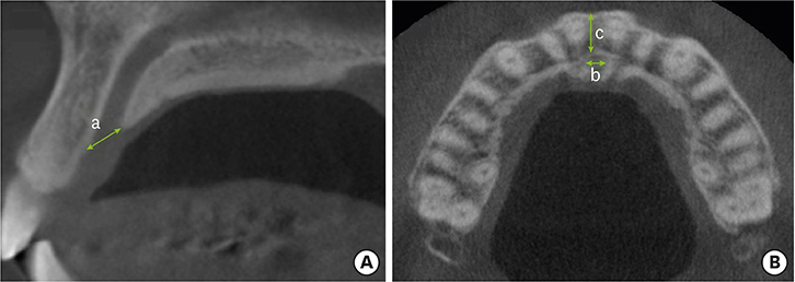
Figure 1 Representative cone-beam computed tomography image at the incisive foramen level, with linear measurements illustrating the anteroposterior (a) and mediolateral (b) diameters of the incisive foramen and the bone width of the buccal wall (c). (A) Sagittal view. (B) Axial view.
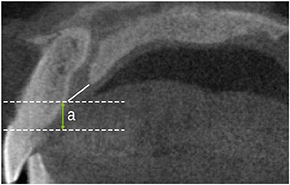
Figure 2 Representative cone-beam computed tomography image at the incisive foramen level, with linear measurements illustrating the height of the alveolar crest to the anterior border of the incisive foramen (a). A funnel-like nasopalatine canal shape is observed.
Cited by 1 articles
-
Quantitative cone-beam computed tomography evaluation of hard and soft tissue thicknesses in the midpalatal suture region to facilitate orthodontic mini-implant placement
Song-Hee Oh, Sae Rom Lee, Jin-Young Choi, Seong-Hun Kim, Eui-Hwan Hwang, Gerald Nelson
Korean J Orthod. 2021;51(4):260-269. doi: 10.4041/kjod.2021.51.4.260.
Reference
-
1. Mecall RA, Rosenfeld AL. Influence of residual ridge resorption patterns on implant fixture placement and tooth position. 1. Int J Periodontics Restorative Dent. 1991; 11:8–23.2. Drake RL, Vogl W, Mitchell AWM. Gray's anatomy for students. 2nd ed. Philadelphia (PA): Elsevier Health Sciences;2009.3. Sarandha DL. Textbook of complete denture prosthodontics. 1st ed. New Delhi: Jaypee Brothers Medical Publishers;2008.4. Glickman GN. Preparation for treatment. In : Cohen S, Burns RC, editors. Pathways of the pulp. St. Louis (MO): Mosby;2002. p. 77–109.5. Mraiwa N, Jacobs R, Van Cleynenbreugel J, Sanderink G, Schutyser F, Suetens P, et al. The nasopalatine canal revisited using 2D and 3D CT imaging. Dentomaxillofac Radiol. 2004; 33:396–402.
Article6. Song WC, Jo DI, Lee JY, Kim JN, Hur MS, Hu KS, et al. Microanatomy of the incisive canal using three-dimensional reconstruction of microCT images: an ex vivo study. Oral Surg Oral Med Oral Pathol Oral Radiol Endod. 2009; 108:583–590.
Article7. Keith DA. Phenomenon of mucous retention in the incisive canal. J Oral Surg. 1979; 37:832–834.8. Liang X, Jacobs R, Martens W, Hu Y, Adriaensens P, Quirynen M, et al. Macro- and micro-anatomical, histological and computed tomography scan characterization of the nasopalatine canal. J Clin Periodontol. 2009; 36:598–603.
Article9. Jacobs R, Lambrichts I, Liang X, Martens W, Mraiwa N, Adriaensens P, et al. Neurovascularization of the anterior jaw bones revisited using high-resolution magnetic resonance imaging. Oral Surg Oral Med Oral Pathol Oral Radiol Endod. 2007; 103:683–693.
Article10. Carlsson GE, Bergman B, Hedegård B. Changes in contour of the maxillary alveolar process under immediate dentures. A longitudinal clinical and X-ray cephalometric study covering 5 years. Acta Odontol Scand. 1967; 25:45–75.
Article11. Pietrokovski J. The bony residual ridge in man. J Prosthet Dent. 1975; 34:456–462.
Article12. Jahangiri L, Devlin H, Ting K, Nishimura I. Current perspectives in residual ridge remodeling and its clinical implications: a review. J Prosthet Dent. 1998; 80:224–237.
Article13. Asumi M, Yamaguchi T, Saito K, Kodama S, Miyazawa H, Matsui H, et al. Are serum cholesterol levels associated with silent brain infarcts? The Seiryo Clinic Study. Atherosclerosis. 2010; 210:674–677.
Article14. Filippi A, Pohl Y, Tekin U. Sensory disorders after separation of the nasopalatine nerve during removal of palatal displaced canines: prospective investigation. Br J Oral Maxillofac Surg. 1999; 37:134–136.
Article15. Edwards CB, Marshall SD, Qian F, Southard KA, Franciscus RG, Southard TE. Longitudinal study of facial skeletal growth completion in 3 dimensions. Am J Orthod Dentofacial Orthop. 2007; 132:762–768.
Article16. Fuentes R, Flores T, Navarro P, Salamanca C, Beltrán V, Borie E. Assessment of buccal bone thickness of aesthetic maxillary region: a cone-beam computed tomography study. J Periodontal Implant Sci. 2015; 45:162–168.
Article17. Hämmerle CH, Tarnow D. The etiology of hard- and soft-tissue deficiencies at dental implants: a narrative review. J Periodontol. 2018; 89 Suppl 1:S291–303.
Article18. Kraut RA, Boyden DK. Location of incisive canal in relation to central incisor implants. Implant Dent. 1998; 7:221–225.
Article19. Salemi F, Moghadam FA, Shakibai Z, Farhadian M. Three-dimensional assessment of the nasopalatine canal and the surrounding bone using cone-beam computed tomography. J Periodontol Implant Dent. 2015; 8:1–7.
Article20. Mardinger O, Namani-Sadan N, Chaushu G, Schwartz-Arad D. Morphologic changes of the nasopalatine canal related to dental implantation: a radiologic study in different degrees of absorbed maxillae. J Periodontol. 2008; 79:1659–1662.
Article21. Jia X, Hu W, Meng H. Relationship of central incisor implant placement to the ridge configuration anterior to the nasopalatine canal in dentate and partially edentulous individuals: a comparative study. PeerJ. 2015; 3:e1315.
Article22. Bornstein MM, Balsiger R, Sendi P, von Arx T. Morphology of the nasopalatine canal and dental implant surgery: a radiographic analysis of 100 consecutive patients using limited cone-beam computed tomography. Clin Oral Implants Res. 2011; 22:295–301.
Article23. Fernández-Alonso A, Suárez-Quintanilla JA, Rapado-González O, Suárez-Cunqueiro MM. Morphometric differences of nasopalatine canal based on 3D classifications: descriptive analysis on CBCT. Surg Radiol Anat. 2015; 37:825–833.
Article24. Tözüm TF, Güncü GN, Yıldırım YD, Yılmaz HG, Galindo-Moreno P, Velasco-Torres M, et al. Evaluation of maxillary incisive canal characteristics related to dental implant treatment with computerized tomography: a clinical multicenter study. J Periodontol. 2012; 83:337–343.
Article
- Full Text Links
-
- Actions
-
Cited
- CITED
-
- Close
- Share
-
- Similar articles
-
- Three-dimensional imaging modalities in endodontics
- A study of incisive canal using a cone beam computed tomography
- Anatomical structure of lingual foramen in cone beam computed tomography
- Foramen transversarium enlargement caused by vertebral artery tortuosity: Diagnosis with cone-beam computed tomography and magnetic resonance angiography
- Accessory mental foramen: A rare anatomical variation detected by cone-beam computed tomography
